Indonesian Journal of Chemistry, 2002, 2 (3), 131-134
Hidenari Inoue
131
N
N
N
N N
N N N
Me
Me Me
Me M
SPECTROSCOPIC AND PHYSICOCHEMICAL METHODS FOR STUDYING
THE INTERACTION OF METALLOPORPHYRIN WITH DNA
Hidenari Inoue
Department of Applied Chemistry, Keio University, 3-14-1 Hiyoshi, Kohoku-ku, Yokohama 223-8522 Japan. E-mail: [email protected]
INTRODUCTION
In recent years studies on the interaction of porphyrin with DNA have received much attention because of the importance in DNA-probing and photodynamic therapy of cancer. A variety of spectroscopic methods, e.g. NMR, ESR, Mössbauer, UV-visible absorption, circular dichroism (CD), magnetic circular dichroism (MCD), IR and Raman spectroscopy, have been employed for studying interactions between porphyrin and DNA. Of these spectroscopic methods, only a few instrumental analytical techniques applicable to an aqueous buffer solution of DNA have been particularly developed to investigate porphyrin-DNA interactions. On the other hand, a number of physicochemical methods, e.g. gel electrophoresis, melting temperature measurements and hydrodynamic methods such as viscosity and sedimentation measurements, have been also used for determining the binding modes of porphyrin to DNA. The present lecture will focus on the application of visible absorption, CD and MCD spectroscopy as well as melting temperature and viscosity measurements to studies of porphyrin-DNA interactions.
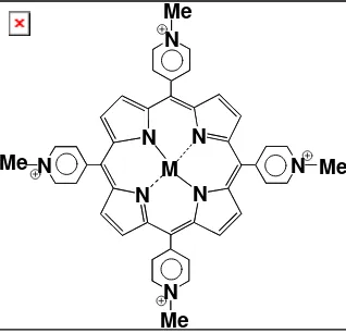
As model metalloporphyrins, we have chosen copper(II) and zinc(II) complexes of meso-tetrakis(N-methylpyridinium-4-yl)porphyrin
(H2TmpyP), meso-tetrakis(1,2-dimethylpyrazo-lium-4-yl)porphyrin (H2PzP) and 5,15-bis(1,3-dimethylimidazolium-2-yl)porphyrin (H2BDMImP). The last two cationic porphyrins have been recently synthesised and characterised in our laboratory[1-3]. The representative structure of these water-soluble metalloporphyrins is shown in Fig. 1. In the present work we have investigated the interaction of these cationic metalloporphyrins with calf thymus DNA and synthetic DNA by the above mentioned spectroscopic and physicochemical methods.
UV-VISIBLE ABSORPTION SPECTROSCOPY
For the purpose of preliminary studies of porphyrin-DNA interaction, so-called spectrophotometric titration of metalloporphyrin is usually performed in a buffer solution by DNA. This rather conventional analytical method provides a lot of information about interactions of metalloporphyrin with DNA. The tendency and magnitude of the peak shift and hypo- or hyper-chromisity in the Soret band is useful for assessing the binding mode and determining the binding constants.
Indonesian Journal of Chemistry, 2002, 2 (3), 131-134
Hidenari Inoue
132
For example, the interaction of meso-tetrakis(1,2-pyrazolium-4-yl)porphyrinato-copper(II) (CuPzP) with synthetic DNA, i.e. poly(dG-dC)2, induces a large spectral shift ( 11 nm) and significant hypochromism (35 %) in the Soret band. The magnitudes of the spectral perturbations are clear evidence for interculative binding. In another case, the spectrophotometric titration of meso-tetrakis(1,2-pyrazolium-4-yl)porphyrin (H2PzP) with CT-DNA was carried out to determine the binding constant of H2PzP to calf thymus DNA (CT-DNA). The binding constant determined by spectro-photometric titration is in the order of 106 M-1 for H2PzP and comparable to that of the corresponding metalloporphyrin. It has been revealed that H2PzP binds to B-form DNA with an affinity similar to that of the well-studied H2TMpyP system.
CIRCULAR DICHROISM (CD) SPECTROSCOPY
The circular dichroism (CD) spectra are indispensable not only for structural analysis of DNA but also for in vivo studies of porphyrin-DNA interaction. This instrumental technique has been developed mainly for structural analysis of chiral compounds and natural products, but it is also a powerful means for stereo analysis of porphyrin-DNA complexes. This instrumental analytical method makes a unique contribution to the stereo analysis, because so-called induced circular dichroism is observed upon coupling of electric dipole transition moments between base pair of DNA and porphyrin. For example, the adduct of
[ 5,15-bis(1,3-dimethylimidazolium-2-yl)-porphyrin-ato] copper(II) (CuBDMImP) to CT-DNA exhibits a negative signal at 396 nm and a positive signal at 413 nm in the Soret region of the circular dichroism (CD) spectrum.
An application of so-called “CD matrix method” to the observed induced CD spectra has revealed that CuBDMImP is intercalated into the 5’CG3’ step of CT-DNA. In the case of intercalation of CuBDMImP into the base pair, one of the positively charged N,N’-dimethylimidazolium rings is located in the minor groove near the phosphate backbone and the other is placed in the major groove near the phosphate back bone.
MAGNETIC CIRCULAR DICHROISM (MCD) SPECTROSCOPY
In many cases achiral porphyrins in D4h symmetry give rise to magnetic circular dichroism(MCD) in the Soret band region. A change in the MCD spectra of porphyrins upon their interaction with DNA brings about variable information on porphyrin-DNA interaction. In the MCD spectra the directly measurable MCD/optical parameter ratio, Δ[θ]MCD
p-t
/εmax, is used for assessing the binding mode, because it is proportional to the excited-state angular momentum, <Lj>. For example, the MCD spectrum
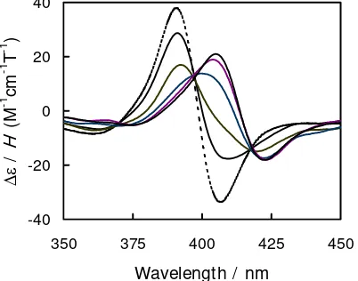
of 5,15-bis(1,3-dimethylimidazolium-2-yl)porphyrin (H2BDMImP) in the phosphate buffer free from CT-DNA exhibits a positive pseudo A-term with a positive peak at 391 nm and a negative peak at 407 nm. In the presence of CT-DNA, both positive and negative peaks shift about 15 nm to the longer wavelength and their intensity (Δε/H) decreases 53% at R([porphyrin]/[DNA in base pair]) = 0.02 (Fig. 2). These changes in the MCD spectra indicate that the excited state angular momentum (Lj), i.e. Δ[θ]MCDp-t/εmax, is reduced 58% from 1.76 to 1.68 (deg cm2 dmol–1 T–1/M–1 cm–1) by the less perturbation to the localized π- and π*-MOs (1a1u, 3a2u and 4eg).
Fig. 2 MCD spectra of free (dashed line) and CT-DNA-bound (solid lines) H2BDMImP. The R ranges from ∞ to 0.01.
350 375 400 425 450
Wavelength / nm
Indonesian Journal of Chemistry, 2002, 2 (3), 131-134
Hidenari Inoue
133
Fig. 3 Plots of the increase in melting temperature (ΔTm) versus the molar ratio of the porphyrin to DNA in base pair (R).
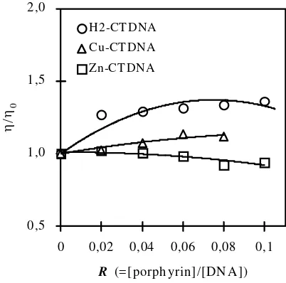
Fig. 4 Plots of the relative viscosity of DNA versus R in the phosphate buffer at pH 6.8 and = 0.2 at 30°C.
MEASUREMENTS OF THERMAL DENATURATION TEMPERATURE
The stability of the DNA double helix influences the melting temperature (Tm) of DNA, while the binding of porphyrin to DNA alters the Tm depending on the strength of interactions. Upon heating DNA in a buffer solution, first the A-T base pairs having two hydrogen bonds are dissociated, second the G-C base pairs with three hydrogen bonds cleaved, and finally the DNA double helix turns into two single stands. It is known that CT-DNA consists of 42 % of G-C base pairs and 52 % of A-T base pairs. Thus, measurements of the Tm of CT-DNA in porphyrin solutions can explore how strong the interaction between porphyrin and DNA
is. For example, the changes in the Tm upon the increasing addition of H2BDMImP, CuBDMImP or ZnBDMImP to the phosphate buffer solution of CT-DNA were measured in the range of R ([porphyrin]/[DNA in base pair]) = 0 ∼ 0.25. Plots of the increase in melting temperature (ΔTm) versus R values are shown in Fig. 3. The Tm of CT-DNA is always increased with the addition of the porphyrins to the CT-DNA buffer solution, indicating that all the porphyrins studied interact with CT-DNA to stabilize the duplex structure. Moreover, the insertion of the metal ions into H2BDMImP leads to an increase in
ΔTm, suggesting that the interaction between porphyrin and CT-DNA is strengthened in the metalloporphyrin more than in the metal-free porphyrin.
0 2 4 6 8 10 12 14 16
0 0,05 0,1 0,15 0,2 0,25
R ( =[p orphyrin] / [DNA ])
Δ
Tm
Zn-CTDNA
Cu-CTDNA
H2-CTDNA
0,5 1,0 1,5 2,0
0 0,02 0,04 0,06 0,08 0,1
R (=[ porph yrin] /[DN A])
η/ η0
H2-CT DNA
Cu-CT DNA
Indonesian Journal of Chemistry, 2002, 2 (3), 131-134
Hidenari Inoue
134
VISCOMETRIC MEASUREMENTS
In the absence of X-ray structural data, a hydrodynamic method such as viscometric measurement is one of the most critical tests for inferring the binding mode of DNA in solution, i.e. intercalation or other binding modes. In general an increase in the relative viscosity is ascribed to a length increase of the DNA double helix due to intercalation. On the other hand, a decrease in the relative viscosity due to the reduction of the effective length of DNA molecules results from the bending of the DNA double helix caused by electrostatic interaction between the anion site of phosphate of DNA and the cation site of porphyrin. The results of viscometric measurements in the buffer solution of CT-DNA are illustrated in Fig. 4, where the molar ratios R of H2BDMImP, CuBDMImP or ZnBDMImP to CT-DNA in base pair are varied in the range of 0.01 and 0.1. The relative viscosity of the buffer solution of CT-DNA is increased with the addition of CuBDMImP, indicating that CuBDMImP is intercalated into CT-DNA.
In contrast, the relative viscosity is decreased with the addition of ZnBDMImP to the buffer solution of CT-DNA. This decrease may be due to the outside binding of ZnBDMImP to CT-DNA. The viscometric consequence that CuBDMImP intercalates into CT-DNA while ZnBDMImP outside binds to CT-DNA is consistent with the conclusion from the visible absorption, induced CD and MCD spectra.
REFERENCES
1. D. H. Tjahjono, T. Akutsu, N. Yoshioka and H. Inoue, Biochim. et Biophys. Acta, 1472, 333-343 (1999).
2. D. H. Tjahjono, T. Yamamoto, S. Ichimoto N. Yoshioka and H. Inoue, J. Chem. Soc.: Perkin Trans. 1, 3077-3081 (2000).