NILAI DIAGNOSTIK DAN PROGNOSTIK DARI
FUNGSI SISTOLIK DAN DIASTOLIK VENTRIKEL
KANAN PADA INFARK MIOKARD INFERIOR
THE DIAGNOSTIC AND PROGNOSTIC VALUE OF RIGHT VENTRICLE SYSTOLIC
AND DIASTOLIC FUNCTION IN INFERIOR MYOCARDIAL INFARCTION
Niniek Purwaningtyas
Cardiologist Staff at Moewardi General Hospital
Korespondensi: dr. Dewi Ayu Paramita, Sp. JP., FIHA. Email: [email protected]
ABSTRAK
Keterlibatan ventrikel kanan (RV) meningkatkan mortalitas dan morbiditas pada infark miokard inferior (MI). Data mengenai keegunaan pencitraan Doppler jaringan berdenyut (TDI) dalam diagnosis disfungsi RV pada
elevasi segmen ST MI (STEMI) jarang. Penelitian ini mengevaluasi signifikansi diagnostik dan prognosis fungsi sistolik dan diastolik RV dibandingkan kriteria diagnostik RVMI elektrokardiografi klasik pada kelompok pasien ini. Pasien berturut-turut dengan STEMI inferior akut dan akut dinilai secara prospektif. RVMI didefinisikan sebagai elevasi segmen ST ≥ 0,1 mV pada timbal V4R. Ekokardiografi dengan TDI dilakukan dalam 24 jam sejak
timbulnya gejala. Dari 31 pasien (usia rata-rata 56,39 ± 9,02 tahun), RVMI ditemukan pada 18 (37%). Analisis multivariat menunjukkan bahwa dua variabel-fungsi sistolik RV dan diastolik, merupakan prediktor independen
prognosis di rumah sakit. Sensitivitas dan spesifisitas fungsi sistolik RV masing-masing 94,4% dan 69,2%.
Sedangkan fungsi diastolik RV masing-masing 44% dan 76,9%. Fungsi sistolik RV memprediksi diagnosis EKG
RVMI dengan sensitivitas dan spesifisitas yang relatif tinggi. Fungsi diastolik RV memprediksi diagnosis EKG RVMI dengan sensitivitas yang relatif rendah namun memiliki spesifisitas tinggi.
Kata kunci: Pencitraan Doppler jaringan, infark miokard RV, infark miokard inferior
ABSTRACT
Right ventricular (RV) involvement increases mortality and morbidity in inferior myocardial infarction (MI). There are sparse data on the usefulness of pulsed tissue Doppler imaging (TDI) in the diagnosis of RV dysfunction in ST segment elevation MI (STEMI). This study evaluate the diagnostic and prognostic significance of RV systolic and diastolic function compared to classical electrocardiographic RVMI diagnostic criteria in this group of patients. Consecutive patients with first, acute, inferior STEMI were prospectively assessed. The RVMI was defined as an ST-segment elevation ≥ 0.1 mV in lead V4R. Echocardiography with TDI was performed within 24 h of the onset of symptoms. Out of 31 patients (mean age 56.39 ± 9.02 years), RVMI was found in 18 (37%). Multivariate analysis showed that two variables—RV systolic and diastolic function, were independent predictors of in-hospital prognosis. Sensitivity and specificity the RV systolic function were 94,4% and 69,2%, respectively. While RV diastolic function were 44% and 76,9%, respectively. RV systolic function predict ECG diagnosis of RVMI with relatively high sensitivity and specificity. RV diastolic function predict ECG diagnosis of RVMI with relatively low sensitivity but with high specificity.
Keywords: tissue Doppler imaging, RV myocardial infarction, inferior myocardial infarction
INTRODUCTION
Right ventricular (RV) myocardial infarction (MI) occurs in 30–50% of patients with inferior MI (Goldstein, 2002). It is caused mainly by proximal right coronary artery (RCA) lesion (Bowers et al., 2002). The RVMI leads to RV dysfunction that increases early mortality and morbidity independently of the degree of left ventricular
dysfunction (Kukla et al., 2006). Rapid, accurate assessment of RV function is of great importance, as it provides not only prognostic information measure
but also allows proper modification of therapy. The
assessment of the RV, but has limited value because of the asymmetric, pyramidal shape of the RV and nonconcentric contraction which makes geometric
assumptions difficult (Rudski et al., 2010). A number of echocardiographic indices have been investigated, including regional contractility, cavity size, myocardial performance index, and tricuspid annular plane excursion (Piestrzeniewicz
et al., 2006). Previous study are available on the usefulness of pulsed wave TDI in the diagnosis of RVMI in patients with inferior MI, but there
are sparse and conflicting data on the usefulness
of RV myocardial velocities derived from TDI in this group of patients (Hsiao et al., 2010). The aim of this study was to evaluate the diagnostic and
prognostic significance of RV systolic and diastolic
function compared to classical electrocardiographic RVMI diagnostic criteria in this group of patients.
METHODS
This study was designed and conducted
prospectively. Consecutive patients with first, acute,
inferior STEMI with standard echocardiographic examination and echocardiography performed within 24 hour of the onset of symptoms, were eligible. The diagnosis of inferior STEMI was based on the European Society of Cardiology
(ESC) criteria: chest pain lasting > 30 min, characteristic ST-segment elevation of ≥ 0.1 mV in
two or more inferior derivation (leads II, III, aVF) on ECG, and an increase in biomarkers: troponin I or creatine kinase (CK)-MB (Van de Werf et al., 2003). Patients with a history of previous MI, pulmonary embolism, chronic obstructive pulmonary disease, documented pulmonary
hypertension, permanent atrial fibrillation, His
bundle branch blocks, moderate or severe valvular diseases, or poor quality echocardiographic imaging were excluded. All patients gave their written consent. The study was approved by the hospital ethics committee.
Standard 12-lead ECG was performed immediately upon arrival at the Emergency Department. Right chest ECG used for RVMI
Standard echocardiographic examination with TDI was performed within 24 hour of the onset of symptoms in all patients. Examinations were performed using Vivid I with phased-array 1.8– 3.6 MHz transducer, harmonic imaging, equipped with TDI technology. Echocardiographers were blinded to clinical and ECG parameters. All measurements were performed according to the recommendations of the American Society of Echocardiography. Measurements of RV and right atrium diameters, fractional area change of RV, change of inferior vena cava diameter during respiration, and assessment of RV wall motion abnormalities were included in standard echocardiographic examination. Left ventricular ejection fraction (LVEF) was calculated according
to modified Simpson’s rule.
For all parameters, descriptive statistics were calculated (mean and SD for continuous variables and frequency tables for categorical variables). Variables were compared using ANOVA, Kruskal-Wallis non-parametrical ANOVA, t-Student test, Mann-Whitney test, chi square test or Fisher exact test where appropriate.
A logistic regression analysis was used to evaluate the predictive value of selected clinical and echocardiographic parameter factors for the
presence of ECG changes specific for RVMI
diagnosis. The included factors were: age, systolic blood pressure, diastolic blood pressure, standard
echocardiographic parameters reflecting RV
function (RV EF, fractional area change of RV, TAPSE, mitral propagation) and TDI parameters (SmRV, EmRV, myocardial performance index).
Model used in the analysis was pre-specified based
on the current knowledge of RV dysfunction. For RV myocardial velocities, values below median were used.
The diagnostic value of parameters in RVMI diagnosis was evaluated by calculating a receiver operating characteristics (ROC) curve. To evaluate
the prognostic significance of RV myocardial
RESULTS AND DISCUSION
The study group consisted of 31 consecutive patients, mean age 56.39 ± 9.02 years with
first, acute inferior STEMI enrolled between 1
November 2014 and 31 December 2014. In 18
patients (37%), first STEMI within 24 h of the
onset of symptoms and with RV localization
was found. All patients fulfilled criteria for
diagnosis of type I MI according to the new MI
definition;8 Exclusion criteria were found in 14
patients: severe aortic valve disease, permanent
atrial fibrillation, severe chronic obstructive
pulmonary disease, history of pulmonary embolism, His bundle branch block, poor quality of standard echocardiographic imaging and lack
of sufficient medical documentation. The clinical
characteristics of the study group are listed in Table 1.
Table 1 Patient characteristics
PARAMETER All MI RV MI (+) RV MI (-)
Age 56.39 ± 9.02 55.89 ± 9,19 57,08 ± 9,09
Systolic Blood Pressure 130,22 ± 30,57 129,05 ± 28,73 119,15 ± 24,48
Diastolic Blood Pressure 80,39 ± 21,18 78,33 ± 20,82 73,23 ± 13,72
TAPSE 1,92 ± 0,56 1,87 ± 0,58 2,00 ± 0,53
S 0,12 ± 0,20 0,11 ± 0,33 0,13 ± 0,31
FAC RV 51,80 ± 15,95 51,27 ± 13,3 52,53 ± 19,6
TDI MPI 480,26 ± 184,12 475,77 ± 18,22 486, 56 ± 18,61
E/A RV 1,15 ± 0,5 1,08 ± 0,46 1,24 ± 0,45
EF RV 50,38 ± 10,47 44,33 ± 8,51 58,76 ± 6,3
Mitral Propagation 53,04 ± 42,02 57,02 ± 5,1 58,01 ± 5,05
S/D 1,15 ± 0,70 1,14 ± 0,51 1,16 ± 0,46
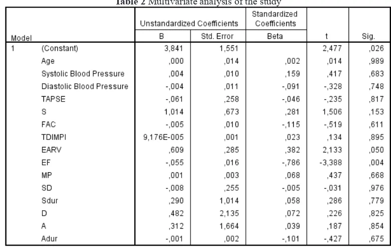
Multivariate analysis showed that two variables—RV systolic and diastolic function, were independent predictors of in-hospital
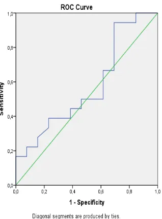
prognosis (table 2). Sensitivity and specificity
the RV systolic function were 94,4% and 69,2%, respectively. While RV diastolic function were
44% and 76,9%, respectively (figure 1 and 2).
Figure 1 ROC curve of diastolic function Figure 2 ROC curve of systolic function
This study showed the high value of RV systolic myocardial function in the diagnosis of acute RVMI in patients with inferior STEMI. In the case of RV, a fundamental role in generating stroke volume is played by the shortening of longitudinal
fibres (Thygesen dan Alpert, 2007). Meluzin et al
showed that a peak systolic velocity of tricuspid annulus correlates well with RVEF measured by MRI (Meluzin et al., 2001). Ueti et al. found a high correlation between RV systolic velocity and RVEF assessed by radionuclide ventriculography (Ueti et al., 2002). Right ventricular ischaemia or infarction can also lead to impairment of diastolic function. Decreased compliance and reduced
filling of RV have been shown (Goldstein, 2002).
Standard echocardiography is the most widely available, semi-quantitative RV assessment modality, but is limited by the complex morphology of the RV and may be further challenged by poor acoustic windows (Kukla
et al., 2006). This technical challenge could be overcome by using TDI with non-geometric indices of RV function. The reproducibility of measurements of RV myocardial velocities was high in this work, and this has also been found by
measurements were not acceptable. Pulse wave TDI allowed simple, rapid, and quantitative measurements.
In our study, RV systolic, and diastolic function were found to be an independent predictor of early, in-hospital prognosis. This supports data showing that RV dysfunction is an independent prognostic parameter in patients with MI. The importance of RV function in the prognosis of various cardiopulmonary disorders is now well understood (Hsiao et al., 2010). Meluzin
et al found that patients with symptomatic heart failure and systolic velocity of tricuspid annulus <
10.8 cm/s exhibited significantly worse event-free
survival (Meluzin et al., 2003). In patients with inferior STEMI, RVMI leads to increased early mortality and morbidity (Kukla et al., 2006).
The major limitation of our study is the lack of a gold standard for the diagnosis of RVMI suitable for the early phase of hospitalisation in a
coronary care units. We chose the ECG definition
of RVMI, as recommended by ESC (Van de Werf et al., 2003), but this definition has its own limitations, mainly in terms of limited specificity
CONCLUSION
RV systolic function predict ECG diagnosis of RVMI with relatively high sensitivity and
specificity. RV diastolic function predict ECG
diagnosis of RVMI with relatively low sensitivity
but with high specificity
ACKNOWLEDGMENT
Thank you for Dewi Ayu Paramita, MD, FIHA for helping this research.
REFERENCES
Bowers TR, O’Neill WW, Pica M, et al. 2002. Patterns of Coronary Compromise Resulting in Acute Right Ventricular Ischemic Dysfunction. Circulation, 106: 1104–1109.
Goldstein J. 2002. Pathophysiology and Management of Right Heart Ischaemia. J Am Coll Cardiol, 40: 841–853.
Hsiao SH, Chiou KR, Huang WC et al. 2010. Right Ventricular Infarction and Tissue Doppler Imaging: Insights from Acute Inferior Myocardial Infarction after Primary Coronary Intervention. Circ J, 74: 2173–2180.
Kukla P, Dudek D, Rakowski T, et al. 2006. Inferior Wall Myocardial Infarction With or Without Right Ventricular Involvement—Treatment and in-Hospital Course. Kardiol Pol, 64: 583–588. Meluzin J, Spinarova L, Bakala J, et al. 2001. Pulsed Doppler Tissue Imaging of the Velocity of
Tricuspid Annular Systolic Motion. A New, Rapid, and Non-invasive Method of Evaluating Right Ventricular Systolic Function. Eur Heart J, 22: 340–348.
Meluzín J, Spinarova L, Dusek L, et al. 2003. Prognostic Importance of the Right Ventricular Function Assessed by Doppler Tissue Imaging. Eur J Echocardiogr, 4: 262–271.
Meluzin J, Spinarova L, Hude P, et al. 2005. Prognostic Importance of Various Echocardiographic Right Ventricular Functional Parameters in Patients with Symptomatic Heart Failure. J Am Soc Echocardiogr, 18: 435–444.
Piestrzeniewicz K, Łuczak K, Piechowiak M, et al. 2006. The Value of Doppler-derived Myocardial Performance Index and Tricuspid Annular Motion in the Evaluation of Right Ventricular Function in Patients with Acute Inferior Myocardial Infarction. Folia Cardiol, 13: 369–378.
Rudski LG, Wyman WL, Afilalo J, et al. 2010. Guidelines for the Echocardiographic Assessment of the Right Heart in Adults: a report from the American Society of Echocardiography Endorsed by the European Association of Echocardiography and the Canadian Society of Echocardiography.
J Am Soc Echocardiogr, 23: 685–713.
Thygesen K and Alpert JS. 2007. White HD; On Behalf of the Joint esc/accf/whf Task Force for the
Redefinition of Myocardial Infarction: Universal Definition of Myocardial Infarction. Eur Heart J, 28: 2525–2538.
Ueti OM, Camargo EE, Ueti Ade A, et al. 2002. Assessment of Right Ventricular Function with Doppler Echocardiographic Indices Derived from Tricuspid Annular Motion: Comparison with Radionuclide Angiography. Heart, 88: 244–248.