EISSN: 2086-4094 DOI: 10.4308/hjb.21.2.53
_________________
∗Corresponding author. Phone: +62-21-8765066/8765067,
Fax: +62-21-8765062, E-mail: [email protected]
Characterization of α-Nitrile Hydratase and Amidase of
Rhodococcus
aff.
qingshengii from Indonesia
AERMA HASTUTY1∗, WIBOWO MANGUNWARDOYO2, BAMBANG SUNARKO1
1Microbiology Division, Research Center for Biology, Indonesian Institute of Sciences-LIPI, Jalan Raya Bogor Km 46, Cibinong 16911, Indonesia
2Department of Biology, Faculty of Mathematics and Natural Sciences, University of Indonesia, Depok 16424, Indonesia
Received June 18, 2013/Accepted February 25, 2014
A study on biotransformation of acetonitrile using Gram-positive bacteria has been conducted. Two isolates of nitrile-degrading bacteria (strain 100A and 100D) were screened from sediments of a contaminated river in Cibinong, West Java. The bacterial isolates were identified as Rhodococcus aff. qingshengii based on molecular phylogenetic
analyses of 16S rRNA sequence. These bacteria were capable to grow on medium containing 100 mM acetonitrile, but unable to grow on medium amended with 25 mM benzonitrile. Analyses using Gas Chromatography (GC) indicated
that R. aff. qingshengii strain 100A and 100D has the ability to produce nitrile hidratase and amidase. The highest
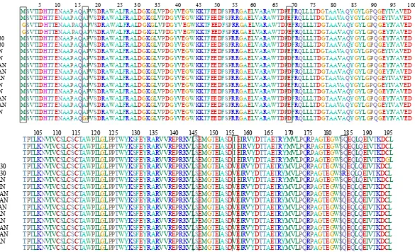
enzyme activity on mineral medium with the addition of 100 mM acetonitrile was 73.49 mmol/min/mL by strain 100A, and 70.52 mmol/min/mL by strain 100D. In addition, the ammonia concentration produced by strain 100A and 100D were 180.20 and 54.10 mM, respectively. These results were supported by molecular characterization using specific primers, where strain 100A and 100D positively contain genes encoding α-nitrile hydratase (α-NHase) and amidase. There was a difference at the first position of amino acid composition of the gene encoding α-NHase between strain 100A (Methionine1) and strain 100D (Glycine1), but the amino acids composition of amidase of both strain were
identical. This is the first report of R. aff. qingshengii as nitrile-degrading bacterium in Indonesia.
Keywords: biotransformation, nitrile, nitrilases, 16S rRNA, Rhodococcus
___________________________________________________________________________
INTRODUCTION
Nitrile hydratase (NHase) (EC 4.2.1.84) and amidase (EC 3.5.1.4) are group of nitrile-converting enzymes that hydrolyze nitriles into the corresponding higher-value amides and acids (Nagasawa & Yamada 1990). NHase and amidase, including other nitrile-converting enzymes, have attracted interest not only because of their role as biocatalyst in the production of solvents, extractans, drug intermediates (chiral synthons), pesticides as well as in the synthesis organic amine, amide, ester, carboxylic acid, aldehydes, ketones, and heterocyclic compounds (Banerjee et al. 2002), but also because nitrilases have advantages over the chemical hydrolysis due to their milder pH and temperature conditions, and the absence of by-products (Nagasawa et al. 2000). For example, the application of NHase from Rhodococcus rhodochrous J1, which is currently used in the production of acrylamide (Nagasawa et al. 1993; Yamada & Kobayashi 1996) and nicotamide (Nagasawa et al. 1988) at industrial
scale. Wyatt and Knowless (1995) reported that NHase has also practical importance as biocatalysts for environmental bioremediation in the removal of nitriles from waste streams.
MATERIALS AND METHODS
Organisms and Growth Condition. Two nitrile-metabolizing bacteria (strain 100A and 100D) were isolated from polluted-river sediment using mineral medium (MM) (Meyer & Schlegel 1983) supplemented with 10-25 and 50-100 mM of acetonitrile. Selected bacteria were grown on nutrient agar (NA) DIFCO and mineral medium (MM) with the following composition (per 1000 mL): 0.4475 g Na2HPO4.2H & Schlegel 1983). The micro-element composed of 0.1 g ZnSO4.7H Chain Reaction (PCR) amplification were carried out in order to obtain genomic DNA for bacterial identification using sequence of 16S rRNA. The genomic DNA from bacteria were obtained from 48 h colonies using the guanidium thiocyanate/ EDTA/Sarkosyl (GES) method as described by Pitcher et al. (1989). The 16S rRNA gene was amplified using the universal primers: 27F (5’-AGAGTTTGATCCTGGCTCAG-3’) and 1500R (5’-GTTACCTTGTTACGACTT–3’). The PCR components were set as follows: 2 μL DNA template (+ 100 ng), 25 μL Go Taq® PCR master mix (Promega), and 20.5 μL PCR-grade water (Sigma). The amplification was carried out using a PCR thermocycler (TaKaRa Shuzo Co., Ltd., Shiga, Japan) with the following condition: 95 oC for 3 min of predenaturation; followed by 30 cycles of 95 oC for 30 sec of denaturation, 50 oC for 30 sec of annealing, 72 oC for 90 sec of extension, and 72 oC for 10 min of final extension. The quality of the PCR products were determined using electrophoresis in 1.5% agarose gel. DNA nucleotides were sequenced by FirstBASE (Malaysia) using the same primer pairs used in the PCR reaction.
Phylogenetic Analysis. Bacteria were identified using phylogenetic analyses. Newly sequences of bacteria generated from 16S rRNA region were aligned with the homologous sequences obtained from NCBI GenBank database (http://www.ncbi. nlm.nih.gov/) using Basic Local Alignment Search Tool (BLAST) available at the GenBank website.
Phylogenetic analysis was performed using MEGA (Molecular Evolutionary Genetics Analysis) v5.1 software (Tamura et al. 2011). Maximum Likelihood (ML) was used in the analyses involving the Tamura Nei model as nucleotide substitution parameter and gap was treated as missing data. Searching of the best phylogenetic trees was done by using a heuristic method with Nearest Neighbor approach Interchange (NNI), an initial tree was generated based on Neighbor Joining analysis (NJ). Phylogenetic tree was evaluated by bootstrap method using 1000 replication.
Bacteria Growth Assay. Bacterial isolates were grown in mineral medium containing nitrile, at 120 rpm. Bacterial growth pattern was observed every 2 h by measuring the optical density (OD). Measurement of microbial growth was carried out every 24 h for 7 days at 436 nm wavelength. pH, acetonitrile concentration, and the formation of transformation products (acetamide, acetic acid, and ammonia) during bacterial growth were measured.
Bacterial Cells Production for Nitrile Biotransformation Assay. A loopful bacteria was inoculated into 50 mL mineral medium in 100 mL Erlenmeyer falsk. 262 µL of 100 mM acetonitrile was added into the medium and incubated at 30 oC for 72 h on the shaker incubator at 120 rpm.
adding 0.25 μL of 5 N HCl. After the enzymatic reaction stopped, samples were neutralized by adding 5 mL of 0.25 N NaOH. Samples were then centrifuged, and the resulting supernatant was taken for determination acetonitrile, acetamide and acetic acid.
Analysis of acetonitrile, acetamide, and acetic acid performed in Gas Chromatography (GC) equipped with Flame Ionization Detector (FID) using a Porapak Q column. Analysis conditions were set as follows: 1 mL sample is injected at 225, 240, and 240 oC of oven, injector, and detector temperature, respectively. N2 was used as a carrier and H2 was used as detectors, and these gases were pumped at 11 mL/min (Sunarko et al. 2000).
Ammonia Concentration Assay. Determination of ammonia was performed using the Nessler method. A total of 10 mL of enzyme solution was added into 0.99 mL of 0.1 N NaOH and 20 mL Nessler reagent. The solution was incubated at 30 oC for 20 min, and then measured using a spectrophotometer at a wavelength of 420 nm (Oliver et al. 1989).
DNA Extraction and PCR Amplification for Nitrilases Gene. DNA extraction and PCR (Polymerase Chain Reaction) amplification were carried out in order to obtain genomic DNA for nitrilases gene characterization. The genomic DNA from bacteria were obtained from 48 h colonies using the guanidium thiocyanate/ EDTA/Sarkosyl (GES) method as described by Pitcher et al. (1989). The NHase gen was amplified using primer pairs of α-NH1-F (5’-GTGAACCAGATGTCAGTAACGATCG-3’) and α-NH1-R (5’-CGCTCAGGCAGTCCTTGGT GACG-3’), and amidase gen was amplified using primer pairs of amd1-F (5’-GTGAAGCCGATCA CATCAGGAGC-3’) and amd2-R (5’-CGGGTACC AATCCCTTACCGTCG-3’) (Brandão et al. 2003). The PCR components and PCR condition were set as previously described in the bacterial identification. The quality of the PCR products were determined using electrophoresis in 1.5% agarose gel. DNA nucleotides were sequenced by FirstBASE (Malaysia) using the same primer pairs used in the PCR reaction.
Phylogenetic Analysis of the α-NHase and Amidase Genes. Nucleotides sequences of α-NHase and amidase of R. aff. qingshengii strain 100A and 100D were aligned with available α-NHase and amidase equences retrieved from GenBank. The phylogenetic analysis of nitrilases genes of selected bacteria was carried out by using the same method as in the identification of bacteria.
RESULTS
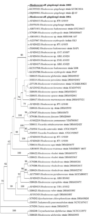
Identification of Bacteria. BLAST result showed that three Rhodococcus species, namely, R. qingshengii, R. baikonurensis, and R. erythropolis were the most homologous bacteria with bacterial isolates strain 100A and 100D, with a maximum identity of sequence homology reaches 99 and 100%, respectively. In the phylogenetic analysis, the type species of these species were included in the analyses together with other Rhodococcus species. The phylogenetic tree generated from the ML analyses of 16S rRNA sequence showed that the genus Rhodococcus is monophyletic with 92% bootstrap support (BS) (Figure 1). Strain 100A and 100D nested in the same clade with R. qingshengii, R. baikonurensis, and R. erythropolis with 83% BS. The clade indicated a close relationship between strain 100A and 100D to those of R. qingshengii, R. baikonurensis, and R. erythropolis. Multiple nucleotide sequence analyses among these isolates showed that there were eight nucleotides differences of R. erythropolis to each of strain 100A and 100D, and there were three nucleotides differences of R. baikonurensis to each of strain 100A and 100D.
The same analyses showed that R. qingshengii was identical to that of strain 100D and only single nucleotide difference of R. qingshengii to strain 100A. Based on the phylogenetic tree and multiple nucleotide sequence analyses, the bacterial strain of 100A and 100D were identified as Rhodococcus aff. qingshengii. Code ‘aff.’ or ‘affinity to’ refers to the closest only nucleotide sequences and phylogenetic affinity of strain 100A and 100D to that of R. qingshengii.
Bacterial Growth Assay. This assay showed that R. aff. qingshengii strain 100A and 100D were capable to grow properly after incubated for 72 h. It was indicated by the increasing of OD value in the mineral medium amended with 100 mM acetonitrile (Figure 2).
Figure 1. The phylogenetic tree from the Maximum Likelihood analyses of 16S rRNA sequence of Rhodococcus aff. qingshengii
was found unpredictable. Range of pH was found about 7-9. During the bacterial growth, there was an increase in enzyme activity of R. aff. qingshengii strain 100A and 100D with 73.49 and 70.52 mmol/ min/mL, respectively at fifth day.
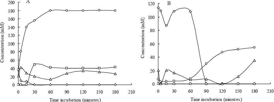
Nitrile Biotransformation Assay. The nitrile biotransformation assay showed that NHase
and amidase enzymes involved during the biotransformation of nitrile by R. aff. qingshengii strain 100A and 100D. In the current study, highest ammonia levels obtained was 180.20 mM produced in the acetonitrile biotransformation by R. aff. qingshengii strain 100A at 50 min incubation (Figure 4). This was supported by faster reduction 0.00
Rhodococcus aff. qingshengii 100A and (B) Isolate Rhodococcus aff. qingshengii 100D. pH, H Total enzyme activity (mmol/mnt/mL), L) OD.
Figure 3. Bacterial growth incubated for 7 days in the mineral medium amended with 100 mM acetonitrile. (A) Isolate Rhodococcus
aff. qingshengii 100A and (B) Isolate Rhodococcus aff. qingshengii 100D. pH, H Total enzyme activity (mmol/mnt/ mL), L) OD.
Figure 4. The nitrile biotransformation assay. (A) Rhodococcus aff. qingshengii 100A (B) Rhodococcus aff. qingshengii 100D. Acetonitrile, Acetic acid, Acetamide, NH3.
level of acetonitrile in the medium containing R. aff. qingshengii strain 100A compared to medium containing R. aff. qingshengii strain 100D.
Gas chromatography analysis showed a reduction in the concentration of acetic acid and acetamide (Figure 4). It was probably due to the acetic acid and acetamide were utilized by R. aff. qingshengii strain 100A and 100D for the synthesis of cells during bacterial growth in biotransformation process. During the biotransformation, acetonitrile concentration in the medium inoculated with R. aff. qingshengii strain 100A started to decrease after 5 min incubation into 9.78 mM. The acetonitrile was completely degraded after 30 min incubation. However, in the medium inoculated with R. aff. qingshengii strain 100D, the acetonitrile was completely degraded after 90 min incubation.
Molecular Characterization of Nitrile Degrading Enzymes (α-NHase and Amidase). NHase consists of two sub-units, α and β, with molecular weight 23 kDa. About 400 bp bands were formed of which indicated the presence of α-NHase gene in the R. aff.
qingshengii strain 100A and 100D. Several strains of R. erythropolis and R. rhodochrous showed high homology to those of α-NHase nucleotide sequence of R. aff. qingshengii strain 100A and 100D with 99-100% similarity (http://blast.ncbi.nlm.nih.gov/ Blast.cgi).
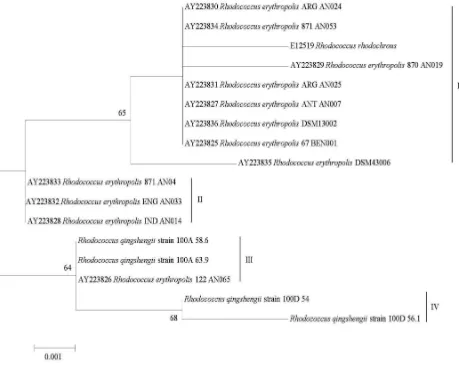
Phylogenetic tree generated from maximum likelihood analyses of α-NHase nucleotide sequence showed that four monophyletic clades were formed (Figure 5). The first clade contains R. rhodochrous and R. erythropolis ARG AN024 (rainforest soil, Argentine), 871 AN053 (sea sediment, Japan), ARG AN025 (rainforest soil, Argentine), ANT AN007 (lake sediment, Antarctic), DSM 13002 (garden soil, Germany), 67 BEN001 (deep-sea sediment, Japan), DSM 43.006 (soil, unknown) with 65% BS. The second clade contains R. erythropolis 871 AN042 (deep-sea sediment, Japan), ENG AN033 (swamp, England) and IND AN014 (mangrove forest, Indonesia). Rhodococcus aff. qingshengii strain 100A nested in the same clade with R. erythropolis 122 AN065 (deep-sea sediment, Japan) with 64%
Figure 5. Phylogenetic tree generated from maximum likelihood analyses of α-NHase nucleotide sequence for R. aff. qingshengii
BS, while two sequences of R. aff. qingshengii strain 100D formed independent clade with 68% BS.
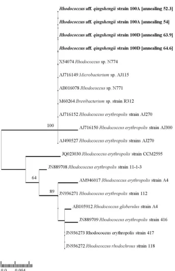
In the electrophoresis analyses of amidase gene of R. aff. qingshengii strain 100A and 100D, about 1900 bp bands were formed. BLAST analyses of amidase nucleotide sequences showed highest similarity with amidase nucleotide sequence belonging to several strains of R. erythropolis (http:// blast.ncbi.nlm.nih.gov/Blast.cgi). Phylogenetic tree generated from ML analyses based on amidase
nucleotide sequences showed that R. aff. qingshengii strain 100A and 100D nested in the same clade with Rhodococcus sp. N774, Microbacterium sp. AJ115, Rhodococcus sp. N771, Brevibacterium sp. strain R312, R. erythropolis strain AJ270, and R. erythropolis strain AJ300 with 100% BS (Figure 6).
DISCUSSION
Bacterial Growth Assay. Inability of these bacteria in degrading benzonitrile was probably due
Figure 6. Phylogenetic tree generated from maximum likelihood analyses of amidase nucleotide sequence for Rhodococcus aff.
to the complexity in benzonitrile chemical bond of which contains cyclic structure (Sulistinah & Riffiani 2011). In general, screening of microbial producing NHase is difficult because of gene regulation of NHase is heavily influenced by amide compounds and few species of bacteria are able to produce NHase using selective media (Kobayashi & Shimizu 1998).
During the growth phase, the pH value of medium was changed to alkaline. This was due to the accumulation of ammonia in the medium during bacterial growth. Suhartono (1989) noted that the presence of extracellular enzyme activities and microbial growth in fermentation process often yields by-products that change the pH of the medium. It is interesting to know that R. aff. qingshengii strain 100A and 100D to the alkaline pH. It is possible that both strains are alkali tolerant, therefore, an increase in the pH value in the medium during the growth phase did not inhibit the growth of R. aff. qingshengii strain 100A and 100D, and did not inhibit the activity of nitrile degrading enzyme. Until now, there is no evidence that the degradation of nitrile compounds by Rhodococcus spp. can be carried out in alkaline conditions. Other than Rhodococcus spp., Sorokin et al. (2007) reported that Natronocella acetinitrilica isolated from soda lakes capable of producing the enzyme NHase, and can grow at pH 10 in medium amended with 3M acetonitrile. Duthaler (1994) stated that the enzymatic hydrolysis reaction of nitrile compounds in alkaline conditions has its own advantages, especially when cyanide compounds contribute in the enzymatic reactions, e.g. Strecker reaction when combined with enzymatic hydrolysis reaction of α-aminonitrile (enantio-selective) will produce α-aminoamida and α-amino acids. This situation caused a decrease in the acetonitrile concentration in the medium because the bacteria will produce enzymes to metabolize the substrate through the process of degradation (Nawaz et al. 1989). During the biotransformation process of acetonitrile by R. aff. qingshengii strain 100A and 100D, amide compounds was produced in the form of acetamide, and carboxylic acid in the form of acetic acid and ammonia. These process involving NHase and amidase enzymes.
In general, the first product formed during the biotransformation process was acetamide.
This product was the result of the acetonitriles biotransformation by NHase into amide compound in the form of acetamide, followed by the formation of acetic acid and ammonia as a result of biotransformation of acetamide by amidase enzyme. The biotransformation of nitrile compounds by bacteria usually takes place in two steps, of which the first reaction involves NHase and amidase, whereas the second reaction involves only nitrilase (Kim et al. 2001). It was supported by the formation of acetic acid, acetamide and ammonia Enzymes (α-NHase and Amidase). Kobayashi et al. (1992) reported that the amino acid sequences of the two sub-units are not interconnected and the structural genes of NHase are usually adjacent in the same operon, although the coding sequence of sub-units of α and β are variable.
Characterization
of
α-NHase
and
Amidase
of
Rhodococcus
af
f.
qingshengii
61
61
5 10 15 20 25 30 35 40 45 50 55 60 65 70 75 80 85 90 95 100 R_qingshengii_100A_58.6 MSVTIDHTTENAAPAQAPVSDRAWALFRALDGKGLVPDGYVEGWKKTFEEDFSPRRGAELVARAWTDPEFRQLLLTDGTAAVAQYGYLGPQGEYIVAVED R_qingshengii_100A_63.9 MSVTIDHTTENAAPAQAPVSDRAWALFRALDGKGLVPDGYVEGWKKTFEEDFSPRRGAELVARAWTDPEFRQLLLTDGTAAVAQYGYLGPQGEYIVAVED R_qingshengii_100D_54 GSVTIDHTTENAAPAQAPVSDRAWALFRALDGKGLVPDGYVEGWKKTFEEDFSPRRGAELVARAWTDPEFRQLLLTDGTAAVAQYGYLGPQGEYIVAVED R_qingshengii_100D_56.1 GSVTIDHTTENAAPAQAPVSDRAWALFRALDGKGLVPDGYVEGWKKTFEEDFSPRRGAELVARAWTDPEFRQLLLTDGTAAVAQYGYLGPQGEYIVAVED AY223836_R_erythropolis_DSM130 MSVTIDHTTENAAPAQAPVSDRAWALFRALDGKGLVPDGYVEGWKKTFEEDFSPRRGAELVARAWTDPDFRQLLLTDGTAAVAQYGYLGPQGEYIVAVED AY223835_R_erythropolis_DSM430 MSVTIDHTTENAAPAQAPVSDRAWALFRALDGKGLVPDGYVEGWKKTFEEDFSPRRGAELVARAWTDPDFRQLLLTDGTAAVAQYGYLGPQGEYIVAVED AY223826_R_erythropolis_122_AN MSVTIDHTTENAAPAQAPVSDRAWALFRALDGKGLVPDGYVEGWKKTFEEDFSPRRGAELVARAWTDPEFRQLLLTDGTAAVAQYGYLGPQGEYIVAVED AY223825_R_erythropolis_67_BEN MSVTIDHTTENAAPAQAPVSDRAWALFRALDGKGLVPDGYVEGWKKTFEEDFSPRRGAELVARAWTDPDFRQLLLTDGTAAVAQYGYLGPQGEYIVAVED AY223827_R_erythropolis_ANT_AN MSVTIDHTTENAAPAQAPVSDRAWALFRALDGKGLVPDGYVEGWKKTFEEDFSPRRGAELVARAWTDPDFRQLLLTDGTAAVAQYGYLGPQGEYIVAVED AY223831_R_erythropolis_ARG_AN MSVTIDHTTENAAPAQAPVSDRAWALFRALDGKGLVPDGYVEGWKKTFEEDFSPRRGAELVARAWTDPDFRQLLLTDGTAAVAQYGYLGPQGEYIVAVED AY223828_R_erythropolis_IND_AN MSVTIDHTTENAAPAQAPVSDRAWALFRALDGKGLVPDGYVEGWKKTFEEDFSPRRGAELVARAWTDPEFRQLLLTDGTAAVAQYGYLGPQGEYIVAVED AY223833_R_erythropolis_871_AN MSVTIDHTTENAAPAQAPVSDRAWALFRALDGKGLVPDGYVEGWKKTFEEDFSPRRGAELVARAWTDPEFRQLLLTDGTAAVAQYGYLGPQGEYIVAVED AY223829_R_erythropolis_870_AN MSVTIDHTTENAAPAQAPVSDRAWALFRALDGKGLVPDGYVEGWKKTFEEDFSPRRGAELVARAWTDPDFRQLLLTDGTAAVAQYGYLGPQGEYIVAVED AY223830_R_erythropolis_ARG_AN MSVTIDHTTENAAPAQAPVSDRAWALFRALDGKGLVPDGYVEGWKKTFEEDFSPRRGAELVARAWTDPDFRQLLLTDGTAAVAQYGYLGPQGEYIVAVED AY223832_R_erythropolis_ENG_AN MSVTIDHTTENAAPAQAPVSDRAWALFRALDGKGLVPDGYVEGWKKTFEEDFSPRRGAELVARAWTDPEFRQLLLTDGTAAVAQYGYLGPQGEYIVAVED AY223834_R_erythropolis_871_AN MSVTIDHTTENAAPAQAPVSDRAWALFRALDGKGLVPDGYVEGWKKTFEEDFSPRRGAELVARAWTDPDFRQLLLTDGTAAVAQYGYLGPQGEYIVAVED E12519_R_rhodochrous MSVTIDHTTENAAPAQGPVSDRAWALFRALDGKGLVPDGYVEGWKKTFEEDFSPRRGAELVARAWTDPDFRQLLLTDGTAAVAQYGYLGPQGEYIVAVED
encoding the amino acid α-NHase also found in respectively. However, R. erythropolis AJ300 encodes Serine338 (S) and Glutamic acid338 (E). Molecular characterization of genes encoding nitrile-degrading enzymes is important, because this method is powerful and more sensitive for further discovery and screening of nitrilases-producing bacteria. With the increasing amount of sequence information obtained from the new gene cloning and sequence genome, it allows the prediction of ‘conserved’ regions to develop specific primer for molecular screening of microorganisms capable of producing α-NHase and amidase, either cultivated or uncultivated. The sequencing results is fundamental for the development of molecular-based screening methods, which is faster and more sensitive than the previous molecular methods using DNA hybridization techniques (Duran et al. 1993).
REFERENCES
Asano Y, Tachibana M, Tani Y, Yamada H. 1982. Purification
and characterization of amidase which participates in nitrile degradation. Agric Biol Chem 46:1175-1181. http://dx.doi. org/10.1271/bbb1961.46.1175
Banerjee A, Sharma R, Banerjee UC. 2002. The nitrile-degrading enzymes: current status and future prospects. Appl Microbiol Biotechnol 60:33-44. http://dx.doi.org/10.1007/ s00253-002-1062-0
Blakey AJ, Colby J, Williams E, O’Reilly C. 1995.
Regio-specific and stereo-Regio-specific nitrile hydrolysis by the nitrile
hydratase from Rhodococcus AJ270. FEMS Microbiol Lett 129:57-61. http://dx.doi.org/10.1111/j.1574-6968.1995. tb07557.x
Brandão PFB, Clapp JP, Bull AT. 2003. Diversity of nitrile hydratase and amidase enzyme genes in Rhodococcus erythropolis recovered from geographically distinct habitats. Appl Environ Microbiol 69:5754-5766. http:// dx.doi.org/10.1128/AEM.69.10.5754-5766.2003
Cahill O. 2004. The biochemical and genetic characterization of nitrile metabolism by the novel isolate R. erythropolis ITCBP [Dissertation]. Ireland: Waterford Institute of Technology, Ireland.
Coffey L, Clarko A, Duggan P, Tambling K, Horgan S, Dowling D, O’Reilly C. 2009. Isolation of identical nitrilase genes from multiple bacterial strains and real-time PCR detection of the genes from soils provides evidence of horizontal gene transfer. Arch Microbiol 191:761-771. http://dx.doi. org/10.1007/s00203-009-0507-6
Duran R, Nishiyama M, Horinouchi S, Beppu T. 1993. Characterization of nitrile hydratase genes cloned by DNA screening from Rhodococcus erythropolis.
Biosci Biotechnol Biochem 57:1323-1328. http://dx.doi. org/10.1271/bbb.57.1323
Duthaler RO. 1994. Recent developments in the stereoselective
synthesis of α-aminoacids. Tetrahedron 50:1539-1650. http://dx.doi.org/10.1016/S0040-4020(01)80840-1
Heald SC, Brandão PFB, Hardicre R, Bull AT. 2001. Physiology, biochemistry and taxonomy of deep-sea nitrile metabolising
Rhodococcus strains. Antonie van Leeuwenhoek 80:169-183. http://dx.doi.org/10.1023/A:1012227302373
Kabaivanova L, Dohreva E, Dimitrov P, Emanullova E. 2005. Immobilization of cells with nitrilase activity from a thermophilic bacteria strain. J Ind Microbiol Biotechnol
32:7-11. http://dx.doi.org/10.1007/s10295-004-0189-7 Kim BY, Kim JC, Lee HH, Hyun HH. 2001. Fed-batch
fermentation for production of nitrile hydratase by
Rhodococcus rhodochrous M33. Biotechnol Bioprocess Eng
6:11-17. http://dx.doi.org/10.1007/BF02942244
Kobayashi M, Nishiyama M, Nagasawa T, Horinouchi S, Beppu T, Yamada H. 1992. Cloning, nucleotide sequence and expression in Escherichia coli of two cobalt-containing nitrile hydratase genes from Rhodococcus rhodochrous J1. Biochim Biophys Acta 1129:23-33. http://dx.doi. org/10.1016/0167-4781(91)90208-4
Kobayashi M, Shimizu S. 1998. Metalloenzyme nitrile hydratase: structure, regulation, and application to biotechnology. Nat Biotechnol 16:733-736. http://dx.doi. org/10.1038/nbt0898-733
Langdahl BR, Bisp P, Ingvorsen K. 1996. Nitrile hydrolysis by Rhodococcus erythropolis BL1, an acetonitrile-tolerant strain isolated from a marine sediment. Microbiology
142:145-154. http://dx.doi.org/10.1099/13500872-142-1-145 Meyer O, Schlegel HG. 1983. Biology of aerobic carbon
monoxide-oxidizing bacteria. Ann Rev Microbiol 37:277-310. http://dx.doi.org/10.1146/annurev.mi.37.100183.001425 Nagasawa T, Mathew CD, Mauger J, Yamada H. 1988. Nitrile
hydratase-catalyzed production of nicotinamide from 3-cyanopyridine in Rhodococcus rhodochrous J1. Appl Environ Microbiol 54:1766-1769.
Nagasawa T, Shimizu H, Yamada H. 1993. The superiority of the third-generation catalyst Rhodococcus rhodochrous J1 nitrile hydratase for industrial production of acrylamide.
Appl Microbiol Biotechnol 40:189-195. http://dx.doi.org/10.1007/ BF00170364
Nagasawa T, Wieser M, Nakamura T, Iwahara H, Yoshida T, Gekko K. 2000. Nitrilase of Rhodococcus rhodochrous J1. Conversion into the active form by subunit association. Eur J Biochem 267:138-144. http://dx.doi.org/10.1046/j.1432-1327.2000.00983.x
Nagasawa T, Yamada H. 1990. Application of nitrile converting enzymes for the production of useful compounds.
Nakasako M, Odaka M, Yohda M, Dohmae N, Takio K, Kamiya N, Endo I. 1999. Tertiary and quaternary structures of photoreactive Fe-type nitrile hydratase from Rhodococcus
sp. N-771: roles of hydration water molecules in stabilizing the structures and the structural origin of the substrate
specificity of the enzyme. Biochemistry 38:9887-9898. http://dx.doi.org/10.1021/bi982753s
Nawaz MS, Chapatwala KD, Wolfram JH. 1989. Degradation of acetonitrile by Pseudomonas putida. Appl Environ Microbiol 55:2267-2274.
Oliver MH, Harison NK, Bishop JE, Cole PJ, Lauren GJ. 1989. A rapid and convenien assay for counting cells cultured in microwell plates: Application for assessment of growth factors. J Cell Science 92:513-518.
Pereira RA, Graham D, Rainey FA, Cowan DA. 1998. A novel thermostable nitrile hydratase. Extremophiles 2:347-357. http://dx.doi.org/10.1007/s007920050078
Pfennig N. 1974. Rhodopseudomonas globiformis sp. n., a new species of the Rhodospirillaceae. Arch Microbiol 100:197-206. http://dx.doi.org/10.1007/BF00446317
Pitcher DG, Saunders NA, Owen RJ. 1989. Rapid extraction of bacterial genomic DNA with guanidium thiocyanate.
Lett Appl Microbiol 8:151-156. http://dx.doi.org/10.1111/ j.1472-765X.1989.tb00262.x
Pratush A, Seth A, Bhalla TC. 2011. Cloning, sequencing, and expression of nitrile hydratase gene of mutant 4D strain of
Rhodococcus rhodochrous PA 34 in E. coli.Appl Biochem Biotechnol 168:465-486. http://dx.doi.org/10.1007/s12010-012-9790-9
Sorokin DY, Van Pelt S, Tourova TP, Takaichi S, Muyzer G. 2007. Acetonitrile degradation under haloalkaline conditions by Natronocella acetinitrilica gen. nov., sp. nov.
Microbiology 153:1157-1164. http://dx.doi.org/10.1099/ mic.0.2006/004150-0
Suhartono MT. 1989. Enzim dan Bioteknologi. Bogor. Departemen Pendidikan dan Kebudayaan, Direktorat Jendral Pendidikan Tinggi Antar Universitas Bioteknologi, Institut Pertanian Bogor.
Sulistinah N, Riffiani R. 2011. Mikroba laut penghidrolisis
senyawa nitril di sekitar Pulau Moti, Ternate. In: Maryanto I, Sutrisno H (eds). Ekologi Ternate. 2nd ed. Jakarta: LIPI Pr. p 301-308.
Sunarko B, Adityarini, Tambunan USF, Sulistinah N. 2000. Isolasi, seleksi dan karakterisasi mikroba pendegradasi asetonitril. Berita Biologi 5:177-185.
Tamura K, Peterson D, Peterson N, Stecher G, Nei M, Kumar S. 2011. MEGA5: molecular evolutionary genetics analysis using maximum likelihood, evolutionary distance, and maximum parsimony methods. Mol Biol Evol 28:2731-2739. http://dx.doi.org/10.1093/molbev/msr121
Wyatt J, Knowles C. 1995. Microbial degradation of
acrylonitrile waste effluents: the degradation of effluents and condensates from the manufacture of
acrylonitrile. Int Biodet Biodegrad 35:227-248. http://dx.doi. org/10.1016/0964-8305(95)00031-Y
Yamada H, Kobayashi M. 1996. Nitrile hydratase and its application to industrial production of acrylamide.