103
Design and construction of DNA vaccine expressing lectin-like
oxidize-LDL receptor-1 (LOX-1) as atherosclerosis vaccine candidate
Valentina Yurina
1, *, Tri Yudani Mardining Raras
2, Achmad Rudijanto
3, Diana Lyrawati
1,
Kusworini Handono
41Department of Pharmacy, 2Department of Biochemistry and Molecular Biology, 3Department of Internal
Medicine, 4Department of Pathology Clinic, Medical Faculty, Brawijaya University, Malang, 65145, East
Java, Indonesia.
Received: August 4, 2017; accepted: September 22, 2017.
Atherosclerosis, a chronic inflammatory disease, is counted as one of the deadliest cardiovascular diseases. Lectin-like oxidized LDL receptor-1 (LOX-1) is the major receptor of the oxidized LDL and the inhibition of LOX-1 can prevent atherosclerosis. The study aimed to predict the epitope of the human LOX-1 protein, to construct the DNA LOX-1 vaccine, and to analyze its expression in the mammalian cell line (HeLa). LOX-1 epitopes were predicted by NetMHC 4.0 Server, NetCTLPan 1.1 Server, PickPocket 1.0 Server, and NetMHC Stab 1.0 Server tools. Plasmids contain LOX-1 epitope were transformed into Escherichia coli DH5α. The correct clone carrying LOX-1 epitope was
verified by sequencing analysis. Subsequently, plasmids were transfected into HeLa cells using lipotransfection method. The mRNA expression was analyzed using qRT-PCR and the protein expression was analyzed using Western blot method. From epitopes prediction tools, human LOX-1 contains two epitopes, FLYSPWWCLAAATLGV and WLWEDGSPLMPHLFRV. Both genes and protein level expression analyses result showed that the LOX-1 mRNA expression in the transfected HeLa cells were higher compared to that in the non-transfected HeLa cells. Our results suggest that pcDNA3.1-LOX-1 is an atherosclerosis DNA vaccine candidate which is recognized in the mammalian cell expression system.
Keywords: atherosclerosis; DNA vaccine; epitope; expression level; HeLa; LOX-1.
*Corresponding author: Valentina Yurina, Pharmacy Department, Medical Faculty, Brawijaya University, Malang, 65145, East Java, Indonesia. Phone: +62 341 551611. Fax: +62 341 564755. Email: [email protected], [email protected].
Introduction
Atherosclerosis is one of the cardiovascular disease that cause fatal complication. This disease has been the leading killer in the world until now [1]. Atherosclerosis is a complex inflammation condition which is initiated by the uptake of the oxidized LDL into the endothelium cells followed by the endothelial dysfunction, releasing of pro-inflammation cytokines, monocytes recruitment and migration into sub-endothelial layer. Attracted monocytes undergo a modification into macrophage, later on LOX-1
mediated the engulfment ox-LDL in macrophage which is followed by the foam cell formation, fatty streak and atherosclerosis plaque development. The rupture of the foam cell plaque causes other fatal complication, such as infarct myocardial and stroke [2, 3].
104
certain immune cascade of this disease [4–6]. Several proteins have been studied as the anti-atherosclerosis vaccine candidate, such as apolipoprotein B [7, 8], interleukin [9], cholesteryl ester transfer protein (CETP) [10–15], and vascular epidermal growth factor receptor-2 (VEGFR-2) [16, 17].
LOX-1 is the major receptor of the ox-LDL in the endothelial cells. Binding of the oxidized LDL to LOX-1 triggers the activation of Nf-kB, secretion of the pro-inflammatory cytokines, and monocytes recruitments and migration into sub endothelial segment. Moreover, the binding of the ox-LDL and the LOX-1 in the smooth muscle cells (SMCs) promote the differentiation and apoptosis of the SMCs. Uptake of the ox-LDL in the macrophage is also mediated by the LOX-1, initiates the foam cells formation. The accumulation of foam cells leads to the fatty streak and plaque development [2, 3]. A previous study stated that the anti-LOX-1 restored the endothelial function in coronary arterioles in atherosclerosis mice [18]. Deletion of LOX-1 is also proved to reduce the atherogenesis in animal models [19]. Therefore, the inhibition of the LOX-1 can prevent the development of the atherosclerosis plaque from the initiation phase. Our previous research showed that the LOX-1 vaccination could lead to the prevention of the foam cell formation and inhibition of inflammation process [20].
The present study aimed to analyze the epitope of the LOX-1 protein using in silico method to predict the antigenicity of the LOX-1 protein. As our knowledge, the LOX-1 has not been studied yet as the atherosclerosis vaccine candidate. Epitope prediction using in silico method is a straight forward method since it saves the time and cost of the vaccine development [21, 22]. The predicted epitope was constructed in the DNA plasmid pcDNA3.1 as the carrier. Subsequently, the plasmid was assayed its ability to be recognized by mammalian expression system (HeLa).
Material and methods
1. Retrieval of the LOX-1 protein sequences
The LOX-1 protein sequence was retrieved from Uniprot (www.uniprot.org) and Protein Data Bank (www.rcsb.org). Data from both source were compared.
2. Identification of the antigenicity of the LOX-1 protein
Identification of the antigenicity of the LOX-1 protein was predicted by using several tools, the first prediction was conducted using Net MHC 4.0 Server [23, 24] based on artificial neural network. The second tool used to predict the T cell epitope of the LOX-1 protein was the Net CTL Pan Server 1.1 [25] based on its binding with MHC class I, C terminal proteasomal cleavage, and TAP (T-cell activating protein) transport efficacy. Other tools used to predict the epitope are the Pick. Pocket 1.1 Server [26] based on the specific position in specific weight matrices; and Net MHC Stab 1.0 server [27] to predict the stability of LOX-1 epitope with various MHC molecules.
3. Construction of the LOX-1 gene
Based on the in silico prediction, the C-type lectin-like domain (CTLD) of the LOX-1 protein contains epitope which can induce the T-cell activation. DNA encoding human CTLD LOX-1 (OLR1) (XM_011520682), 369 base pairs (bp), was constructed and inserted into the pcDNA3.1 vector (Invitrogen, San Diego, California, USA) between the restriction sites Nhe1 and EcoRV to generate pcDNA3.1-LOX-1 (pLOX-1). The start codon, Kozak sequence (ACCATGG), and stop codon were also added. For expression analysis, the enhanced green fluorescence protein (eGFP) encoding gene (714 bp) was fused downstream of the CTLD LOX-1 encoding gene (369 bp) resulting pcDNA3.1-GFP-LOX-1 (pGFP-LOX-1). Plasmids were transformed into Escherichia coli DH5α (kindly provided by Widodo, Ph.D., Biology
105
analyses. Restriction analysis was performed by using NheI and EcoRV restriction enzymes (Jena BioScience, Jena, Thuringia, Germany). The nucleotide sequence of the LOX-1 coding region was also confirmed by nucleic acid sequencing.
4. Cell culture and DNA transfection
The expression of LOX-1 was performed in mammalian cells. HeLa cells were transfected with pLOX-1 and pGFP-LOX-1 using lipotransfection method. Exponentially growing HeLa cells were seeded at 105 cells per well in
24-well plates in 300 ml of DMEM supplemented with 5% FCS (without antibiotics). The cells were transfected 24 h later by using LipofectAMINE 2000 (Invitrogen, San Diego, California, USA) at a
ratio of lipid (μl) / DNA (μg) of 1: 0.5 as described
in the protocol. Briefly, 0.5 μl of LipofectAMINE
2000 was diluted into 25 μl of serum-free media;
at the same time, 1 μg of plasmid DNA was also diluted into 25 μl of serum-free media and left to equilibrate for 5 min. The DNA and LipofectAMINE 2000 dilutions were mixed by pipetting, and complexes were allowed to form for 25 min at room temperature. After 25 min, the complex mixture (50 μl) was carefully added to the cells and mixed gently, and transfection was allowed to proceed at 37°C, in 5% CO2 for 3
h. After 3 h, 1 ml of DMEM supplemented with 5% FCS was added to each well, and the cells were returned to the incubator. The transient expression of the fusion GFP-LOX-1 protein was observed on 6, 12, 18, and 24 h post transfection using Olympus fluorescence microscope with the 100x and 200x magnification respectively. After 24 h, the cells were harvested and the total RNA and protein were isolated (Jena Bioscience, Jena, Thuringia, Germany).
5. LOX-1 mRNA expression analysis
The expression analysis was performed by using quantitative reverse transcriptase real time PCR (qRT-PCR) technology on CFX96TM Real Time PCR Detection System (Bio Rad, Hercules, California, USA) to analysis the LOX-1 mRNA expression using KAPA SYBR® FAST Universal One-Step qRT-PCR kit (Sigma-Aldrich, St. Louis, MO, USA). For detection of LOX-1 and
GFP-LOX-1, the sequences of the primers used were as follows: LOX-1 forward primer: 5’
AACTGTTACCTATTTTCCTCGGG 3’, LOX-1 reverse
primer: 5’ACTGTGCTCTTAGGTTTGCC3’,
GFP-LOX-1 forward primer: 5' CATGGGCCCTTTAACTGGGA 3', and GFP-LOX-1 reverse primer: 5' CTCAGGTAGTGGTTGTCGGG
3’. For the qRT-PCR, 10 μl of qPCR master mix, an 200 nM concentration of each primer, 75 ng
of the total RNA sample, 0.4 μl of reverse transcriptase were mixed and adjusted to a total volume of 20 μl with RNase-free water. Standard conditions were used for the qRT-PCR (30 min at 42°C, 5 min at 95°C, and then 40 cycles of 20 sec at 95°C, 20 sec at 54°C, 45 sec at 72°C). For each qRT-PCR, a no-template reaction was included as negative control and a no reverse transcriptase reaction was also included to check the genomic DNA contamination. Each sample was tested in
triplicate and β-actin was used as relative measurement expression analysis. The expression of LOX-1 and GFP-LOX-1 gene was
calculated through normalization with human β -actin gene as endogenous control.
6. LOX-1 protein expression analysis
Total protein was isolated 6, 12, 18, and 24 h after transfection using cytoplasmic extraction buffer (HEPES 10 mM pH 7.9, KCl 10 mM, EDTA 0.1 mM, Triton X-100 1%, protease inhibitors). LOX-1 protein expression analysis was conducted by using Western blot method. HeLa cell lysates were subjected to denaturing SDS-PAGE electrophoresis and were transferred onto nitrocellulose membrane for 2 h. Polyclonal anti-LOX-1 antibody (Biorybt, Cambridge, UK) was used as detecting antibody. Subsequently the alkaline phosphatase labelled secondary antibody (Santa Cruz, Dallas, Texas, USA) was added. After final washed, the substrate was applied to the membrane and the observed bands were analyzed.
Results
106
Figure 1.A. Restriction analysis of plasmid pLOX-1 and pGFP-LOX-1 was performed in DNA gel agarose electrophoresis. DNA marker (Lane 1); restriction of the pLOX-1 and pGFP-LOX-1 with single enzyme resulted in single bands, 6,554 bp (Lane 7) and 5,821 bp (Lane 9), respectively. Restriction of the pLOX-1 and pGFP-LOX-1 with double enzymes resulted in bands with the correct empty plasmid size (5,428 bp) (lane 7 and lane 10). Insert of the pGFP-LOX-1 was observed in 1,116 bp (lane 7) while the pLOX-1 insert could not be observed (lane 10). Notes: 1: DNA marker, 2: uncut pcDNA3.1, 3: pcDNA3.1 digest with NheI, 4: pcDNA3.1 digest with NheI and EcoRV, 5: uncut 1, 6: 1 digest with NheI, 7: pLOX-1 digest with NheI and EcoRV, 8: uncut pGFP-LOX-pLOX-1, 9: pGFP-LOX-pLOX-1 digest with NheI, 7: pGFP-LOX-pLOX-1 digest with NheI and EcoRV. B. BLAST result from pLOX-1 sequenced with specific LOX-1 forward and reverse primer. Both sequences showed similarity with human OLR1.
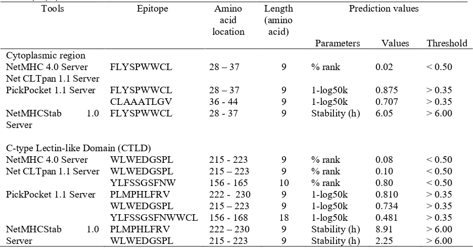
Table 1. Epitope prediction result.
Tools Epitope Amino
acid location
Length (amino acid)
Prediction values
Parameters Values Threshold Cytoplasmic region
NetMHC 4.0 Server FLYSPWWCL 28 – 37 9 % rank 0.02 < 0.50
Net CLTpan 1.1 Server
PickPocket 1.1 Server FLYSPWWCL
CLAAATLGV 28 – 37 36 - 44 9 9 1-log50k 1-log50k 0.875 0.707 > 0.35 > 0.35
NetMHCStab 1.0
Server FLYSPWWCL 28 - 37 9 Stability (h) 6.05 > 6.00
C-type Lectin-like Domain (CTLD)
NetMHC 4.0 Server WLWEDGSPL 215 - 223 9 % rank 0.08 < 0.50
Net CLTpan 1.1 Server WLWEDGSPL
YLFSSGSFNW 215 – 223 156 - 165 10 9 % rank % rank 0.10 0.80 < 0.50 < 0.50 PickPocket 1.1 Server PLMPHLFRV
WLWEDGSPL YLFSSGSFNWWCL
222 - 230 215 – 223 156 - 168
9 9 18
1-log50k 1-log50k 1-log50k
0.810 0.734 0.481
> 0.35 > 0.35 > 0.35
NetMHCStab 1.0
107
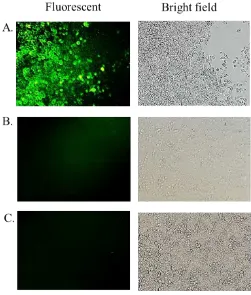
Figure 2. Transient expression of the fusion GFP-LOX-1 in HeLa cells observed 18 h post transfection.pGFP-LOX-1 transfected HeLa cells showed fluorescence (A) while the HeLa transfected with pcDNA3.1 without GFP-LOX-1 (B) as negative control and non-transfected Hela cells (C) did not show any fluorescence. Magnification 100x.
From two databases which were compared, we can find similar amino acid sequences of the LOX-1 protein (Uniprot ID P78380 and Protein Data Bank ID 1PYX). The whole amino acid sequence (length: 273 amino acids) was used to predict its antigenicity. Our antigenicity studies revealed that human LOX-1 contains conserved T cells reactive regions (Table 1). The epitopes which were predicted are FLYSPWWCLAAATLGV and WLWEDGSPLMPHLFRV. From these prediction results, the CTLD of the LOX-1 protein is prospective as the vaccine candidate.
2. pLOX-1 and pGFP-LOX-1 construction
pLOX-1 and pGFP-LOX-1 were analyzed using restriction, PCR, and sequencing analysis. Restriction analysis was carried out by plasmid digestion with the single (NheI) and double enzymes (NheI and EcoRV) (Figure 1). Single enzyme (NheI) digestion showed a band in
108
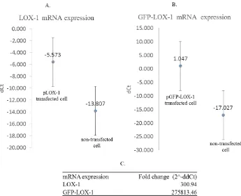
Figure 3. The mRNA expression of LOX-1 and GFP-LOX-1 in HeLa cells. The mRNA expression was calculated as dCt that presented the target gene
expression normalized to β-actin. LOX-1 and GFP-LOX-1 mRNA expression level were higher in transfected cells compare to the non-transfected Hela cells, 300 times and 275,813 times, respectively. Notes: A. LOX-1 mRNA expression of pLOX-1 transfected and non-transfected HeLa cells, B. GFP-LOX-1 mRNA expression of pGFP-LOX-1 transfected and non-transfected HeLa cells, C. fold change expression was calculated as 2^-ddCt.
3. Increased LOX-1 and GFP-LOX-1 expression in transfected HeLa cell lines
The expression of eGFP-LOX-1 in Hela cells was able to be visualized under fluorescence microscope while the HeLa transfected with empty plasmid pcDNA3.1 and the non-transfected HeLa cells did not show fluorescence signals (Figure 2). The highest fluorescence intensity was observed in 18 h post transfection.
In our study, the mRNA expression levels of LOX-1 and GFP-LOX-LOX-1 were quantified with the help of the qRT-PCR. The mRNA levels of LOX-1 and GFP-LOX1 were compared with that of β-actin mRNA. As predicted, the mRNA levels of LOX-1 and GFP-LOX-1 in transfected cells were higher than those in non-transfected cells (Figure 3). mRNA expression level of pLOX-1 transfected HeLa was 300 times higher than that in the non-transfected HeLa cells, while the GFP-LOX-1 was
109
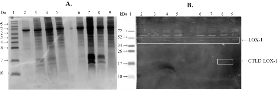
Figure 4. Detection of LOX-1 by SDS-PAGE (A) and Western Blot (B) analysis. Expressed proteins on the 6, 12, 18, and 24 h post transfection (A: lane 2-5) from HeLa cells; expressed proteins on the 6, 12, 18, 24 h post transfection (A: lane 6-9) from HeLa+ pLOX-1 cells. Cell extracts were analyzed by Western blotting using an anti-LOX-1, all of the Hela cells extracts that were expressed on the 6, 12, 18, and 24 h post transfection (B: lane 2-5) and Hela+pLOX-1 cells extracts that were expressed on the 6, 12, 18, 24 h post transfection (B: lane 6-9) produced bands in about 45 kDa, showing a mature form of the LOX-1 protein. The lane 8 on B showed Hela+pLOX1 expressed CTLD LOX-1 with the size about 14 kDa. This band showed that CTLD LOX-1 only expressed in pLOX-1 transfected Hela cells, that was detected 12 h post transfection.
Discussion
In this study, we designed epitope based vaccine using epitope prediction tolls. Epitope prediction used in silico study is a new method that is applied to increase the effectiveness and safety of vaccines. Through epitope prediction, produced vaccine is expected to work more specifically on the target antigen and to reduce the risk of side effects, particularly the risk of hypersensitivity [22, 28]. To evaluate the antigenicity of the LOX-1 protein, we used several tools, Net MHC 4.0 Server, Net CTL Pan Server 1.1, Pick. Pocket 1.1 Server, and Net MHC Stab 1.0 server. The tools identified two epitopes of human LOX-1, FLYSPWWCLAAATLGV and WLWEDGSPLMPHLF RV. The first epitope located in the cytoplasmic region while the second epitope presented in the C-type lectin-like domain (CTLD) LOX-1. CTLD is the most important domain in LOX-1 which plays roles in the binding activity of LOX-1 with its ligands. Modification of several amino acids in this domain in fact reduced the binding activity of the LOX-1 protein [29, 30]. From our data, it suggested that LOX-1 protein is prospective to be developed as DNA vaccine candidate which can induce the T cell activation.
Despite the promising results of the LOX-1 epitope as a vaccine candidate, there are concerns that the vaccine will be recognized as auto-antigen and tolerated by T cells. Several other studies that were conducted to overcome the problem suggested that dose optimization and delivery system are required to increase the immune response [31, 32]. Other studies showed the use of vaccine along with Salmonella typhimurium AroA- as a live vector successfully induced a T cell response that inhibited atherosclerosis [16, 17, 33].
Based on our epitope prediction result, we construct the DNA vaccine using pcDNA3.1 carrying CTLD encoding gene (LOX-1 epitope). Constructed plasmids were successfully transformed into host cells and characterized by the ability of host cells to grow in the selection media LB agar containing ampicillin. Characterization of plasmids was conducted using enzyme restriction and sequencing analyses.
non-110
oncogenic, non-immunogenic, and easily manufactured. Liposomes facilitate entry of DNA through the cell membrane via endocytosis. This process was mediated by chlatrin and caveolae [34]. Although it has been proven effective, some studies indicate the toxic side effect of the liposome to the host cells. Different cell types have different resistance - depending on the toxicity of lipid [35, 36]. From our optimization result, the optimum DNA and lipid composition
is 1 μg DNA: 0.5 μl lipid.
Measurement of mRNA level 24 h post transfection showed that the transfected Hela cells had higher LOX-1 mRNA expression than non-transfected HeLa (p < 0.05). The pcDNA3.1 contains CMV promoter. Zarrien et al. not the endogenous protein expressed by HeLa cells. This result supports our hypothesis that the LOX-1 DNA vaccine can be recognized and expressed by the mammalian cells.
Surprisingly, from Western blot result (Figure 4), LOX-1 was expressed either in the transfected as well as in non-transfected HeLa cells. LOX-1 is an endogenous protein expressed by the HeLa. Previous research by Hirsch et al. suggested the correlation of the LOX-1 expression with the cancer cell growth. Lipid metabolism related genes are highly expressed in cancer cells, including HeLa, demonstrating the importance of the lipid metabolism during cellular transformation. Lipid metabolism activation has major roles in tumorigenesis. LOX-1 is one of the lipid metabolism related protein which has been known to be upregulated in the cancer cell lines [38]. LOX-1 expression is vital to sustain tissue integrity and its increasing expression is correlated with cancer cell invasion [38, 39]. Although the mRNA level of the LOX-1 in transfected HeLa cells is much higher than that in non-transfected HeLa cells, in the translational
level, the expression of LOX-1 of the transfected and non-transfected cells were not significantly different (p > 0.05). This result suggested that the post transcription regulation of the OLR1 gene may play a main role in controlling the LOX-1 protein level.
All transfected and non-transfected HeLa cells expressed LOX-1 protein in their mature form with the size was about 45 kDa. The molecular mass of the human LOX-1 is 30.939 Da. However, the glycosylation process produces the 45 kDa mature LOX-1 [32]. The CTLD LOX-1 with the size about 14 kDa was only observed in the pLOX-1 transfected HeLa cells, which suggested that the pLOX-1 could be recognized by the mammalian cells.
Our results suggest that optimization of the LOX-1 promoter is needed in this DNA vaccine development for higher expression efficiency. A rational approach to overcome the low-level expression of the antigen is to use intron as a combination with the promoter, such as the combination of CMV promoter and Intron A [40]. The Intron A induces the antigen level expression through its enhancer activity. It has Nuclear Factor 1 (NF1) that acts as the transcription binding sequence. Intron A also increases the mRNA stability and mRNA transport from nucleus to the cytoplasm, therefore, enhances the translation process [41, 42]. Other study modified the CMV promoter and combine the promoter with chicken β-actin promoter (CAG) or woodchuck hepatitis virus post-transcriptional response element (WPRE), an RNA sequence that escalates the mRNA stability and extra nucleus transport [43]. Another approach to increase the antigen expression is by reducing the bacterial element which has been confirmed to improve the transfection efficiency and antigen expression [44, 45].
Conclusion
111
WLWEDGSPLMPHLFRV. pLOX-1 can be recognized by mammalian expression system. While this study only examined the expression of LOX-1 in mammalian cells, we expect that similar expression could occur in animal model.
Acknowledgments
This research was funded by Ministry of Health, Republic of Indonesia through Riset Binaan Ilmu dan Teknologi Kedokteran (Risbin Iptekdok) 2016 Grant. The authors thank Widodo Ph.D., Faculty of Mathematics and Natural Sciences, Brawijaya University, for kindly providing us with the E. coli DH5α strain and for valuable
discussion regarding the research results. We also thank Professor Fatchiyah Ph.D. for her brilliant suggestions during the research, Tarina Widaningrum S.Si, MP. and Suci Megasari S.Si. MP. for their excellent technical assistance during the works.
Reference
1. Mozaffarian D, Benjamin EJ, Go AS, Arnett DK, Blaha MJ, Cushman M de FS, et al. 2015. Executive Summary: Heart Disease and Stroke Statistics--2015 Update: A Report From the American Heart Association. Circulation. 131: 434–441. 2. Packard RRS, Libby P. 2008. Inflammation in atherosclerosis: atherosclerosis. J Am Coll Cardiol. 64. 25.
5. Jan M, Meng S, Chen NC, Mai J, Wang H, Yang XF. 2010. Inflammatory and autoimmune reactions in atherosclerosis and vaccine design informatics. J Biomed Biotechnol. doi:10.1155/2010/459798 Nilsson J. 2008. Treatment with apo B peptide vaccines inhibits atherosclerosis in human apo B-100 transgenic mice without inducing an increase in peptide-specific antibodies. J Intern Med. 264(6):563–570. doi: 10.1111/j.1365-2796.2008.01995.x 9. Tissot AC, Spohn G, Jennings GT, Shamshiev A, Kurrer MO,
Windak R, et al. 2013. A VLP-based vaccine against
interleukin-1α protects mice from atherosclerosis. Eur J Immunol.
43(3):716–722. Doi:10.1002/eji.201242687.
10. Mao D, Kai G, Gaofu Q, Zheng Z, Li Z, Jie W, et al. 2006. Intramuscular immunization with a DNA vaccine encoding a 26-amino acid CETP epitope displayed by HBc protein and containing CpG DNA inhibits atherosclerosis in a rabbit model of atherosclerosis. Vaccine. 24 (23): 4942–4950. doi:10.1016/j.vaccine.2006.03.082
11. Liaw YW, Lin CY, Lai YS, Yang TC, Wang CJ, Whang-Peng J. 2014. Vaccine Targeted at CETP Alleviates High Fat and High Cholesterol Diet-Induced Atherosclerosis and Non-Alcoholic Steatohepatitis in Rabbit. PLoS One. 9(12).e111529. doi:10.1371/journal.pone.0111529.
12. Gaofu Q, Jun L, Xiuyun Z, Wentao L, Jie W, Jingjing L. 2005. Antibody against cholesteryl ester transfer protein (CETP) elicited by a recombinant chimeric enzyme vaccine attenuated atherosclerosis in a rabbit model. Life Sci. 77 (21) :2690–2702. doi:10.1016/j.lfs.2005.05.037
13. Rittershaus CW, Miller DP, Thomas LJ, Picard MD, Honan CM, Emmett CD, et al. 2000. Vaccine-Induced Antibodies Inhibit CETP Activity In Vivo and Reduce Aortic Lesions in a Rabbit Model of Atherosclerosis. Arterioscler Thromb Vasc Biol. 20 (9) :2106–2112.
14. Davidson MH, Maki K, Umporowicz D, Wheeler A, Rittershaus C, Ryan U. 2003. The safety and immunogenicity of a CETP vaccine in healthy adults. Atherosclerosis. 169(1):113–120. doi:10.1016/S0021-9150(03)00137-0
15. Salazar-González JA, Rosales-Mendoza S. 2013. A perspective for atherosclerosis vaccination: Is there a place for plant-based vaccines? Vaccine. 31(10):1364–1369.
16. Hauer AD, Van Puijvelde GHM, Peterse N, de Vos P, van Weel V, van Wanrooij. 2007. Vaccination against VEGFR2 attenuates initiation and progression of atherosclerosis. Arterioscler Thromb Vasc Biol. 27(9):2050–2057. doi: 10.1161/ATVBAHA.107.143743
17. Petrovan RJ, Kaplan CD, Reisfeld RA, Curtiss LK. 2007. DNA vaccination against VEGF receptor 2 reduces atherosclerosis in LDL receptor-deficient mice. Arterioscler Thromb Vasc Biol. 27(5):1095–1100. doi: 10.1161/ATVBAHA.106.139246. 18. Adianingsih OR, Tamara F, Putri AP, Nurkhairina A, Albaar TM,
Arthamin MZ, Yurina, V. 2016. Lectin-like Oxidized LDL Receptor-1 (LOX-1) Protein Vaccination Reduces Inflammation and Attenuates Atherosclerosis Progression in Atherogenic-Diet Wistar Rats. Int Cardiovasc Res J. 10:159–164.
19. Xu X, Gao X, Potter BJ, Cao JM, Zhang C. 2007. Anti-LOX-1 rescues endothelial function in coronary arterioles in atherosclerotic ApoE knockout mice. Arterioscler Thromb Vasc Biol.27(4):871–877.
20. Mehta JL, Sanada N, Hu CP, Chen J, Dandapat A,Sugawara F. 2007. Deletion of LOX-1 reduces atherogenesis in LDLR knockout mice fed high cholesterol diet. Circ Res. 100 (11):1634–1642. doi: 10.1161/CIRCRESAHA.107.149724. 21. Patronov A, Doytchinova. 2013. T-cell Epitope Vaccine Design
by Immunoinformatics. Open Biol. 3. 120139. http://dx.doi.org/10.1098/rsob.120139 2013.
22. Correia BE, Bates JT, Loomis RJ, Baneyx G, Carrico C, Jardine JG, et al. 2014. Proof of principle for epitope-focused vaccine design. Nature. 507(7491):201–206.
23. Andreatta M, Nielsen M. 2015. Gapped sequence alignment using artificial neural networks: Application to the MHC class i system. Bioinformatics. 32(4):511–517.
24. Nielsen M, Lundegaard C, Worning P, Lauemøller SL, Lamberth K, Buus S, Brunak S, Lund O. 2003. Reliable prediction of T-cell epitopes using neural networks with novel sequence representations. Protein Sci. 12(5):1007–1017.
112
26. Zhang H, Lund O, Nielsen M. 2009. The PickPocket method forpredicting binding specificities for receptors based on receptor pocket similarities: Application to MHC-peptide binding. Bioinformatics. 25(10):1293–1299.
27. Jørgensen KW, Rasmussen M, Buus S, Nielsen M. 2014. NetMHCstab - Predicting stability of peptide-MHC-I complexes; impacts for cytotoxic T lymphocyte epitope discovery. Immunology. 141(1):18–26.
28. Flower DR, Phadwal K, Macdonald IK, Coveney PV, Davies MN. 2010. T-cell epitope prediction and immune complex simulation using molecular dynamics: state of the art and persisting challenges. Immunome Res. 6 (Suppl 2): S4. 29. Tate S. 2006. Structure and mode of ligand recognition of the
oxidized LDL receptor, LOX-1. Functional and Structural Biology on the Lipo-network. 661 (2): 179–198.
30. Park H, Adsit FG, Boyington JC. 2005. The 1.4 Å crystal structure of the human oxidized low-density lipoprotein receptor lox-1. J Biol Chem. 280(14): 13593–13599.
31. Husseiny MI, Rawson J, Kaye a, Nair I, Todorov I, Hensel M, Kandeel F, Ferreri K. 2014. An oral vaccine for type 1 diabetes based on live attenuated Salmonella. Vaccine. 32(20):2300– 2307.
32. Shi X, Niimi S, Ohtani T, Machida S. 2001. Characterization of residues and sequences of the carbohydrate recognition domain required for cell surface localization and ligand binding of human lectin-like oxidized LDL receptor. J Cell Sci. 114:1273– 1282.
33. Hauer AD, Habets KLL, van Wanrooij EJ, de Vos P, Krueger J, Reisfeld R, et al. 2009. Vaccination against TIE2 reduces atherosclerosis. Atherosclerosis. 204(2):365–371.
34. Cui S, Zhang S, Chen H, Wang B, Zhao Y, Zhi D. 2012. The Mechanism of Lipofectamine 2000 Mediated Transmembrane Gene Delivery. Engineering. 04.172–175. doi:10.4236/eng.2012.410B045.
35. Felgner PL, Gadek TR, Holm M, Roman R, Chan HW, Wenz M. et al. 1987. Lipofection: a highly efficient, lipid-mediated DNA-transfection procedure. Proc Natl Acad Sci USA. 84 (21) :7413– 7417.
36. Adil MM, Erdman Z, Kokkoli E. 2014. Transfection mechanisms of polyplexes, lipoplexes, and stealth liposomes in α5β1 integrin bearing DLD-1 colorectal cancer cells. Langmuir. 30(13):3802–3810. dx.doi.org/10.1021/la5001396
37. Zarrin AA, Malkin L, Fong I, Luk KD, Ghose A. 1999. Comparison
of CMV, RSV, SV40 viral and Vα1 cellular promoters in B and T
lymphoid and non-lymphoid cell lines. Biochimica et Biophysica. 1446: 135–139.
38. Rizzacasa B, Morini E, Pucci S, Murdocca M, Novelli G, Amati F. 2017. LOX-1 and Its Splice Variants: A New Challenge for Atherosclerosis and Cancer-Targeted Therapies. Int J Mol Sci. 290. doi:10.3390/ijms18020290.
39. Hirsch H, Iliopoulos D, Joshi A, Zhang Y, Savina A, Bulyk M, et al. 2010. A transcriptional signature and common gene networks link cancer with lipid metabolism and diverse human diseases. Cancer Cell. 17(4):348–361.
40. Li L, Saade F, Petrovsky N. 2012. The future of human DNA vaccines. J Biotechnol. 162(2):171–182.
41. Garmory HS, Brown K A, Titball RW. 2003. DNA vaccines: improving expression of antigens. Genet Vaccines Ther. 1:2. 42. Wang S, Farfan-Arribas DJ, Shen S, Chou THW, Hirsch A, He F,
et al. 2006. Relative contributions of codon usage, promoter efficiency and leader sequence to the antigen expression and immunogenicity of HIV-1 Env DNA vaccine. Vaccine. 24(21):4531–4540.
43. Garg S, Oran AE, Hon H, Jacob J. 2004. The hybrid cytomegalovirus enhancer/chicken beta-actin promoter along with woodchuck hepatitis virus posttranscriptional regulatory
element enhances the protective efficacy of DNA vaccines. J Immunol. 173(1):550–558.
44. Faurez F, Dory D, Le Moigne V, Gravier R, Jestin A. 2010. Biosafety of DNA vaccines: New generation of DNA vectors and current knowledge on the fate of plasmids after injection. Vaccine. 28(23):3888–3895.