ORIGINAL ARTICLE
Factors important for efficacy of stereotactic body radiotherapy of
medically inoperable stage I lung cancer. A retrospective
analysis of patients treated in the Nordic countries
PIA BAUMANN
1, JAN NYMAN
2, INGMAR LAX
1, SIGNE FRIESLAND
1,
MORTEN HOYER
3, SUZANNE REHN ERICSSON
4, KARL-AXEL JOHANSSON
2,
LARS EKBERG
5, ELISABETH MORHED
4, MERETE PALUDAN
3, LENA WITTGREN
5,
HENRIK BLOMGREN
1& ROLF LEWENSOHN
11
Radiumhemmet Karolinska University Hospital, Division of Oncology and Hospital Physics, Sweden,2Sahlgrenska University Hospital Gothenburg, Department of Oncology and Radiation Physics, Sweden,3Aarhus University Hospital, Division of Oncology and Medical Physics, Denmark,4University Hospital, Uppsala, Department of Oncology and Radiology, Sweden and5Malmo¨ University Hospital, Division of Oncology and Hospital Physics, Sweden
Abstract
We reviewed results of SBRT treatment of 138 patients with medically inoperable stage I NSCLC treated during 1996
2003 at five different centres in Sweden and Denmark. Mean age was 74 years (range 5690) with 69 men and 72 women. SBRT was delivered using a 3D conformal multifield technique and a stereotactic body frame. Doses delivered were 3048 Gy (65% isodose at the periphery of planning target volume, PTV) in 24 fractions. Equivalent dose in 2 Gy fractions (EQD2) was in the range of 50100 Gy. Mean gross tumour volume (GTV) was 39 cm3(2436), and planning target
volume was 101 cm3(11719). Overall response rate (CR, PR) was 61% (84/138). SD was noted in 36% (50/138). During
a median follow-up period of 33 months (1107), 16 (12%) local failures occurred, ten of which also included distant
metastases. Local failure was associated with tumour size, target definition and central or pleura proximity. Distant metastases occurred in 25% (35/138) of the patients. Ninety-one (65%) patients died during follow-up of which 55 patients (60%) died of other causes than lung cancer. Three- and 5-year overall survival was 52 and 26% respectively. Lung cancer specific 3- and 5-year overall survival was 66 and 40% respectively. Fifty nine percent (83/138) of the patients had no side effects. Fourteen patients experienced grade 34 toxicity according to radiation therapy oncology group (RTOG). EQD2
(/v.s.B/55.6 Gy) showed a statistically significant benefit survival for the higher doses. SBRT for stage I NSCLC results in
favourable local control not inferior to fractionated RT and with acceptable toxicity.
For patients with Stage I (T12, N0, M0) non-small
cell lung cancer (NSCLC), surgical resection re-mains the treatment of choice, resulting in local control rates of 80%100% and overall survival rates
of 50%97% after 5 years[15]. For patients with severe co-morbidity, e.g. chronic obstructive pul-monary disease (COPD), cardiovascular disease (CVD) or other malignancies, surgical resection is often not feasible or involves excessive risks. The alternative treatment for these patients has been conventional fractionated external radiotherapy (RT) [69] with local control rates of 4070% and 5-year survival rates of 530% [1013]. The most
common site of relapse of early stage NSCLC treated with conventional RT is local failure[10]. Several studies report that large tumour size and insufficient dose of radiation are the main reasons for poor local control [7,9,10,12,1417]. Early stage lung cancer (T12, N0, M0) is usually not con-sidered a systemic disease from diagnosis, and prophylactic nodal irradiation is not recommended [9,14]. Increasing the dose, with conventional RT methods, for better tumour control is not always feasible in these patients because of restricted respiratory function and an associated risk of in-creased pulmonary toxicity. However, with improved
Correspondence: Rolf Lewensohn, Department of Oncology, Radiumhemmet, Karolinska University hospital, SE171 76 Stockholm, Sweden. Tel:/46 8
5177 3188. Fax:/46 8 5177 100. E-mail: [email protected]
(Received 16 June 2006; accepted 5 July 2006)
ISSN 0284-186X print/ISSN 1651-226X online#2006 Taylor & Francis
DOI: 10.1080/02841860600904862
Acta Oncol Downloaded from informahealthcare.com by 188.237.180.55 on 05/20/14
methods including higher geometrical accuracy of dose delivery, escalation of the dose to the target with acceptable toxicity may be obtained.
Immobilisation is an essential component when using stereotactic body radiotherapy (SBRT) for high accuracy and reproducibility of dose delivery. For extracranial targets internal motion must be considered. Based on promising results with intra-cranial stereotactic radiotherapy [1821] a method for SBRT was developed [2224]. This method has been in clinical use since 1991 and has during the last decade migrated to other radiotherapy centres in various countries. The four cornerstones of this method are: 1) Stereotactic methodology for target localization and treatment set-up, 2) CT verification in order to directly verify the position of the tumour in the stereotactic coordinate system, 3) Heteroge-neous dose distribution in the target, in order to increase the probability to kill the most resistant tumour cells anticipated to be localized to the central parts of the tumour, 4) Hypofractionation, in order to prevent repopulation of the tumour as well as increasing cost effectiveness and improving the convenience for the patient. The method is further-more based on the use of abdominal compression in order to reduce the tumour motion with breathing [24,25]. By reducing the motion of the target and increasing the geometrical accuracy, SBRT allows smaller margins.
The use of hypofractionation, with a decrease of overall treatment time to one week compared to more than six weeks for conventional radiotherapy is of great importance, not only to prevent repopula-tion of tumour cells during therapy, but also to prevent treatment interruptions due to high grade toxicity in this category of medically inoperable patients and subsequently reduced survival rates [26].
Several earlier studies on thoracic and non-thor-acic tumours with SBRT demonstrate high local control rates and a mild panorama of side-effects [22,23,27]. The majority of patients experienced no side-effects at all whereas a minority of cases showed clinical pneumonitis, rib fracture and thoracic pain. In these studies patients with inoperable early stage lung cancer who received treatment with SBRT seemed to have less side-effects, better local control of the tumour and at least equivalent numbers of five-year survival rates, compared to patients treated with conventional radiotherapy. The set up using the SBRT methodology has been utilized in all centres participating (4 in Sweden and 1 in Denmark in the present study). The 141 patients with early stage lung cancer (T12, N0, M0) in the present study
form one of the larger cohorts of stage I NSCLC patients at present treated with SBRT. Together with
the variation in dose and fractionation this permits analysis of dose dependence and impact of tumour volume on local control and survival.
Material and methods
One hundred and forty one (72 women and 69 men) consecutive patients with inoperable non-small cell lung cancer (NSCLC) stage I (T1T2N0M0)
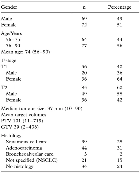
received treatment with SBRT between 1996 to 2003 at four institutions (Stockholm, Gothenburg, Uppsala, Malmo¨) in Sweden and one institution in Denmark (Aarhus) (Table I). The patients were considered inoperable mainly due to poor lung function, severe cardiovascular disease or other malignancies (Table II). Four percent (5/141) of the patients refused to undergo surgery. The median tumour diameter was 37 mm (1090) and the tumours were mainly localized in the periphery of the lung. At the time of treatment the mean age was 74 years (range 5690). Patient information was retrieved from medical files, x-ray films and from the national registries of death causes.
All patients underwent CT-scans in the diagnostic work up and in 76% (107/141) of the patients tumour diagnosis was histologically confirmed with either bronchial lavage, biopsy or sputum cytology (Table I). Risk of fatal pneumothorax rendered biopsy unfeasible in many of the patients with severe pulmonary dysfunction. An experienced thoracic
Table I. Patient and tumour characteristics.
Gender n Percentage
Median tumour size: 37 mm (1090) Mean target volumes
PTV 101 (11719) GTV 39 (2436)
Histology
Squamous cell carc. 39 28 Adenocarcinoma 44 31 Broncheoalveolar carc. 3 2 Not specified (NSCLC) 21 15
No histology 34 24
Acta Oncol Downloaded from informahealthcare.com by 188.237.180.55 on 05/20/14
radiologist evaluated the CT-scans. If no cytology was available an increase in size of the lung lesion with ‘‘malignant appearance’’ between the two latest CT-scans, in an interval of approximately 3 months, together with no evidence of any extra pulmonary tumour, set the basis for assigning the lesion as a lung cancer. Bone scintigraphy, brain and abdominal CT-scans or PET were not routinely used to search for distant metastases.
All patients were treated with SBRT in the Elekta Stereotactic Body Frame (SBF) with 6 MV from a linear accelerator. Fluoroscopy of the thorax was performed with the patient positioned in the SBF. Movements of the diaphragm were examined during normal breathing. If the movements were more than 9/5 mm, pressure was applied on the upper abdo-men, using the diaphragm control device, in order to minimize the diaphragmatic movements, to control the cranio-caudal movements of the tumour [24,25]. A CT-scan, with the patient in the SBF, was done at first treatment occasion to control reproducibility of the tumour in the stereotactic coordinate system (should be within 5 mm in the transversal plane and 10 mm in the longitudinal direction). Reproduci-bility of the position of the target in the stereotactic coordinate system has been found to be 3.7 mm in the transversal plane and 5.7 mm in the longitudinal plane [24]. Based on previous experience a margin of 510 mm (transversal plane) and 10 mm (long-itudinal plane) was added around the Clinical target volume (CTV) to define the planning target volume (PTV). Verification CT-scans for image guidance were performed prior to the first treatment. If reproducibility was unclear, repeated CT-scans were done before the second/third treatments.
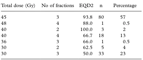
Dose was specified to the periphery of the PTV. The dose to the central parts of the PTV was generally 140%150% of the specified dose, using
dose plans with inhomogeneous dose distributions. Different fractionation schemes were used (Table III), ranging from 2 to 4 fractions with a dose/ fraction of 10 to 20 Gy given two to three days apart in general. A total dose of 30 to 48 Gy was given. The three-dimensional dose planning was based on CT slicesB/5 mm within the PTV, taken under free
shallow breathing. Five to nine non-coplanar or coplanar beams were used. The beams were shaped with the help of multi-leaf collimators to conform to the projection of the PTV in different directions of the beams.
For the subsequent radiological evaluation after SBRT, the response was considered complete (CR) if there was no visible tumour, partial (PR) if the cross sectional tumour area was reduced with at least 50% and stable disease (SD) if there was less than 50% decrease or less than 25% increase in this parameter. Local failure was defined as /25% increase of cross sectional tumour area.
Statistical analysis
Kaplan-Meier algorithms calculating survival and log-rank test evaluating the equality of the curves were used. Cross tabulation andx2test were used in comparative analysis.
Results
There are some limitations in the response evalua-tion in this retrospective study, since patients, were followed according to local tradition with CT-scans performed at different time points. Response is therefore based on CT-scans performed in a period of 0.589.3 months (median 16.3) post therapy, and should therefore be regarded as ‘‘best response’’. The overall response rate (CR 33%, PR 28%) was 61% (84/138). Forty-five percent of the T1 and 24% of the T2 tumours showed CR response. Stable disease (SD) was noted in 36% (50/138). Of the T1 tumours 23% and of the T2 tumours 44% showed SD. Local tumour progression was seen in four patients (3%) who all had large T2 tumours. Of the 141 patients three patients were not possible to evaluate or lost to follow-up and hence all evalua-tions were made on 138 patients. The median follow-up period was 33 months (range 1107).
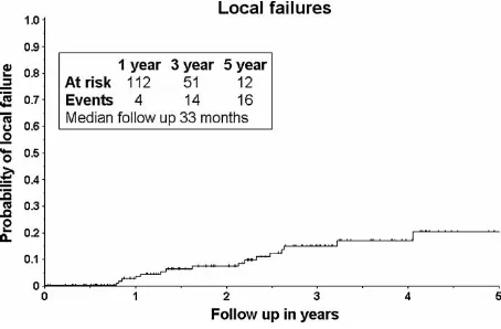
An important aim using RT in early LC is to obtain local control. Out of the 138 evaluable patients entered into the study 16 patients (12%) had a local failure within the observation time. Time
Table II. Reasons for inoperability.
n Percentage
COPD 78 55
CVD 25 18
COPD/CVD 21 15
Other malignancies 14 10 Other compromising diseases 3 2
Abbreviations: CVD: Cardio vascular disease, COPD: Chronic obstructive pulmonary disease.
Table III. Distribution of doses, fractions and EQD2 (at the periphery of PTV).
Total dose (Gy) No of fractions EQD2 n Percentage
45 3 93.8 80 57
Acta Oncol Downloaded from informahealthcare.com by 188.237.180.55 on 05/20/14
to failure varied between 1049 months (median
17.8). Within 36 months 13/16 local failures oc-curred. We could see a decline in local failure and a plateau at three years with no local failures appearing after four years (Figure 1). It is interesting to understand to which extent local failures are isolated or combined with regional or distant metastases. Ten of the 16 local failures had additional metastatic disease (Figure 2). Overall, 44 patients with pro-gressive disease were encountered during the follow-up time of which only 4 (3%) appeared as local failure without evidence of distant metastasis (Figure 2). Local failure was more common among T2 (13%) compared with T1 tumours (3%). Best response (CR and PR) did not correlate with risk of local failure. While there was no relation between CR and survival in T1 tumours, CR in T2 tumours are predictive of better survival as compared to what is seen with PR/SD cases (pB/0.0361). In T2 tumours a similar relation and p-value was found between CR and general failure (pB/0.0361).
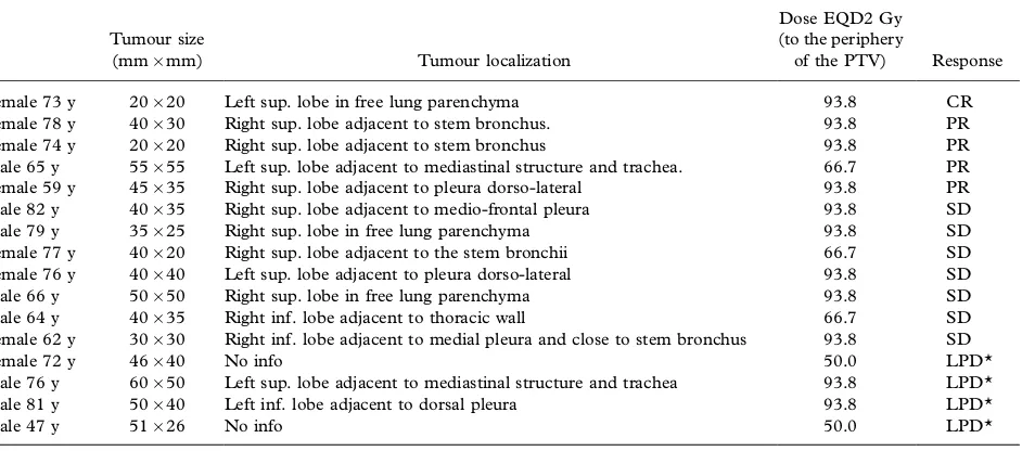
Some interesting observations on factors related to local failure can be made. In our material the majority of local failures (11/14) were seen in tumours in proximity to, and possible contact with, centrally located risk organs or pleura (Table IV).
We addressed the issue of tumour size as a predictive factor of risk of local failure with our SBRT approach on stage I tumours. Cases with T1 tumours had significantly less risk for local failure than T2 tumours (pB/0.0067) with failures appear-ing later after treatment of T1 than T2 disease (Figure 3). In a similar way it was possible to use GTV as a measure of tumour size and divide the tumours by their median GTV (26 cm3) with a statistically, significant difference in risk of local failure (pB/0.0015).
Since there is a variation of the doses delivered to the tumours in our material we were interested in
the impact of dose on local control. Mean doses to GTV calculated as EQD2 (equivalent dose in 2 Gy fractions) (a/b/10) were within the range of 65205 Gy. Within this dose range we found no
general correlation between dose and local control or overall survival rates. The EQD2 at the periphery of the PTV ranged from 50 to 100 Gy. We divided the patient material into two groups at EQD2/ and B/55.6 Gy and analyzed the frequencies of local failures and survival. This cut-off point was chosen with reference to an earlier study where a clearly measurable difference in rates of local failure was seen for the high dose group, with BED/100 Gy to isocenter. BED/100 Gy to isocenter will give BED/67 Gy to the periphery of PTV with the dose distributions we have used. That in turn will give EQD2/55.6 Gy to the periphery of PTV. They also compared dose groups where both groups received doses above BED/100 Gy to the isocenter (converted to our dose distributions; 55.6 Gy to the periphery of the PTV) and could not find any difference [39].
We could, however, not find any difference in risk of local failure between the groups B/and /55.6 Gy
Figure 1. Probability of local failure among the 138 stage I NSCLC cases. Patients at risk for local failure and number of cases with local failure are given for time periods up to four years.
Figure 2. Failure pattern of the 44 disease progressions encoun-tered during follow-up of the 138 stage I NSCLC cases.
Figure 3. Probability of local failure as related to T-stage of the 138 patients with stage I NSCLC.
Acta Oncol Downloaded from informahealthcare.com by 188.237.180.55 on 05/20/14
in EQD2, but there was a statistical significant advantage in survival for the group with higher doses than 55.6 Gy (pB/0.0018) (Figure 4).
With the current deficiencies in assessing meta-static disease and thus assigning a correct stage, many patients will carry metastatic disease already at the outset of treatment. In 25% (35/138) of the patients distant metastases occurred. Between T1-and T2-tumours no difference in metastatic pattern was noted. We found no difference in survival between T1 and T2 tumours. Considering T1 and T2 tumours in two groups, one with and one without cytological diagnosis none of the two groups showed a survival difference or risk of local failure with respect to T-status. There was no change in incidence of general failure after 3 years of follow-up (Figure 5). Ninety one (65%) of the patients died during follow-up and among them 60% (55/91) died
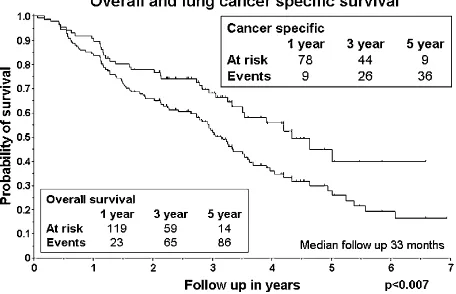
of other causes than lung cancer. Seven patients (8%) died with loco-regional lung cancer present (Table V). Both local and regional disease and distant metastases were more frequent as cause of death in T2 as compared to T1 tumours (Table V). Overall survival was 52% and 26% after 3 and 5 years of follow-up (Figure 6). Lung cancer specific survival was 66% and 40% after 3 and 5 years (Figure 6). Local failure free survival was 55% and 29% after 3 and 5 years. Total failure free survival was 56% and 36% after 3 and 5 years. Women had a slightly better overall 3- and 5-year survival rate than men, namely of 58% and 31% as compared to 46% and 21%. These differences were, however, not statistically significant.
An important issue during the development of SBRT as a treatment option is to register side effects
Table IV. Relapse pattern of 138 stage I NSCLC tumours treated by SBRT.
Id
Tumour size
(mm/mm) Tumour localization
Dose EQD2 Gy (to the periphery
of the PTV) Response
Female 73 y 20/20 Left sup. lobe in free lung parenchyma 93.8 CR
Female 78 y 40/30 Right sup. lobe adjacent to stem bronchus. 93.8 PR
Female 74 y 20/20 Right sup. lobe adjacent to stem bronchus 93.8 PR
Male 65 y 55/55 Left sup. lobe adjacent to mediastinal structure and trachea. 66.7 PR
Female 59 y 45/35 Right sup. lobe adjacent to pleura dorso-lateral 93.8 PR
Male 82 y 40/35 Right sup. lobe adjacent to medio-frontal pleura 93.8 SD
Male 79 y 35/25 Right sup. lobe in free lung parenchyma 93.8 SD
Female 77 y 40/20 Right sup. lobe adjacent to the stem bronchii 66.7 SD
Female 76 y 40/40 Left sup. lobe adjacent to pleura dorso-lateral 93.8 SD
Male 66 y 50/50 Right sup. lobe in free lung parenchyma 93.8 SD
Male 64 y 40/35 Right inf. lobe adjacent to thoracic wall 66.7 SD
Female 62 y 30/30 Right inf. lobe adjacent to medial pleura and close to stem bronchus 93.8 SD
Female 72 y 46/40 No info 50.0 LPD*
Male 76 y 60/50 Left sup. lobe adjacent to mediastinal structure and trachea 93.8 LPD*
Male 81 y 50/40 Left inf. lobe adjacent to dorsal pleura 93.8 LPD*
Male 47 y 51/26 No info 50.0 LPD*
*Local progressive disease.
Figure 4. EQD2 (equivalent dose in 2 Gy fractions) to the periphery of PTV divided in 2 groups 5/55.6 Gy and /55.6
Gy and overall survival of the 138 stage I NSCLC cases.
Figure 5. Time to total failure for the 138 stage I SSCLC cases. Failures include local, regional and distant metastasis. Patients at risk for failure and number of cases with failure are given for one, three and five year time periods.
Acta Oncol Downloaded from informahealthcare.com by 188.237.180.55 on 05/20/14
and understand its underlying causes. Toxicity was mild (according to information in medical records) and 60% (83/138) of the patients had no side effects. The most common side effects were skin rash, costal fracture, cough and radiological pneumonitis/fibrosis without clinical symptoms. In ten patients atelec-tases occurred. Fourteen patients experienced grade 34 toxicity, according to RTOG, where pain in thorax was the major cause (Table VI). More information on toxicity was not possible to retrieve from patient records.
Discussion
When using RT, with curative intent in early stage NSCLC read out parameters for the efficacy of treatment are local control and survival. In the present retrospective study of medically inoperable stage I NSCLC analysis of local control as assessed during the follow-up time (median 33 month), showed that only 12% of the tumours failed locally, a figure similar to other SBRT studies on this group of patients [22,2835]. In a recent review of 18
papers published 19882000, reporting results with
conventional RT, the incidence of local failure of stage I tumours was 40% (6.470%) [10]. Local failure has been reported up to at least 3 years after RT treatment of stage I NSCLC [29,36,37]. This is similar to our finding with SBRT. We found a decline in the number of patients with risk for local
failure at a time point around 3 years after SBRT. In previous studies using SBRT, the rate of distant metastases was about 20% [29,32,33,38,39], similar to our results of 25% (35/138). Despite extensive pre-treatment staging procedures including PET and CT of the brain, the rate of distant metastases reported during follow-up still remains at 20% [27]. This is important to understand when evaluat-ing survival since even if we can achieve local tumour control, the problem with distant metastases still remains unresolved.
The biological effect of the high doses and extreme hypofractionation of the tumour in SBRT treatment still needs to be explored. While CR and PR implicate cell death and are defined as response, the meaning of SD remains obscure but may mirror either inhibition of tumour cell growth or substitu-tion of tumour by fibrosis. All these response situations may of course harbour tumour cells with potential to recur with time. Except for the problem with radiation fibrosis the time of response may strongly vary between cases. In spite that we envisage all these difficulties it is interesting to see that in our study CR in T2 tumours are predictive of better survival as compared to what is seen with PR/SD cases. The exact importance of CR as a predictor of survival thus has to await prospective trials with improved diagnostic tools. We did not find any correlation between best response and local or general failure. One apparent explanation is that we have no T2 cases with CR among patients with local relapse.
In the present study tumour size is an important determinant of local control. Tumour size as a prognostic factor for SBRT has only been addressed in a few studies. In the study by Onishi et.al., a difference in local control as well as survival was demonstrated between patients with T1 and T2 tumours [39]. The reason for the increased pre-valence of local failure in proximity to central
Table V. Cause of death.
T1 T2 Total
% of expired cases
% of all cases
Distant metastases 9 20 29 32 20 Loco reg. disease, only 1 6 7 8 5 Comorbidity 26 29 55 60 40
Figure 6. Overall and lung cancer specific survival of the 138 stage I NSCLC cases. Patients at risk for failure and number of cases with failure are given for one, three and five year time periods.
Table VI. Toxicity pattern.
Total Grade 34
No side effects 83 Lung fibrosis 21
Skin rash 12
Lung atelectasis 10 2
Cough 9
Rib fracture 8 2
Esophagitis 5
Pleural exudates 4
Pneumonitis 1 1
Thoracic pain 6 4
Pneumonia 1 1
Nausea 1
Decreased lung function 2 2 Decreased performance status 2 2
Acta Oncol Downloaded from informahealthcare.com by 188.237.180.55 on 05/20/14
structures or pleura remains obscure. The difficulties in separating tumour tissue from central structures in the work-up of an ideal dose plan could be one reason for the preponderance of failures at these locations. Another contributing factor is that the dose planning in some cases with central structures close to target was made in a way such that parts of PTV and even CTV received a lower dose. A third factor is that respiration related movements of the target, especially when abdominal pressure is not used, may result in an inadequate covering of the target during irradiation. A new method to reduce the insecurity of respiratory related tumour motion could be gating.
With conventional RT, dose escalation studies have shown that to obtain local control, radiation doses of more than 100 Gy in 2 Gy fractions are required. Due to normal tissue toxicity the max-imum tolerated dose has been set to approximately 84 Gy [40]. A recent prospective dose escalating study of stage I to III NSCLC (106 pts with 35 T1
T3N0M0) showed a positive dose-response relation-ship concerning survival and local control in the dose range of 63103 Gy [17]. To deliver these high
doses the investigators had to reduce the risk of severe toxicity, especially pneumonitis, by individua-lizing the prescribed dose based on the amount of normal lung volume irradiated. Five year overall survival in the dose interval of 6369 Gy was 4%, for
7484 Gy 22% and for 92103 Gy 28% [17]. These figures are interesting as they are similar to those emerging from our experience with SBRT in stage I NSCLC.
When comparing different studies, the BED formalism is a way of understanding the efficacy of the hypofractionated large dose deliveries used in SBRT. If we first consider what has been reported so far most studies unfortunately consist of small patient materials that do not permit full dose-effect analysis. However, one Japanese multi center study of 245 patients collected from 13 different institu-tions reported by Onishi et al. has been published. In the Japanese study, 87 patients (36%) were consid-ered operable but treated by SBRT, total radiation doses varied between 1875 Gy given in 122
fractions. Twenty seven of the patients were treated with conventional RT reaching total doses of 3044
Gy prior to SBRT. In principle this makes Onishis?
material difficult to interpret with regard to dose relationships compared to our study were our 141 medically inoperable patients were treated in a conformal way with a small range of different doses and fractionation schemes. In the Japanese study the majority of the tumours were T1 whereas most tumours in the present study were T2. The Onishi study reports a dose-response relationship on local
control and survival divided by 100 Gy to the isocenter. Increasing the dose to a BED over 120 Gy did not give any extra local control or survival benefit. BED100 Gy to the isocenter is comparable to BED 67 Gy to the periphery of the PTV, with the dose distributions used here. The equivalent dose in 2 Gy fractions (EQD2,a/b/10) will be 55.6 Gy to the periphery of the PTV. In spite of the differences between our studies as mentioned above we obtained the same result as Onishi when we divided the material into two groups with an EQD2/ and B/55.6 Gy, resulting in a statistical significant survival gain for the group receiving more than 55.6 Gy (pB/0.0018). The group receiving an EQD2B/55.6 Gy mainly consisted of T2 tumours. Consequently, we were also interested to understand if there is a difference within the T2 group with respect to dose. Looking at T2 tumours that received doses in EQD2 higher or lower than 55.6 Gy we still reached a statistical significant difference in survival favouring higher doses (pB/0.0486). Even if we could see an advantage for higher doses with respect to survival we were not able to see that patients who received lower doses had the highest rates of local failure as reported in some dose escalation studies [29,36,37].
The 5-year overall survival rate of 26% in this retrospective study was similar to what has been reported with conventional RT. Earlier SBRT stu-dies have a follow-up time insufficient to report 5-years survival, and instead mainly present 2-years survival of 6479% [2729,38]. In the study of Onishi et al. (with 36% of operable patients), however, a 5-year overall survival of 47% is reported. The high fraction of patients in the Onishi et al. study that were operable (36%) most probably form a group with a higher life expectancy than in our study on medically inoperable cases. Onishi et al. reported a 5-year overall survival for operable T1 to be as high as 90% and for T2 84%. The data presented by Uematsu et al. 2001 supports this with similar results in a smaller group of 29 operable patients who refused surgery, with a 3-year overall survival rate of 86%. These results are equivalent to what can be achieved with surgery alone with a 5-year overall survival rate of 79% to 82% [1,41]. Most of the patients in our analyses were in poor medical condition with an estimated limited survival despite local control of their cancer. Several factors are important to consider when analysing survival data of this group. First, the ‘‘observation only’’ cases have a poor outcome with a 3-year survival of approximately 20% (1419 months in median
survival) most probably related to both the lung cancer and intercurrent disorders like chronic ob-structive pulmonary disease (COPD) [42,43]. In the
Acta Oncol Downloaded from informahealthcare.com by 188.237.180.55 on 05/20/14
absence of adequate control groups the impact on survival of RT to the primary tumour of these cases remains unclear. We report that 60% (55/91) of all deaths were not primarily caused by lung cancer. Interestingly there are studies on operable COPD cases which have similar survival as cases without COPD [44]. It should, however, be remembered that inoperable cases form a group of patients that most probably show more severe COPD and cardio vascular disease (CVD) impairing survival. There-fore for studies relating to effect on survival in this patient group the issue of adequate controls should be considered in future studies.
A very important issue is to understand dose-limiting toxicity using SBRT for thoracic lesions. Analysing lung toxicity by clinical assessment is doubtful since symptoms like dyspnoea, related to reduced lung function is already present in the patient group, of the present study, at the outset of treatment. Accordingly clinical evaluation and espe-cially the retrieval of information on this point from patient records are unreliable. Also, since objective spirometry post treatment was not performed in a consistent way, the effect of SBRT on lung function in this patient group will have to await prospective trials. The data collected from patient records do, however, present a picture of low incidence of severe toxicity. In general other toxicities were mild in our material and similar to results reported by others [27,29,39,40]. Patients with atelectases as treatment side effect, carried tumours located more centrally or near a stem bronchus. Costal fractures were mainly non clinical and healed without any sequel. No life threatening toxicity was seen.
In conclusion SBRT represents a promising and effective treatment option with high local control rates and low toxicity for patients with inoperable early stage lung cancer. Compared with conventional fractionated RT the results with SBRT obtained in the present and in other studies seem to give similar long term survival but better local control [1013]. In this study we have shown that local failures during 1107 months of follow-up were 12%. The tumour size (T-stage) and volume were significantly corre-lated with the risk of local failure. The radiation dose calculated in equivalent dose in 2 Gy fractions (EQD2) at the periphery of PTV had an impact on survival but not on local failure rates.
References
[1] Chang MY, Sugarbaker DJ. Surgery for early stage non-small cell lung cancer. Semin Surg Oncol 2003;21(2):7484. [2] Ginsberg RJ, Shepherd FA. Surgery for small cell lung
cancer. Semin Radiat Oncol 1995 Jan;5(1):4043. [3] Martini N, Bains MS, Burt ME, Zakowski MF, McCormack
P, Rusch VW, et al. Incidence of local recurrence and second
primary tumors in resected stage I lung cancer. J Thorac Cardiovasc Surg 1995 Jan;109(1):1209.
[4] Naruke T, Goya T, Tsuchiya R, Suemasu K. Prognosis and survival in resected lung carcinoma based on the new international staging system. J Thorac Cardiovasc Surg 1988 Sep;96(3):4407.
[5] Mountain CF. Revisions in the international system for staging lung cancer. Chest 1997 Jun;111(6):17107. [6] Ono R, Egawa S, Suemasu K, Sakura M, Kitagawa T.
Radiotherapy in inoperable stage I lung cancer. Jpn J Clin Oncol 1991 Apr;21(2):1258.
[7] Jeremic B, Classen J, Bamberg M. Radiotherapy alone in technically operable, medically inoperable, early-stage (I/II) non-small-cell lung cancer. Int J Radiat Oncol Biol Phys 2002 Sep 1;54(1):119130.
[8] Sirze´n F, Kjellen E, So¨renson S, Cavallin-Sta˚hl E. A systematic overview of radiation therapy effects in non-small cell lung cancer. Acta Oncol 2003;42(56):493515. [9] Cheung PC, Mackillop WJ, Dixon P, Brundage MD, Youssef
YM, Zhou S. Involved-field radiotherapy alone for early-stage non-small-cell lung cancer. Int J Radiat Oncol Biol Phys 2000 Oct 1;48(3):70310.
[10] Qiao X, Tullgren O, Lax I, Sirzen F, Lewensohn R. The role of radiotherapy in treatment of stage I non-small cell lung cancer. Lung Cancer 2003 Jul;41(1):111.
[11] Dosoretz DE, Galmarini D, Rubenstein JH, Katin MJ, Blitzer PH, Salenius SA, et al. Local control in medically inoperable lung cancer: an analysis of its importance in outcome and factors determining the probability of tumor eradication. Int J Radiat Oncol Biol Phys 1993 Oct 20;27(3):50716.
[12] Kaskowitz L, Graham MV, Emami B, Halverson KJ, Rush C. Radiation therapy alone for stage I non-small cell lung cancer. Int J Radiat Oncol Biol Phys 1993 Oct 20;27(3):51723.
[13] Robertson JM, Ten Haken RK, Hazuka MB, Turrisi AT, Martel MK, Pu AT, et al. Dose escalation for non-small cell lung cancer using conformal radiation therapy. Int J Radiat Oncol Biol Phys 1997 Mar 15;37(5):107985.
[14] Krol AD, Aussems P, Noordijk EM, Hermans J, Leer JW. Local irradiation alone for peripheral stage I lung cancer: could we omit the elective regional nodal irradiation? Int J Radiat Oncol Biol Phys 1996 Jan 15;34(2):297302. [15] Sibley GS, Jamieson TA, Marks LB, Anscher MS, Prosnitz
LR. Radiotherapy alone for medically inoperable stage I non-small-cell lung cancer: the Duke experience. Int J Radiat Oncol Biol Phys 1998 Jan 1;40(1):14954. [16] Bradley J. A review of radiation dose escalation trials for
non-small cell lung cancer within the Radiation Therapy Oncol-ogy Group. Semin Oncol 2005 Apr;32(2 Suppl 3):S1113. [17] Kong FM, Ten Haken RK, Schipper MJ, Sullivan MA, Chen M, Lopez C, et al. High-dose radiation improved local tumor control and overall survival in patients with inoper-able/unresectable non-small-cell lung cancer: long-term results of a radiation dose escalation study. Int J Radiat Oncol Biol Phys 2005 Oct 1;63(2):32433.
[18] Alexander E 3rd, Loeffler JS. The case for radiosurgery. Clin Neurosurg 1999;45:3240.
[19] Flickinger JC, Loeffler JS, Larson DA. Stereotactic radio-surgery for intracranial malignancies. Oncology (Huntingt) 1994 Jan;8(1):816; discussion 86, 94, 978.
[20] O’Neill BP, Iturria NJ, Link MJ, Pollock BE, Ballman KV, O’Fallon JR. A comparison of surgical resection and stereo-tactic radiosurgery in the treatment of solitary brain metas-tases. Int J Radiat Oncol Biol Phys 2003 Apr 1;55(5):1169 76.
Acta Oncol Downloaded from informahealthcare.com by 188.237.180.55 on 05/20/14
[21] Zabel A, Milker-Zabel S, Thilmann C, Zuna I, Rhein B, Wannenmacher M, et al. Treatment of brain metastases in patients with non-small cell lung cancer (NSCLC) by stereotactic linac-based radiosurgery: prognostic factors. Lung Cancer 2002 Jul;37(1):8794.
[22] Blomgren H, Lax I, Na¨slund I, Svanstro¨m R. Stereotactic high dose fraction radiation therapy of extracranial tumors using an accelerator. Clinical experience of the first thirty-one patients. Acta Oncol 1995;34(6):86170.
[23] Blomgren H, Go¨ransson J, Kraepelien T, Nilsson B, Na¨slund I, Svanstro¨m R et al. Radiosurgery for tumours in the body: Clinical experience using a new method. J Radio-surgery 1998;1:6374.
[24] Lax I, Blomgren H, Na¨slund I, Svanstro¨m R. Stereotactic radiotherapy of malignancies in the abdomen. Methodolo-gical aspects. Acta Oncol 1994;33(6):67783.
[25] Lax I, Larsson D, Na¨slund I. Extracranial stereotactic radiosurgery of localized targets. J Radiosurgery 1998;1:13548.
[26] Jeremic B, Shibamoto Y, Milicic B, Dagovic A, Nikolic N, Aleksandrovic J, et al. Impact of treatment interruptions due to toxicity on outcome of patients with early stage (I/II) non-small-cell lung cancer (NSCLC) treated with hyperfractio-nated radiation therapy alone. Lung Cancer 2003 Jun;40(3):31723.
[27] Zimmermann FB, Geinitz H, Schill S, Grosu A, Schratzen-staller U, Molls M, et al. Stereotactic hypofractionated radiation therapy for stage I non-small cell lung cancer. Lung Cancer 2005 Apr;48(1):10714.
[28] Nagata Y, Negoro Y, Aoki T, Mizowaki T, Takayama K, Kokubo M, et al. Clinical outcomes of 3D conformal hypofractionated single high-dose radiotherapy for one or two lung tumors using a stereotactic body frame. Int J Radiat Oncol Biol Phys 2002 Mar 15;52(4):10416.
[29] Timmerman R, Papiez L, McGarry R, Likes L, DesRosiers C, Frost S, et al. Extracranial stereotactic radioablation: results of a phase I study in medically inoperable stage I non-small cell lung cancer. Chest 2003 Nov;124(5):194655. [30] Uematsu M, Shioda A, Tahara K, Fukui T, Yamamoto F,
Tsumatori G, et al. Focal, high dose, and fractionated modified stereotactic radiation therapy for lung carcinoma patients: a preliminary experience. Cancer 1998 Mar 15;82(6):106270.
[31] Uematsu M, Shioda A, Suda A, Fukui T, Ozeki Y, Hama Y, et al. Computed tomography-guided frameless stereotactic radiotherapy for stage I non-small cell lung cancer: a 5-year experience. Int J Radiat Oncol Biol Phys 2001 Nov 1;51(3):66670.
[32] Wulf J, Hadinger U, Oppitz U, Thiele W, Ness-Dourdoumas R, Flentje M. Stereotactic radiotherapy of targets in the lung and liver. Strahlenther Onkol 2001 Dec;177(12):64555.
[33] Lee SW, Choi EK, Park HJ, Ahn SD, Kim JH, Kim KJ, et al. Stereotactic body frame based fractionated radiosurgery on consecutive days for primary or metastatic tumors in the lung. Lung Cancer 2003 Jun;40(3):30915.
[34] Onimaru R, Shirato H, Shimizu S, Kitamura K, Xu B, Fukumoto S, et al. Tolerance of organs at risk in small-volume, hypofractionated, image-guided radiotherapy for primary and metastatic lung cancers. Int J Radiat Oncol Biol Phys 2003 May 1;56(1):12635.
[35] Jeremic B, Hennig M, Zimmermann FB. Predictors of radiation pneumonitis after radiotherapy in lung cancer. Int J Radiat Oncol Biol Phys 2005 Jan 1;61(1):302. [36] McGarry RC, Papiez L, Williams M, Whitford T,
Timmer-man RD. Stereotactic body radiation therapy of early-stage non-small-cell lung carcinoma: phase I study. Int J Radiat Oncol Biol Phys 2005 Nov 15;63(4):10105.
[37] Wulf J, Baier K, Mueller G, Flentje MP. Dose-response in stereotactic irradiation of lung tumors. Radiother Oncol 2005 Oct;77(1):837.
[38] Hof H, Herfarth KK, Munter M, Hoess A, Motsch J, Wannenmacher M, et al. Stereotactic single-dose radio-therapy of stage I non-small-cell lung cancer (NSCLC). Int J Radiat Oncol Biol Phys 2003 Jun 1;56(2):33541. [39] Onishi H, Araki T, Shirato H, Nagata Y, Hiraoka M, Gomi
K, et al. Stereotactic hypofractionated high-dose irradiation for stage I non-small cell lung carcinoma: clinical outcomes in 245 subjects in a Japanese multiinstitutional study. Cancer 2004 Oct 1;101(7):162331.
[40] Bradley J, Graham MV, Winter K, Purdy JA, Komaki R, Roa WH, et al. Toxicity and outcome results of RTOG 9311: a phase I-II dose-escalation study using three-dimensional conformal radiotherapy in patients with inoperable non-small-cell lung carcinoma. Int J Radiat Oncol Biol Phys 2005 Feb 1;61(2):31828.
[41] Smythe WR. Treatment of stage I non-small cell lung carcinoma. Chest 2003 Jan;123(1 Suppl):181S187S. [42] McGarry RC, Song G, des Rosiers P, Timmerman R.
Observation-only management of early stage, medically inoperable lung cancer: poor outcome. Chest 2002 Apr;121(4):11558.
[43] Cardinale RM, Wu Q, Benedict SH, Kavanagh BD, Bump E, Mohan R. Determining the optimal block margin on the planning target volume for extracranial stereotactic radio-therapy. Int J Radiat Oncol Biol Phys 1999 Sep 1;45(2):51520.
[44] Iwasaki A, Shirakusa T, Enatsu S, Maekawa S, Yoshida Y, Yoshinaga Y. Surgical treatment for lung cancer with COPD based on the Global Initiative for Chronic Obstructive Lung Disease (GOLD). Thorac Cardiovasc Surg 2005 Jun; 53(3):1627.
Acta Oncol Downloaded from informahealthcare.com by 188.237.180.55 on 05/20/14