VOL. 3, NO. 1, pp. 69 – 73, January, 2013
BZLF1 Expression of EBV is correlated with PARP1 Regulation
on Nasopharyngeal Carcinoma Tissues
Wahyu Nur Laili Fajri1, Achmad Rofi’i2, Fatchiyah1*
1
Biology Departement, Faculty of Mathematics and Natural Sciences, Brawijaya University, Malang, Indonesia 2
Ulin Hospital, Banjarmasin, Indonesia
ABSTRACT
Nasopharyngeal carcinoma (NPC) is a cancer that arises inside of the nasopharyngeal mucosa and nasopharynx. Epstein Barr Virus (EBV) has been shown to be associated with the development of NPC. Latent Membrane Protein 1 (LMP1) function include to activate BamHI -Z Leftward Reading Frame 1 (BZLF1)-EBV. The infection tissues respond to the expansion of EBV infection by activating Poly(ADP-ribose)Polymerase-1 (PARP1). The objective of this study are to observe the expression of BZLF1 and to determine the PARP1 regulation in NPC tissues. This study used three kind of tissue slides, there are non-keratinizing carcinoma, undifferentiated carcinoma, and polyp. Tissue slides were stained by HE used for characterization of the necrotic cells such as pyknosis cells, karyorrhexsis cells, and karyolysis cells. Tissues slides were analyzed by immunohistochemical (IHC) assays using EBV BZLF1-FITC and anti-PARP1-Rhod. The percentage of necrotic cells and the expression intensity of BZLF1 and PARP1 were analyzed using statistical analysis (P-value < 0.05). This study also used correlation and regression analysis. The research showed that the number of karryorhexis cells higher than pyknosis cells and karyolysis cells in NPC and polyp tissues. The high expression intensity of BZLF1 induced the increase of expression intensity of PARP1, and the expression intensity of BZLF1 and PARP1 are not correlated with percentage of necrotic cell. Interestingly, all tissues showed the increased number of karyolysis cells but the number of pyknosis cells and karryorrhexis cells were remain low. The intensity of expression indicated that the BZLF1 induce PARP1 to rep air DNA damage against EBV infection.
Keywords: BZLF1, CLSM, EBV, nasopharyngeal carcinoma, PARP1
INTRODUCTION
NPC is a cancer that arises in the epithelial tissues that covers lining of surface, inner mucosa and crypts of nasopharynx. Features of cancer were included rapid growth of cells, in-creased cell turn-over, invasive growth, metas-tasis, vascular or lymphatic channel invasion for malignant lesions [1-2]. EBV is a human herpesvirus, and be a mayor risk for NPC. Most cells and blood of patients have a part of EBV. All herpes viruses have two phases life cycle, involved latency and lytic (reactivation or replication) phase.
EBV in a latent infection is associated with the expression of nine latent proteins. Latent
*
Corresponding author: Fatchiyah
Biology Department, Faculty of Mathematics and Natural Sciences, Brawijaya University, Malang, Indonesia 65145
membrane Protein 1 (LMP1) is one of the latent proteins, and major EBV oncoprotein. This protein functions include for metastasis, apoptotic resistance, immune modulation and toactivate BamHI-Z Leftward Reading Frame 1 (BZLF1)-EBV, which can inhibit p53 to induce apoptotic resistance, metastasis, and immune modulation. BZLF1 is one of immediate-early (IE) transcription factors, that are responsible for the expression of packing proteins of EBV. BZLF-EBV functions to induce lytic infection in EBV infected cell [3-4].
and this may induce a decrease in NAD+ and ATP levels which promotes cell dysfunction and cell death by necrosis [6].
Correlation among PARP1, BZLF1-EBV, and necrotic percentage in NPC tissue are still unclear. The objective of this study is to ob-serve the expression of BZLF1 and determine the PARP1 regulation in nasopharyngeal carci-nomas tissues.
MATERIALS AND METHODS
Three kinds of histopathological tissue slides, non-keratinize carcinoma, undifferen-tiated carcinoma, and polyp was used in this study. All of these tissues slides were collected from Ulin Hospital, Banjarmasin, South Kali-mantan. NPC tissues were obtained from biop-sy which was done for early prediction of NPC patients. Methods and protocol of this study was approved by the Ethical Research Com-mission of Brawijaya University (Indonesia).
The number of necrotic cells and the expression intensity of BZLF1 and PARP1 of each histopathological tissue slides were obser-ved. This study used two kind of staining methods, hematoxylin eosin (HE) and immu-nohistochemical (IHC) assays. HE staining was conducted at Ulin Hospital, South Kalimantan. Histological slides were stained by HE used for characterization of the necrotic cells such as pyknosis (nuclear shrinkage) cells, karyorrhex-sis (nuclear fragmentation) cells, and karyolykaryorrhex-sis (nuclear fading) cells. The necrotic cells obser-ved using a BX-53 microscope (Olympus) with CellSens Standard software at the Biosains Laboratory, Brawijaya University, Malang.
Tissues slides were analyzed by IHC, modified from [7] by using EBV-BZLF1 Mouse anti-EBV monoclonal antibody (Life-span Biosciences) (1:1000) against Goat anti-mouse IgG-FITC (Santa Cruz Biotechnology) (1:1500) and anti-PARP1 antibody (MC-10) (1:1500) against Goat anti-mouse IgG labeled Rhodamin (Santa Cruz Biotechnology)(1:1500). The expression intensity of BZLF1 and PARP1 from fluorescent colour was observed by Confocal Laser Scanning Microscope (Olympus) with Olympus FluoView software
version 1.7α at the Central Laboratory of Life
Sciences (LSIH). Visualization of expression of BZLF1 and PARP1 was performed on three fields of view of each slide. Measurement of the expression intensity of each field of view is taken from 5 points. Observations and ANOVA test (P-value < 0.05). This study used correlation analysis to test the statistical significance of association between variables, and regression analysis to identify the effect of each variable.
RESULTS AND DISCUSSION
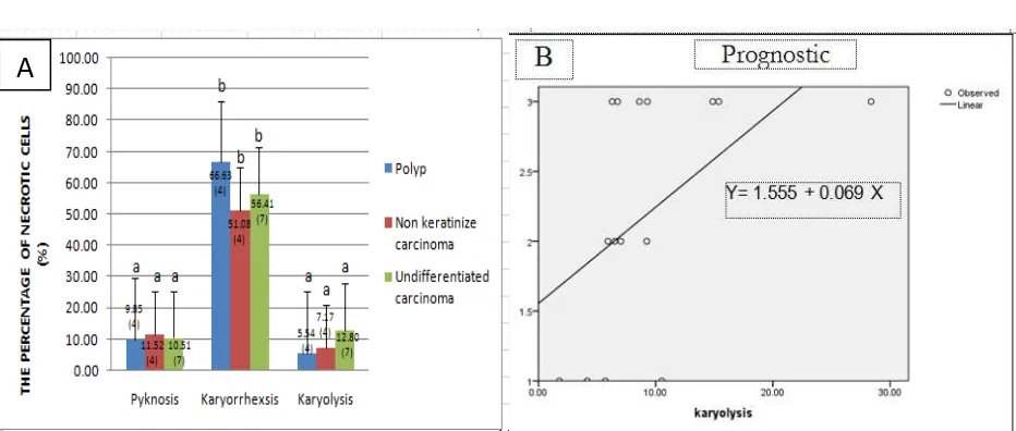
The result of this study showed that karyorrhexsis cells more commonly found than pyknosis cells and karyolysis cells. All tissues (non keratinize carcinoma, undifferentiated carcinoma, and polyp) showed the increased number of karyolysis cells, but the number of pyknosis cells and karryorrhexis cells were remain low (Fig. 1).
The high percentage of karyorrhexsis cells in all tissues indicates that the nucleus was damage and PARP1 failed to repair damaged cells, therefore the tissue target has a me-chanism of cell death by necrosis.
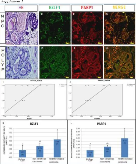
This result was similar with previous keratinize carcinoma, undifferentiated carci-noma. The expression intensity of BZLF1 and PARP1 at polyp tissue were remain low (Fig. 2 (supplement 1)). Nevertheless, the results of statistical analysis showed that the intensity of expression of BZLF1 and PARP1 was not significantly different in polyp, non keratinize carcinoma and undifferentiated carcinoma. The high intensity of expression of BZLF1-NPC showed that the infection has reached the stage of EBV lytic phase.
Figure 1. The percentages of necrotic cells (pyknosis cells, karyorrhexsis cells, and karyolysis cells) (A), and the result of regression analysis of karyolysis cells (B).
BZLF1 is a transcription factor immediate-early (IE) than BRLF1, which is responsible for the expression of proteins required in the form PARP1-NPC than PARP1 in polyps was due to infection of EBV in NPC conditions will acti-vate PARP1 which is protection of cells against DNA damage. PARP1 is involved in initiation of DNA damage response mechanisms, inclu-ding DNA repair, cell cycle arrest and apoptosis [10].
A number of PARP1 inductions are included
gamma radiation, TNFα, alkylating agents,
UVB, and free radicals. After the cells exposed to induction agents of PARP1, the PARP1 will bound directly to DNA damage and have auto modification thus forming polymer poly (ADP-Ribose) long, branched at the target protein,
using the substrate NAD+, which affects the
depletion of NAD+ intracellular [11].
PARP1 is the first cellular protein identified in the mechanism of apoptosis [5]. Activation of caspase-3 (ACA-3) break down proteins, inclu-ding poly (ADP Ribose) polymerase-1 (PARP1) is important in DNA repair, causing by apop-tosis. Over activation of PARP1 led to
deplet-ion of NAD+ and ATP, and causing necrosis
[11].
The severity of disease in polyp is lower than in the NPC. It was shown that the expression intensity of PARP1 in polyp lower than in NPC. The expression intensity of BZLF1 tends to be lower than PARP1. This condition occurred because in the normal condition PARP1 were synthesized even in small amounts, whereas BZLF1 will be expressed when the infection of EBV reaches to the lytic stage.
PARP1 is responsible for post-translational modification of proteins in response to many endogenous and environmental genotoxic agents. PARP and poly (ADP-ribosyl)ation to be something important in the regulation of many cellular processes such as DNA repair, cell death, chromatin functions and genomic stability [5].
CONCLUSIONS
The result of this study showed that the
highest number of necrotic cells was
karryorhexsis cells in all tissues. Interestingly, all tissues (non keratinize carcinoma, undifferentia-ted carcinoma, and polyp) showed that the increased number of karyolysis cells, but the number of pyknosis cells and karryorrhexis cells were remain low. The high expression intensity of BZLF1 induced the increasing of expression intensity of PARP1, but not correlated with percentage of necrotic cell. The intensity of expression indicated that the BZLF1 induce PARP1 to repair DNA damage against EBV infection.
ACKNOWLEDGEMENT
We thanks to Dr. Sri Widyarti and Dr. Estri Laras Arumingtyas from Biology Department, Brawijaya University for reviewing this paper, and also to Praba Sanjaya Putra, S. Si for assisting to use CLSM analysis.
REFERENCES
1. Jain, D, Parkash V, Li M, Gill J, Crouch J, Howe G, Tallini G (2000) Epstein-Barr virus RNA detection and glandular differentiation in nasopharyngeal carcinoma: report of 2 cases. Archives of pathology & laboratory medicine, 124 (9): 1369-72.
2. Idikio, HA (2011) Human Cancer
Classification : A systems biology-based model integrating morphology, cancer stem cells, proteomics, and genomics. Cancer. 107-115. 3. Ning S (2011) Innate immune modulation in
EBV infection. Herpesviridae. 2 (1): 1.
4. Zhang, QIN, Gutsch D, Kenney S, Carolina, N,
Hill C (1994) Functional and physical
interaction between p53 and BZLF1:
Implications for epstein-barr virus latency. Microbiology. 14 (3): 1929-1938.
5. Herceg, Z, Wang ZQ (2001) Functions of poly (ADP-ribose) polymerase (PARP) in DNA repair, genomic integrity, and cell death. Mutation Research. 477: 97-110.
6. Cepeda, V, Fuertes MA, Castilla J, Alonso C, Quevedo C, Soto M, Perez JM (2006) Poly (ADP-Ribose) Polymerase-1 (PARP-1) inhibitor in cancer chemotherapy. Recent Patens on Anti-Cancer Drug Discovery. 1 (1): 39-53.
7. Bancroft, JD, Gamble M (2002) Theory and practice of histological techniques. Churchill Living Stone. Elsevier. 105: 121.
8. Zhang, QIN, Hong YU, Dorsky D, Holley-guthrie E, Zalani S, Elshiekh NALY, Kiehl A, Le T, Kenney S (1996) Functional and physical interactions between the epstein-barr virus (EBV) proteins BZLF1 and BMRF1 : Effects on EBV transcription and lytic replication. Microbiology. 70 (8): 5131-5142.
9. Gelardi, M, Marilena T, Michele C, Gaspare B, Maria LF, Agata C, Maria AC, Pasquale C. (2006) Epstein-barr virus induced cellular changes in nasal mucosa. Journal of Virology, 3 (6): 1-6.
10. Masutani, M, Nozaki T, Nishiyama E, Tachi Y, Suzuki H, Nakagama H, Wakabayashi K (1999) Function of poly (ADP-ribose) polymerase in response to DNA damage : Gene-disruption
study in mice. Molecular and Cellular
Biochemistry. 149-152.
11. Sairanen, T, Szepesi R, Saksi J, Paetau A, Lindsberg PJ (2009) Neuronal caspase-3 and PARP-1 correlate differentially with apoptosis
Supplement 1