Effect of Dexamethasone Administration toward Hematopoietic
Stem Cells and Blood Progenitor Cells Expression on BALB/c
Mice
Wira Eka Putra1*, Aris Soewondo2, and Muhaimin Rifa’i1,2
1
Graduate Program of Biology, Brawijaya University, Jl. Veteran 65145 Malang, Indonesia
2
Department of Biology, Brawijaya University, Jl. Veteran 65145 Malang, Indonesia
Corresponding email: [email protected]
Received 7 October 2015; Accepted 13 January 2016; Published online for edition September-December 2015
ABSTRACT
Hematopoiesis plays important roles on maintaining body homeostasis. Dexamethasone (Dex) known as synthetic glucocorticoids and widely affect many regulations i.e. immune system, inflammation, reproduction and all aspects of metabolism. Generally Dex were used as therapeutic agents to ameliorate many kind of diseases including cancer and auto-immune diseases. The aims of this research were to investigate the effect of Dex administration toward expression of hema-topoietic stem cells and blood progenitor cells. Two weeks old BALB/c mice were used and treated with 3 groups of Dex administration i.e. untreated-mice, 0.5 mg/kg BW, and 10 mg/kg BW. Mice were sacrificed on day-7 after administration of Dex. The bone marrow stromal cells were isolated, and then followed by flow cytometry analysis. The result showed that, Dex administration with dose 0.5 mg/kg BW significantly increase the expression of hematopoietic stem cells, CD34+, chemokines, SDF-1+, erythroid progenitor cells, Gr-1-TER-119+, granulocytes, Gr-1+, and B progenitor cells, B220+.
Key words: BALB/c mice, blood progenitor cells, dexamethasone, flow cytometry, hematopoietic stem cells
INTRODUCTION
Blood cells formation and development occur in bone marrow, called hematopoiesis. Traditionally, hematopoiesis consists of two lineage groups, lymphoid and myeloid. Hematopoiesis plays important roles on maintaining body homeostasis. Several researches reported that hematopoiesis disorder are causing harmful diseases i.e. anemia, lymphoma, myelodysplasia, myeloproliferative, leukemia and other metabolic disorders [1]. All type of blood cells derivate from hematopoietic stem cells, which have pluripotent and self-renewal characteristic. Then, it allow them to initiating self-proliferation or differentiation to lineage committed progenitors that influenced by some intrinsic factors like Bmi1-p53 and PI3K signaling pathways [2].
known as a factor that caused massive cell death and cell cycle arrest in malignant cells from the lymphoid lineage [4].
The aims of this research were to investigate the effect of Dex administration toward expression of population number of hematopoietic stem cells and blood progenitor cells, which abundant and produced on bone marrow [5]. We used several parameters in this research, they are hematopoietic stem cells, CD34+, chemokines, SDF-1+, erythroid lineage cells, Gr-1-TER-119+, granulocytes, Gr-1+, and B lineage cells, B220+.
EXPERIMENT
This experiment was conducted on September 2014 to June 2015 in Animal Anatomy and Physiology Laboratory, Department of Biology, Faculty of Mathematics and Natural Sciences, Brawijaya University. All procedures were performed in this experiment has been accepted by the Research Ethics Committee of Brawijaya University with the number 441-KEP-UB.
Research design
These experiments were used two weeks old BALB/c mice and maintained in animal facility. There were 3 treatments of Dex administration with six replications i.e. untreated-mice (C), 0.5 mg/kg BW (D1), 10 mg/kg BW (D2). The administration of Dex performed by intraperitoneal technique and continued by exploring the effect of Dex administration on day-7 after injection.
Bone marrow isolation
Dex-induced mice were terminated and dissected to collect femur and tibia. Both femur and tibia were washed in PBS solution and flushed them by syringe with needle into matrix to isolate bone marrow. Squeezed-bone marrows were diluted into 15 ml polypropylene tube with PBS until 10 ml. The suspension centrifuged on 2,500 rpm on 10oC during 5 minutes. Supernatant were discarded, then homogenize 1 ml of PBS suspended pellet.
Flow cytometry analysis
Flow cytometry analysis was performed by using FACS CaliburTM flow cytometer (BD-Biosciences, San Jose, CA) and BD-Cell Quest ProTM software. Monoclonal antibodies used for research i.e. FITC-conjugated anti-mouse CD 34, PE/Cy5-conjugated anti-mouse SDF-1, FITC-conjugated anti-mouse Gr-1 (clone RB6-8C5) antibodies were purchased from BD Biosciences Pharmingen (San Diego, CA), biotin-conjugated anti-mouse TER-119 (clone TER-119) purchased from eBioscience Inc. (San Diego, CA) and PE-conjugated anti-mouse B220 (clone RA3-6B2). Antibodies were provided by Muhaimin Rifa’i (Chief of Animal Anatomy and Physiology Laboratory).
Statistical analysis
RESULTS AND DISCUSSIONS
Dexamethasone increase population number of hematopoietic stem cells, CD34+
Hematopoietic stem cells are the origin form of the other blood cells. The growth and development of hematopoietic stem cells were affected by growth factors. In this experiment, we found the relative number of hematopoietic stem cells, CD34+ that analyzed by flow cytometry around 11.51% (C), 17.11% (D1) and 13.78% (D2) (Figure 1A). In addition, we also calculate the absolute number of hematopoietic stem cells, CD34+ around 1.55x10⁶ cells (C), 9.99x10⁶ cells (D1) and 1.98x10⁶ cells (D2) (Figure 1B). Based on these results, the data improved that Dex administration with dose 0.5 mg/kg BW significantly (p<0.05) increase the absolute number of hematopoietic stem cells, CD34+.
Figure 1. The population number of hematopoietic stem cells, CD34+. (A) Flow cytometry analysis showed the
relative number of hematopoietic stem cells, CD34+ were increased in dose 0.5 mg/kg BW of Dex treatment. (B) The bars are calculation of the absolute number of hematopoietic stem cells, CD34+ in the bone marrow of BALB/c mice after treatment. It shown the absolute number of hematopoietic stem cells, CD34+ were increased in dose 0.5 mg/kg BW of Dex treatment. Data are mean±SD in each group with p-value<0.05. (C: untreated mice, D1: 0.5 mg/kg BW of Dex, D2: 10 mg/kg BW of Dex).
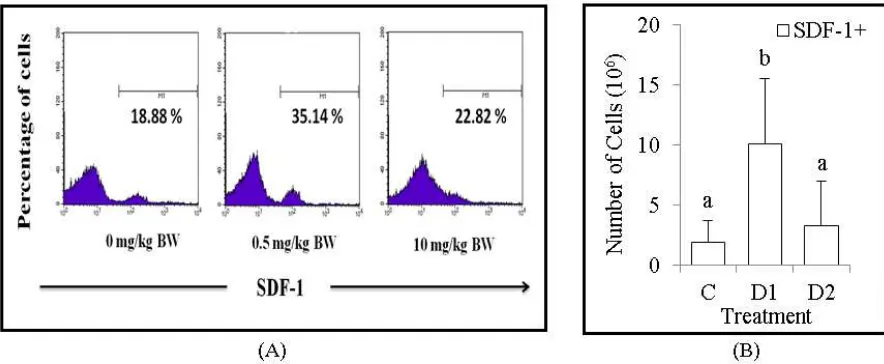
Dexamethasone increase population number of chemokines, SDF-1+
Stromal cell derived factor-1 (SDF-1) is a kind of chemokine that has important role on regulate growth and development of hematopoietic stem cells. In this experiment, we found the relative number of chemokines, SDF-1+ that analyzed by flow cytometry around 18.88% (C), 35.14% (D1) and 22.82% (D2) (Figure 2A). In addition, we also calculate the absolute number of chemokines, SDF-1+ around 1.91x10⁶ cells (C), 10.1x10⁶ cells (D1) and 3.28x10⁶ cells (D2) (Figure 2B). Based on these results, the data improved that Dex administration with dose 0.5 mg/kg BW significantly (p<0.05) increase the absolute number of chemokines, SDF-1+.
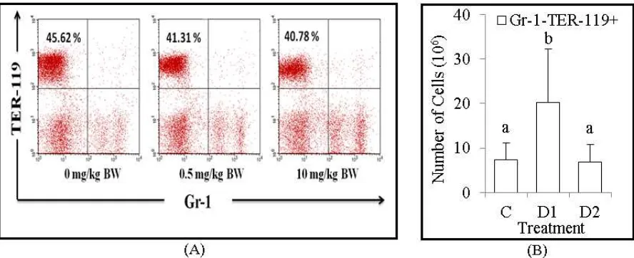
Figure 3. The population number of erythroid progenitor cells, Gr-1-TER-119+. (A) Flow cytometry analysis showed the relative number of erythroid progenitor cells, Gr-1-TER-119+ were decreased in dose 0.5 mg/kg BW and 10 mg/kg BW of Dex treatment. (B) The bars are calculation of the absolute number of erythroid progenitor cells, Gr-1-TER-119+ in the bone marrow of BALB/c mice after treatment. It shown the absolute number of erythroid progenitor cells, Gr-1-TER-119+ were increased in dose 0.5 mg/kg BW of Dex treatment. Data are mean±SD in each group with p-value<0.05. (C: untreated mice, D1: 0.5 mg/kg BW of Dex, D2: 10 mg/kg BW of Dex).
Dexamethasone increase population number of erythroid progenitor cells, Gr-1- TER-119+
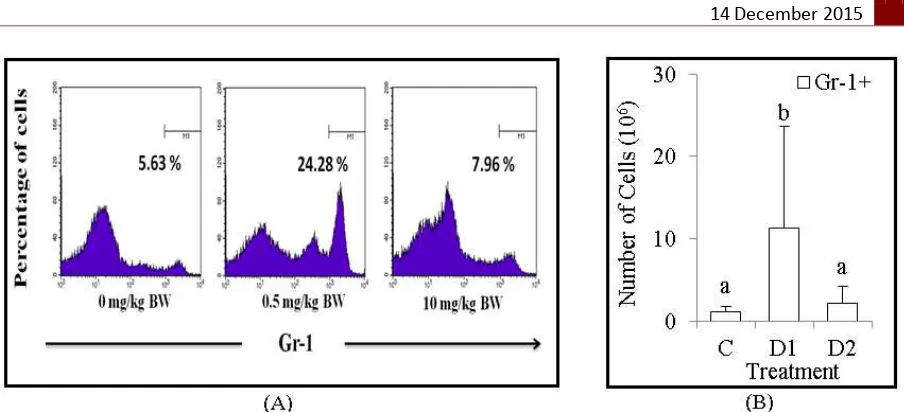
Figure 4. The population number of granulocytes, Gr-1+. (A) Flow cytometry analysis showed the relative number of granulocytes, Gr-1+ were increased in dose 0.5 mg/kg BW of Dex treatment. (B) The bars are calculation of the absolute number of granulocytes, Gr-1+ in the bone marrow of BALB/c mice after treatment. It shown the absolute number of granulocytes, Gr-1+ were increased in dose 0.5 mg/kg BW of Dex treatment. Data are mean±SD in each group with p-value<0.05. (C: untreated mice, D1: 0.5 mg/kg BW of Dex, D2: 10 mg/kg BW of Dex).
Dexamethasone increase population number of granulocytes, Gr-1+
Granulocytes are kind of granulated-leukocytes, which have ability to eliminate the antigens by phagocytosis. Granulocytes consist of neutrophil, eosinophil and basophil. In this experiment, we found the relative number of granulocytes, Gr-1+ that analyzed by flow cytometry around 5.63% (C), 24.28% (D1) and 7.96% (D2) (Figure 4A). In addition, we also calculate the absolute number of granulocytes, Gr-1+ around 1.19x10⁶ cells (C), 11.26x10⁶ cells (D1) and 2.12x10⁶ cells (D2) (Figure 4B). Based on these results, the data improved that Dex administration with dose 0.5 mg/kg BW significantly (p<0.05) increase the absolute number of granulocytes, Gr-1+.
Dexamethasone increase population number of B progenitor cells, B220+
B progenitor cells are kind of lymphoid progenitor family, which develop and mature in bone marrow. In this experiment, we found the relative number of B progenitor cells, B220+ that analyzed by flow cytometry around 26.87% (C), 29.42% (D1) and 14.04% (D2) (Figure 5A). In addition, we also calculate the absolute number of B progenitor cells, B220+ around 6.09x10⁶ cells (C), 19.26x10⁶ cells (D1) and 3.98x10⁶ cells (D2) (Figure 5B). Based on these results, the data improved that Dex administration with dose 0.5 mg/kg BW significantly (p<0.05) increase the absolute number of B progenitor cells, B220+.
Basically, Dex is a kind of synthetic glucocorticoid which has an anti-inflammatory and immune-suppressive effects, then it could suppress the response of the immune system by inhibiting the secretion of pro-inflammatory cytokines such as TNFα and IL-6 [6,7]. Based on Galon et al. (2002), glucocorticoid does not fully provide suppressive effects, but it could provide stimulant effects for several cells that associated with the immune system i.e. increase the population of erythroid progenitor proliferation and prevent apoptosis in neutrophils. In addition, Dex may increase the synthesis of pro-inflammatory mediators such as cytokines and chemokines which act as a growth factor of hematopoietic stem cells [8].
Figure 6. Chemical structure of dexamethasone [9]
Both lymphoid and myeloid lineages mainly develop from hematopoietic stem cells [10,11]. During hematopoiesis, it develops and differentiates to committed lineages affected by hematopoietic growth factors that mainly in the form of glycoprotein. Commonly, hematopoietic growth factors secreted by several types of cells i.e. T cells, monocytes, macrophages and bone marrow stromal cells. T cells, monocytes and macrophages have role to secreting growth factor in the form of cytokines such as IL-1 and TNFα, then secreted cytokines will trigger the other cells to secrete another type of growth factors like GM-CSF, G-CSF, M-CSF and IL-6 [12].
In this study, we found that Dex with dose of 0.5 mg/kg BW significantly increase the population of the absolute number of hematopoietic stem cells, CD34+. Another research showed that Dex has protective effect toward hematopoietic stem cells that treated by chemotherapies. Dex was identified as a small molecule that known as stimulating factor to increase quiescent hematopoietic stem cells on the bone marrow. The population of hematopoietic stem cells was increased by the number of Angiopoietins-1 (Ang-1) on the bone marrow. Ang-1 has roles to induce phosphorylation and make a complex with Tie-2 as tyrosine kinase receptor on hematopoietic stem cells. Based on past research, Dex could increase the population of Ang-1 on the bone marrow then possibly bind with Tie-2 to modulate proliferation of quiescent hematopoietic stem cells [13].
monocytes, megakaryocytes, platelets and progenitor cells from bone marrow [16,17]. Chemokines, SDF-1 have role to induce migration and cells homing for both hematopoietic stem cells and hematopoietic progenitor cells (HPSCs) by interaction to form G complex protein with its receptor, CXCR4 that expressed by stromal cells [16,18,19]. Other researches explained that mice with SDF-1 deficiency have fewer myeloid lineage cells than normal mice on its bone marrow. SDF-1 is also known as chemo-attractant for both lymphocytes and monocytes, and also help to enhancing B-cell progenitor proliferation [16]. Furthermore, in this study showed that the absolute number of chemokines, SDF-1+ that expressed on bone marrow were significantly increased at dose 0.5 mg/kg BW. There is positive correlation between SDF-1 and its receptor, CXCR4 expression on the bone marrow. Dex was known to increase CXCR4 expression on CD4 T cells, eosinophil, and monocytes. Dex administration with high consent-ration could decrease SDF-1 secretion on bone marrow stem cells [18]. Based on Kollet et al. (2013), that another glucocorticoid like corticosterones increase the secretion of SDF-1 on low dose [20]. While based on Zubkova et al. (2005), the enhancement of SDF-1 secretion is caused by the increasing of gene expression that responsible for SDF-1 regulation [21].
Erythropoiesis is erythrocytes formation process that begins with the development of hematopoietic stem cells. Furthermore, in this study showed that the absolute number of erythroid progenitor cells, Gr-1-TER-119+ that expressed on bone marrow were significantly increased at dose 0.5 mg/kg BW. The molecular mechanism of Dex on erythropoiesis until now is not clear. According to Konto-Ghiorghi (2013), glucocorticoids were known to enhance the colony formation of erythrocytes and increase cell proliferation [22]. Besides that, Dex also increase the Janus Kinase 2 (JAK2) gene, which is the most important on erythropoiesis regulation. In addition, Dex increase the expression of suppressor of cytokine signaling-1 (SOC 1), which has function in regulating the amount of hematocrit and the total of erythrocyte precursors [23].
B cell is a kind of lymphoid lineage that develops and matures in bone marrow. During development stage, B220 as molecule surface of B progenitor cells will be expressed [30]. B cells have responsible to control the antigen invasion to body by synthesize the antibodies and work together with other immune cells such as T cells, dendritic cells and macrophages to eliminate the antigens [31]. In this experiment we found that Dex administration with dose 0.5 mg/kg BW significantly increase the absolute number of B progenitor cells, B220+. This data explained that Dex could be stimulating factor that induce B progenitor cells population to proliferate and develop to mature B cells. The increasing of B cells, B220+ expression improve that there are some development and normalize of B cells itself [32]. On the other hand, T cells could increase and normalize population of granulocytes and B cells [33,34]. B progenitor cells development and proliferation affected by CD4 T cells that secrete IL-2 which responsible for survival and proliferation of B-cells [31]. Gruver-Yates & Cidlowski (2013), improved that treatment with high dose of Dex could increase apoptotic rate on B cells [35]. Graversen et al. (2012), revealed that high dose of Dex could block the proliferation and life time of B cells [26]. The effect began shortly after glucocorticoid receptors activation that triggers apoptotic rate and anti-inflammation effects. Glucocorticoids signaling increase the expression of pro-apoptotic gene, Bim that activate pro-apoptotic protein, Bax/Bak. It worked by shatter the potential membrane of mitochondria. As the result, it will excrete cytochrome c and several apoptotic proteins that lead to activation of caspase 9 and caspase 3 [36]. Muhaimin Rifa'i. The authors would like to thank Aris Soewondo and Muhaimin Rifa'i who have reviewed and gave much suggestion for this work.
REFERENCES
[7] Webster-Marketon, Jeanette, Burnsides, Corry, Alexander, Balint, Cosmar & Philips., J. Inflam. Res., 2012, 89.
[8] Galon J., Franchimont D., Hiroi N., Frey G., Boettner A., Ehrhart-Bornstein M.,
[9] INCHEM, Dexamethasone (WHO Food Additives Series 33). http://www.inchem.org/ documents/jecfa/jecmono/v33je09.htm, Accessed 04/05/2015.
[10] Weissman I. L., Cell, 2000,100, 157-168.
[11] Lemischka I., Rev Clin Exp Hematol, 2001, 5,15-25.
[12] Hoffbrand A. V., Pettit J. E., and Moss P. A. H., Hematologi, 2005, EGC Press, Jakarta. [13] Kim H., Ji-Young C., Jung M. L., Yoon S. P., Hwal S., Hae-Ryong S., Sangmee A. J.,
and Inho J., FEBS Letters, 2008, 582 (23-24), 3509–14.
[14] Askari A. T., Unzek S., Popovic Z. B., Goldman C. K., Forudi F., Kiedrowski M., Rovner A., Ellis S. G., Thomas J. D., Di Corleto P. E., Topol E. J., and Penn M. S,
Lancet, 2003, 362(9385), 697–703.
[15] Lapidot T. and Petit I., Exp. Hematol, 2002, 30(9), 973–981.
[16] Lataillade, Jean-Jacques, Clay D., Dupuy C., Rigal S., Jasmin C., Bourin P., and Marie-Caroline Le Bousse-Kerdilès, Blood, 2000, 95(3), 756–68.
[17] Gazitt Y. and Liu Q., Stem Cells, 2001, 19 (1), 37–45.
[18] Kim S. W., Ha-Yon K., Hyo-Jin L., Hwan-Jung Y., Samyong K., and Deog-Yeon J.,
Leukemia Lymphoma, 2009, 50 (7), 1163–73.
[19] Abraham M., Beider K., Wald H., Weiss I. D., Zipori D., Galun E., Nagler A., Eizenberg O., and Peled A., Leukemia, 2009, 23 (8),1378–88.
[20] Kollet O., Vagima Y., D’Uva G., Golan K., Canaani J., Itkin T., and Gur-Cohen S.,
Leukemia, 2013, 27 (10), 2006–15.
[21] Zubkova I., Mostowski H., and Zaitseva M., J. Immunol., 2005, 175 (4), 2321–30. [22] Konto-Ghiorghi Y. J Pediatrics Child Care, 2013, 1(1), 5.
[23] Narla A., Dutt S., and McAuley J. R., Blood, 2011, 118(8), 2296-2304. [24] Putra WE., Soewondo A., and Rifa'i M., J. Biotropika, 2015, 3(1), 42-45.
[25] Hodrea J., The glucocorticoid deksametasone programs human dendritic cells for enhanced phagocytosis of apoptotic neutrophils and inflammatory response, Doctoral School of Molecular Cell and Immune Biology, University of Debrecen, Hungary, 2011.
[26] Graversen J. H., Svendsen P., Dagnæs-Hansen F., Dal J., Anton G., Etzerodt A., Petersen M. D., Christensen P. A., Møller H. J., and Moestrup S. K., Mol. Ther., 2012, 20 (8).
[27] Drewniak A., Van Raam B. J., Geissler J., Tool A. T. J., Mook O. R. F., Van Den B. T. K., Baas F., and Kuijpers T. W., Blood, 2009, 113 (23), 5979-98.
[28] Stroncek D. F., Yau Y. Y., Oblitas J., and Leitman S. F., Transfusion, 2001, 41(8),
![Figure 6. Chemical structure of dexamethasone [9]](https://thumb-ap.123doks.com/thumbv2/123dok/2861250.1694396/6.595.217.382.331.450/figure-chemical-structure-dexamethasone.webp)
