Effect of Curcuma longa L. extract on the AP1 expression in rat cochlear
fibroblasts under noise conditions
Tengku Siti Hajar Haryuna
1*, Ramsi Lutan
1, Faathir Agung Ainul Taufika
1,
Ratna Anggraeni
2,
Tengku Siti Harilza Zubaidah
31. Department of Otorhinolaryngology, Faculty of Medicine, University of North Sumatera, Medan 20155, Indonesia 2. Department of Otorhinolaryngology, Faculty of Medicine, Padjadjaran University, Bandung 45363, Indonesia 3. Department of Ophthalmology, Faculty of Medicine, University of North Sumatera, Medan 20155, Indonesia
Abstract: Noise-induced cellular stress can cause damage to fibroblasts within the cochlear supporting tissues and lateral wall. In the present study, we aimed to evaluate the role of curcumin as the safe and effective therapeutic agent in the prevention and treatment of this condition according to the expression of activator protein-1 (AP1). A total of 24 Rattus norvegicus were randomly divided into four groups (n = 6). Group 1: control; group 2: noise (+); group 3: noise (+), 50 mg/day curcumin (+); group 4: noise (+), 100 mg/day curcumin (+). All groups (except for group 1) were subjected to a sound pressure level (SPL) of 100 dB for 2 h/day during 2 weeks. Curcumin used in this study was derived from Curcuma longa L. (Turmeric), and it was orally administered for 2 weeks. All samples were immunohistochemistrically examined for the expression of AP1 in cochlear fibroblasts. The results showed that there were significant differences for the AP1 expression (P<0.05) among all groups, except for between groups 1 and 3, or between groups 1 and 4. Our data proved that curcumin was potentially effective in the prevention and treatment of damage of fibroblasts within the cochlear supporting tissues and lateral wall due to the decreased AP1 expression following noise exposure.
Keywords: Noise, Curcumin, AP1, Fibroblast, Cochlea
CLC number: R961 Document code: A Article ID: 1003–1057(2016)9–690–05
Received: 2016-03-30, Revised: 2016-04-25, Accepted: 2016-05-15.
Foundation items: Daftar Isian Pelaksanaan Anggaran (DIPA) 2014 (Grant No. 4806/UN5.1.R/KEU/2014) Universitas Sumatera Utara.
*Corresponding author. Tel.: +628126061694, +6282167578676,
E-mail: [email protected]
http://dx.doi.org/10.5246/jcps.2016.09.077
1. Introduction
Immoderate noise is increasingly dealt with in many aspects of everyday life and predominantly found in the developing and industrial countries with poor
hearing conservation[1,2]. According to recent global
estimates released by the World Health Organization (WHO, 2012), there are 360 million people worldwide (more than 5% of the world‟s population) with disabling
hearing loss[3]. Excessive noise exposure, both long
-term
and a single exposure to an extremely intense sound,
can damage the auditory system, leading to noise-induced
hearing loss (NIHL)[2].
Noise exposure is able to negatively affect all three areas of the cochlea, the organ of Corti, the lateral wall
and the spiral ganglion neurons (SGN). Most studies on NIHL have concentrated on the sensory hair cells in the organ of Corti where auditory transduction takes place. However, there is increasing awareness that the SGN and lateral wall of the cochlea are also negatively
affected by noise[4]. Within the cochlear lateral wall, the
spiral ligament is situated between the otic capsule and the stria vascularis (medially), which are predominantly composed of connective tissue elements, including extracellular material and cells from mesenchymal origin. Fibroblasts with stress fibers (tension fibroblasts) containing contractile proteins are discovered in the tissue that anchors the spiral ligament to the lateral aspect of the basilar membrane (BM), suggesting that the spiral ligament is capable of producing and/or
regulating BM tension[5].
The molecular mechanisms that adjust the balance of
cell death and cell survival in the inner ear are not well
that mitogen-activated protein kinase (MAPK), a stress
-activated member of the MAPK family, may play a crucial role. It serves as an importing necessary signaling protein that links activity at the cell membrane to
downstream signaling in the nucleus[4]. The MAPK
family consists of three subfamilies: the extracellular
signal-regulated kinases (ERKs), the c-Jun N-terminal
kinases (JNKs), and the p38. Activator protein-1 (AP1)
is phosphorylated by MAPKs[6]. AP1 is a potent stress
response transcription factor and able to trigger the de novo expression of many transcription factors or
adaptor proteins, aiding the matrix metalloproteinase-9
(MMP-9) promoter activity[7]. MMPs represent a large
family of calcium-dependent zinc-containing
endopepti-dases that play a role in the tissue remodeling and degradation of the extracellular matrix, including collagens, elastins, gelatin, matrix glycoproteins and
proteoglycan[8].
For years, many researchers have made efforts to use
natural plant-derived compounds as potential therapeutic
agents for a variety of diseases in humans[9]. Curcumin,
a yellow pigment extracted from the rhizomes of Curcuma longa Linnaeus, is a major component of turmeric originated from Asia and commonly used as
a spice and food-coloring agent. Curcuminoids refer
to a group of phenolic compounds present in turmeric, which are chemically related to its principal ingredient
curcumin[10]. The composition of curcuminoids is
approximately 70% curcumin (curcumin I), 17%
demethoxycurcumin (curcumin II), 3% bis-demethoxy
-curcumin (-curcumin III) and 10% cyclo-curcumin
(curcumin IV)[9].
The role of curcumin in the prevention and treatment
of noise-induced fibroblast damage within the cochlear
supporting tissues and lateral wall mechanism has never been investigated. In the present study, we aimed to assess whether higher dose of curcumin (100 mg/day) exerts more beneficial effects on inhibition of AP1 pathway compared with low dose of curcumin (50 mg/day).
2. Material and methods
Male Wistar strain white rats Rattus norvegicus
(150–250 g, 8–12 weeks of age) were used in this study. The study was conducted in the standardized laboratory, which has complete equipment and sufficient experience in the maintenance of experimental animals, in the Laboratory of Biochemistry Faculty of Medicine, University of Airlangga (Surabaya, Indonesia). This experimental study was also approved by Health Research Ethical Committee of North Sumatera c/o Medical School, Universitas Sumatera Utara (Medan, Indonesia).
Curcumin was derived from Curcuma longa L.
(Turmeric), and it was identified by Dr. Rer. Nat. M. Yuwono, MS., Apt. (sheet number No. 2036/SA/XII/2012) at Assessment Service Unit Faculty of Pharmacy Airlangga University. The curcumin content level was 28.1%±1.0% w/w compared with standard, and it was suspended in 0.5% carboxymethyl cellulose. Afterwards, the suspension was directly administered to the stomach of rat via nasogastric tube, once a day for 2 weeks. The 24 Rattus norvegicus samples were divided into four groups. Group 1: the control group; group 2: noise (+); group 3: noise (+), 50 mg/day curcumin (+); group 4: noise (+), 100 mg/day curcumin (+). The given dose of noise exposure was 100 dB SPL for 2 h during 2 weeks.
After 2 weeks, the rats were sacrificed by ether inhalation, and necropsy procedure on temporal bone of rats was performed. All samples underwent standard tissue processing with fixation in buffered formaldehyde, followed by dehydration in graded alcohol solutions. Subsequently, they were embedded in paraffin blocks, serially cut into 4 µm thick sections, and placed on glass slides. Representative sections were stained with hematoxylin and eosin (H&E). Immunohistochemical staining was carried out to examine the expression of AP1.
peroxidase activity was blocked with 3% hydrogen
peroxide in absolute methanol. Non-specific binding of
the secondary antibody was prevented by incubation with
10% non-immune serum (0.25% Triton X-100 in PBS).
C-Fos antibody (SANTA CRUZ, sc-271243) served as the
primary antibody, and it was separately applied to each specimen and incubated in a humidified chamber. After rinsing with PBS, sections were incubated with biotinylated secondary antibody, and then the sections were washed once more and incubated with a horseradish
streptavidin-peroxidase conjugate. Next, a substrate of
chromogen solution (3,3‟-diaminobenzidine
tetrahydro-chloride) was added. This reaction involved peroxidase catalysis of the substrate and conversion of the chromogen to a brown deposit that marked the antigen. The final steps included counterstaining with H&E and application of coverslips.
The samples in each slide were examined by three investigators, and the fibroblasts within the cochlear supporting tissues and lateral wall which expressed AP1 in all fields were manually calculated with hand counter. The expression of AP1 was quantitatively calculated for the average distribution of fibroblasts with single nucleus
expressing AP1 (showing brown-colored cytoplasm).
The data were processed using the Statistical Package
for the Social Sciences (SPSS) one-way analysis of
variance (ANOVA), and a P value of less than 0.05
was considered as statistically significant.
3. Results
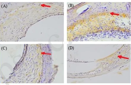
Immunohistochemistry analysis showed that the AP1
expression was increased in the noise-exposed group
(Fig. 1B) compared with other groups. The curcumin
-treated groups showed lower density of the brown
color, and less AP1-expressing fibroblasts than the
noise-exposed group (Fig. 1C–1D).
Table 2 reveals that there were significant differences in the AP1 expression (P<0.05) among all groups, except
for between control group and 50 mg curcumin-treated
group, or between control group and 100 mg curcumin
-treated group. A dose of 100 mg curcumin per day significantly decreased the AP1 expression compared with a dose of 50 mg curcumin per day.
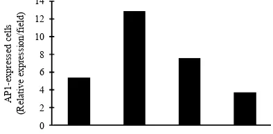
The AP1 expression was increased in group 2 (noise
-exposed group), and it was decreased in curcumin
-treated groups (groups 3 and 4). In general, a dose of 100 mg curcumin per day reduced the AP1 expression compared with a dose of 50 mg curcumin per day (Fig. 2).
Figure 1. The expression of AP1 in each group (1000× zoom):
(A) Group 1; (B) Group 2; (C) Group 3; (D) Group 4. The white
arrow indicates the expression of AP1 in cochlear fibroblastsmarked
by the brown color.
(A) (B)
(C) (D)
Table 1. Inhibitory effect of curcumin on the AP1expression in each
group (primary data).
Compounds Group
Curcumin (Control)I (Noise)II (Noise+C50)III (Noise+C100)IV
1 3 11 10 4
2 6 15 9 3
3 3 10 6 4
4 8 14 8 2
5 4 15 5 5
6 8 12 7 4
(I) Group (J) Group Mean difference Std. error Sig. Group 1 Group 2 –7.500 1.103 .000*
Group 3 –2.167 1.103 .381 Group 4 1.667 1.103 .879 Group 2 Group 3 5.333 1.103 .001*
Group 4 9.167 1.103 .000*
Group 3 Group 4 3.833 1.103 .014*
Table 2. ANOVA test in regard of the AP1 expressions.
*Denotes statistically significant.
4. Discussion
Noise-induced fibroblast damage within cochlear
supporting tissues and lateral wall can be caused by many pathways, and the underlying mechanisms remain unclear. In 2013, Haryuna et al. have proved on rat model that noise inflicts fibroblast damage within cochlear supporting tissues and lateral wall viewed due
to the increased expression of Hsp-70, and curcumin
can significantly prevent such a damage[11]. However,
noise influence on AP1 expression and role of curcumin
in the prevention and treatment of noise-induced
fibroblast damage have never been conducted.
Both physiological and pathological stimuli (growth factors, stress signal, infections and oncogenic stimuli)
can regulate AP1 activity[12]. In this study, we found
that the AP1 expression was significantly increased in
cochlear fibroblasts in noise-exposed group (group 2)
compared with the control. MAPKs, consisting of three subfamilies (ERK, JNK and p38), are crucial signal transducers, and they regulate distinct cellular functions and can be activated by phosphorylation in cytoplasm and translocated into nucleus, where they stimulate
phosphorylation of AP1[6].
AP1 is confirmed as fundamental transcriptional factor
for MMP-9 expression[13]. MMPs contribute to tissue
remodeling and degradation of extracellular matrix, including collagens, elastins, gelatin, matrix glycoproteins,
proteoglycan[8]. Collagen is an important structural
component of cochlear lateral wall, and its most plentiful type (type II) is an extracellular matrix material and
sub-epithelial connective tissue of inner ear which can
be also detected in BM[14].
Wang et al. reported that AP1 acts as an important modulator in regulating inflammation prompted by pathogens (Group B streptococcus, Herpes simplex
virus 1, Helicobacter pylori)[15]. Moghadamtousi found
that AP1 is critical for transcriptional regulation of
high-risk human papillomaviruses (HPVs)[16]. Mishra et al.
reported a fundamental role of AP1 transactivation in
severe lesions during oral carcinogenesis[17].
JNK activation inflicted by cellular stress results in
c-Jun activation, and its translocation from cytoplasm
to nucleus is vital for AP1 activity[18,19]. Curcumin may
interfere with multiple cell signaling pathways, and it apparently inhibits AP1 binding via its action on
mitogen-activated protein kinase kinase-1 (MEKK1)-JNK
pathway, glutamate-induced JNK phosphorylation, c-Jun
phosphorylation, AP1 binding activation and excitotoxicity
in a concentration-dependent manner[20,21].
In the present study, we proved that curcumin was able to suppress AP1 expression in cochlear fibroblasts. Aggarwal et al. (2006) stated that curcumin inhibits AP1
and NF-kβ activation induced by tumor promoters[22].
The c-Jun domain of AP1 is a positive regulator of
cyclin D1 expression, contributing to tumorigenic
phenotype stimulation[23]. Jurenka (2009) reported
curcumin‟s great potential as a therapeutic agent for
various inflammatory diseases[24]. Farhangkhoee (2006)
found that curcumin treatment of diabetic rats reduces the damage of oxidative DNA and protein, which is
mediated by decreased activation of redox-sensitive
transcription factors, NF-kβ and AP1[25].
In this study, we found that a dose of 100 mg/day curcumin could significantly decrease the expression
of AP1 compared with its half-dose (50 mg/day).
The expression of AP1 in groups 3 and 4 was not significantly different from the control group, indicating that higher dose of curcumin was able to prevent AP1 activation, and thereby its expression was found to be nearly similar from control.
Figure 2. The mean of AP1 expression in each group.
5. Conclusions
Curcumin is considered to be a safe and effective therapeutic agent in the prevention and treatment for
the noise-exposed damage of fibroblasts within the
cochlear supporting tissues and lateral wall. Moreover, the study thus provided more insights into the mechanism of curcumin against AP1, and we showed that curcumin could inhibit AP1 signaling pathways. Moreover, our study also served as a scientific basis for its usage in the traditional systems of medicine for the management of NIHL in the future.
Acknowledgements
The authors are deeply grateful to Daftar Isian Pelaksanaan Anggaran (DIPA) Universitas Sumatera Utara 2014 (Grant No. 4806/UN5.1.R/KEU/2014) for the financial support. The authors also would like to thank Biochemisty Laboratory, Faculty of Medicine, Universitas Airlangga Surabaya; Anatomic Pathology Laboratory, Faculty of Medicine, Dr. Soetomo General Hospital Surabaya; Biochemistry Laboratory, Faculty of Medicine, Universitas Brawijaya Malang, for providing equipment and scientific apparatus.
References
[1] Harmadji, S.; Kabullah, H. Folia Medica Indonesiana. 2004, 40, 171–174.
[2] World Health Organization. Hearing loss due to recreational exposure to loud sounds: a review. 2015. 1–32. This article can be found online at http://apps.who.int/iris/bitstream/ 10665/154589/1/9789241508513_eng.pdf?ua=1
[3] Tan, W.J.T.; Thorne, P.R.; Vlajkovic, S.M. World J. Otorhinolaryngol. 2013, 3, 89–99.
[4] Jamesdaniel, S.; Hu, B.; Kermany, M.H.; Jiang, H.; Ding, D.; Coling, D.; Salvi, R. J. Proteomics. 2011, 75, 410–424. [5] Raphael, Y.; Altschuler, R.A. Brain Res. Bull. 2003, 60,
397–422.
[6] Eriksson, M. dissertation, University of Helsinki. 2005. [7] Gordon, G.M.; Ledee, D.R.; Feuer, W.; Fini, M.E. J. Cell
Physiol. 2009, 221, 402–411.
[8] Verma, R.P.; Hansch, C. Bioorg. Med. Chem. 2007, 15, 2223–2268.
[9] Trujillo, J.; Chirino, Y.I.; Molina-ijon, E.; Anderica-Romero, A.C.; Tapia, E.; Pedraza-Chaverri, J. Redox Biol. 2013, 1, 448–456.
[10] Yadav, S.K.; Sah, A.K.; Jha, R.K.; Sah, P.; Shah, D.K. Pharmacogn Rev. 2013, 7, 42–46.
[11] Haryuna, T.S.H.; Lutan, R.; Purnami, N.; Ma‟at, S.; Riawan, W. International J. Life Sci. Pharm. Res. 2013, 3, 29–35.
[14] Buckiova, D.; Popelar, J.; Syka, J. Exp. Gerontol. 2006, 41, 296–302.
[18] Collett, G.P.; Campbell, F.C. Carcinogenesis. 2004, 25, 2183–2189.
[19] Watabe, M.; Ito, K.; Masuda, Y.; Nakajo, S.; Nakaya, K. Oncogene. 1998, 16, 779–787.
[20] Chen, R.W.; Qin, Z.H.; Ren, M.; Kanai, H.; Chalecka -Franaszek, E.; Leeds, P.; Chuang, D.M. J. Neurochem. 2003, 84, 566–575.
[21] Anand, P.; Sundaram, C.; Jhurani, S.; Kunnumakkara, A.B.; Aggarwal, B.B. Cancer Lett. 2008, 267, 133–164. [22] Aggarwal, B.B.; Bhatt, I.D.; Ichikawa, H.; Ahn, K.S.; Sethi,
G.; Sandur, S.K.; Sundaram, C.; Seeram, N.; Shishodia, S. “Curcumin-biological and medicinal properties,” in Tumeric: the genus curcuma, P. N. Ravindran, N. Babu, K. Sivaraman, editors, Florida, CBC Press. 2007, 297–368,
[23] Wilken, R.; Veena, M.S.; Wang, M.B.; Srivatsan, E.S. Mol. Cancer. 2011, 10, 1–19.
[24] Jurenka, J.S. Altern Med. Rev. 2009, 14, 141–153. [25] Farhangkhoee, H.; Khan, Z.A.; Chen, S.; Chakrabarti, S.
ESPEN J. 2006, 3, 1–8.