Corresponding author: [email protected]
The effect of mitomycin-c in keloid
fibroblast cultures
Ishandono Dachlan1* Teguh Aryandono2, Mae Sri Hartati Wahyuningsih3, Hardyanto
Soebono4,Yohanes Widodo Wirohadidjojo4
1Plastic and Reconstructive Surgery Division, Department of Surgery, Faculty of Medicine,
Universitas Gadjah Mada/Dr. Sardjito Hospital, Yogyakarta, Indonesia, 2Surgical Oncology
Division, Department of Surgery, Faculty of Medicine, Universitas Gadjah Mada/Dr. Sardjito Hospital, Yogyakarta, Indonesia, 3Department of Pharmacology and Therapy,
Faculty of Medicine, Universitas Gadjah Mada, Yogyakarta, Indonesia, 4Department of
Dermatology and Venereology, Faculty of Medicine, Universitas Gadjah Mada/Dr. Sardjito Hospital, Yogyakarta, Indonesia
DOI: http://dx.doi.org/10.19106/JMedSci004803201603
ABSTRACT
Keloid occurs due to hyperactivity of keloid ibroblast (KF) in proliferation, migration,
collagen deposition, together with low rates of collagen degradation. These are under
the responsibility of TGF-β. Mitomycin C (MC) is used for treating keloid by a topical application during surgery at the level of 0.02% to 0.08%. Unfortunately, the lowest
effective level of MC for keloid has not been determined yet. We aimed to determine
the lowest effective level of MC in the suppression of KF activities. Various levels of MC diluted in growth medium were administered on KF that were isolated from six patients.
After 24 hours and 72 hours of incubation, cellular proliferation, collagen deposition, cellular migration and level of TGF-β, were analyzed. Application of 120 uM MC on KF
culture for 24 hours could signiicantly reduce TGF-β production from 1265.74 ± 274.81 pg/mL to 265.17 ± 12.20 pg/mL; proliferation index from 100% to 84.01 ± 12.91%;
inhibit cellular migration to 64.38 ± 3.66%; but reduce collagen depositions from 100%
to only 91.13 ± 10.19%. The lowest MC level is on 30 uM or equal with 0.001%. In conclusion, the lowest level of MC can suppress the activities of KF is 0.001%.
Moreover, due to low activity in inhibiting collagen deposition, MC would be better as an
adjuvant drug for keloid surgery.
ABSTRAK
Keloid timbul karena proliferasi, migrasi, dan sintesis kolagen oleh ibroblas keloid (FK) secara berlebihan diikuti dengan rendahnya degradasi kolagen. Semua itu terjadi karena pacuan TGF-β. Olesan mitomycin C (MC) 0,02% sampai 0,08% digunakan untuk
mengobati dan mencegah kekambuhan keloid yang dioperasi. Sayangnya, dosis terendah yang masih efektif belum pernah diteliti. Penelitian ini bertujuan untuk menentukan
kadar terendah MC yang masih efektif untuk mengobati keloid. MC dilarutkan dalam medium pertumbuhan dalam berbagai kadar dan diberikan pada biakan FK yang diisolasi dari material keloid enam orang pasien. Setelah inkubasi 24 jam dan 72 jam, proliferasi, migrasi, timbunan kolagen dan kadar TGF-β, dianalisis dan dibandingkan. Pemberian 120
274,81 pg/mL menjadi 265.17 ± 12.20 pg/mL; indek proliferasi dari 100% menjadi
84,01 ± 12.91%; menghambat migrasi sampai 64,38 ± 3.66%, tetapi daya hambat timbunan kolagen hanya dari 100% menjadi 91.13 ± 10.19%. Kadar MC terendah yang masih efektif adalah 30 uM atau setara dengan 0.001%. Kadar MC di bawah itu tidak lagi efektif. Kesimpulan, kadar MC terendah yang masih efektif sebesar 0,001% dan karena daya hambat timbunan kolagen yang rendah, MC sebaiknya digabung dengan operasi
keloid.
Keywords: keloid ibroblast – mitomycin-c - TGF-β – deposition – proliferation
INTRODUCTION
Keloid is a ibroproliferative benign tumor that found only on human skin with characteristics in persistent overgrowth of ibrous tissues during wound healing leading to the formation of over scar tissues more than original wound size, and can invade surrounding normal tissue as a crab-claw of keloid lesion.1,2 This overgrowing is due
to keloid ibroblast (KF) proliferation and producing collagen excessively3-5 together
with low activity of extracellular matrix metalloproteinase (MMP).6,7 KF also have a
characteristic of mesenchymal stem cells and can migrate to surrounding normal tissues to produce enlargement of ibrous tissues.8
Moreover, one of various growth factors that have an important role in keloid behavior is transforming growth factor-β (TGF-β).9
This growth factor can induce Wnt/β-catenin signaling,10 and over-expression of Wnt is
parallel with collagen deposition in both keloid tissues and KF culture.11,12 Wnt/β catenin
signaling pathway is not only responsible for collagen production but it also has an important role in inducing transformation of human dermal microvascular endothelial cells to become KF.12
Mitomycin-C (MC) is an anti-cancer commonly used for bladder cancer and others, including as the preventing agent for pterygium by topical application intra-operatively.13 Interestingly, the target of
this agent is pterygial body ibroblasts that produce ibrotic tissues under stimulation of connective tissue growth factor (CTGF) initiated by TGF-β.14 Similar with it, MC
is also used in keloid lesion by topical application intra-operatively to suppress KF activities. Talmi et al15 apply MC to the wound of keloid excision and resulting in the complete disappearance of keloid lesion. MC can prevent keloid recurrence up to 80% when applied topically to the base of shave-removing keloid surgery.16 The side effects
of topical application of 0.02% MC (equal with 598 uM) among patients with pterygium is low, mild, and reversible.13 Application of
MC intra-operatively is ranged from 0.02% to 0.08% (equal with 2393 uM), but whether 0.02% as the lowest effective level of MS is still unknown. In order to know the lowest level of MC in topical-application for keloid surgery, we reported the effect of various low level MC on KF culture, therefore, the best level of MC in keloid surgery may be determined.
MATERIAL AND METHODS
Isolation and culture of KF
The core of keloid materials was obtained from six patients underwent keloid debulking surgery with informed concerns. About 2 cm3
of each material was thinly sliced into 2-3 mm3 pieces and cultured by explants method
(DMEM, Gibco®, USA) containing 10% fetal bovine serum (FBS, Gibco®,USA) and 1% penicillin/streptomycin (Gibco®, USA) at 37°C and 5 % CO2. The spindle-shaped cells that outgrown from the explants then were subcultured until passage 4.
Experiments
Stock MC-preparation
MC (molecular weight = 334.33) was purchased from Biochem Pharmaceutical Industry Ltd., Mumbai, India under license
from Kyowa Hakko Kogyo, Japan. After dissolving 10 mg MC in 25 mL sterile water (molarity = 1.1964mM), further dilution with DMEM was performed until stock MC in the level of 600 uM. Referring to the indings of Wang et al.,17 on human dermal ibroblasts, we began to treat KF with 120 uM MC.
Design and treatment
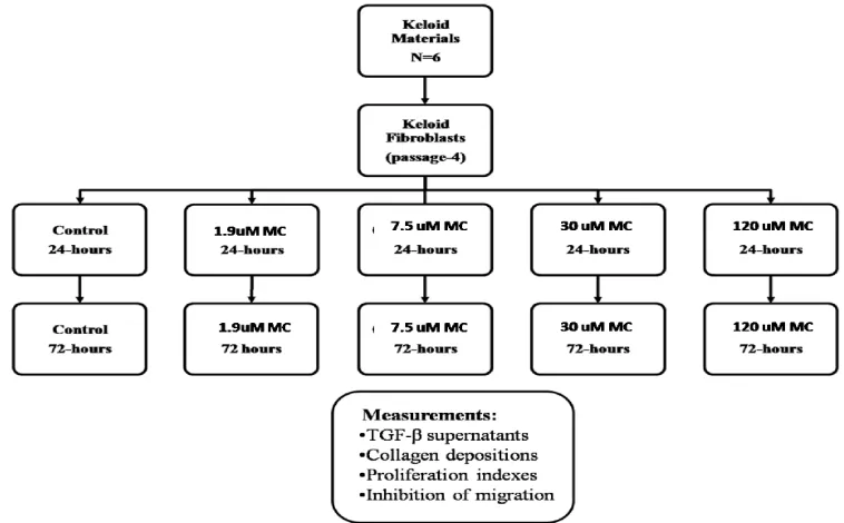
All of the experiments were then conducted in the following scheme:
FIGURE 1. Preparation and measurement scheme
The experiment was carried out with KF reseeding in wells of 200 uL cell suspension containing 103 cells/mL. After 24 hours of incubation to let cells attached on the bottom of wells, various treatments carried out in accordance with a written scheme on above. All of the procedures were in triplicate.
Measurement of variables
Proliferation index
Cellular viabilities were measured using
MTT
considered as 100% of the proliferation index, therefore, the proliferation index of various treated groups was counted as: (OD of treated group / OD of paired control) x 100 %.
Ability to deposite collagen
Collagen deposition was measured by insoluble collagen of Sirius red (purchased from Sigma-Aldrich, Steinheim, Germany) assay according to Taskiran et al.,18 the OD
Sirius red-bound collagen represented the amount of insoluble collagen and it was read using spectrometer at 570 nm of wavelength. The OD of the control group was considered as 100%ability to deposit collagen.The ability of various treated groups was counted as: (OD of treated group / OD of paired control) × 100 %.
Inhibition of cellular migration
Cellular migration assay was performed based on method Liang et al.,19 briely, after
serum starvation,all bottom of wells were linearly scratched with the blunt tip of a 32G sterile needle through the center of the well bottom. After cultivation with various media and incubations, the cells were then stained with Meyer’s Haematoxylin and microscopic photo images were taken using a Moticam-350 camera (China) in JPG format. Both blue color’s pixels of ibroblasts along the scratch line and white color’s pixels of empty space can be measured using Adobe-Photoshop. Migration rate was counted as: (blue color pixel of KF along the scratch line / total pixel along the scratch line ) × 100%. Inhibition of migration of treated group was counted as
migration rate of control minus migration rate of paired treated group.
TGF-β level
The level of TGF-β in the supernatant of various groups was measured using human TGF-β 1 ELISA kit purchased from Bender MedSystem (Burlingame, USA). The OD of each well was read using spectrometer at 570 nm of wavelength and the level of TGF-βwas counted using standard curve obtained from a correlation between various standardized TGF-β with their OD.
Statistics
All data are presented as a mean ± standard error. We used one -ay analysis of variance (ANOVA) followed by LSD to analyze the migration rate data and Friedman test followed by Wilcoxon as post hoc test to analyze the proliferation index and collagen deposition data. The level of statistical signiicance was accepted at p<0.05.
RESULTS
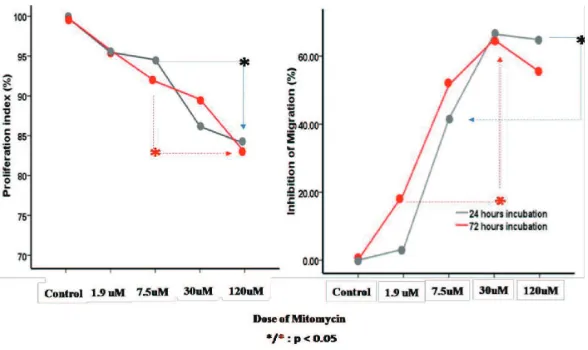
FIGURE 2. Effect of MC on proliferation and cellular migration
FIGURE 3. The effect of MC on TGF-β and collagen deposition
DISCUSSION
Various keloid treatments, starting from surgery to intralesional corticosteroid and various anti-cancers such as luorouracil,20
bleomycin,21 MC,15,16,22 or a combination of
them have been performed.23 MC has been
chosen because it can inhibit normal skin ibroblast proliferation, induce ibroblast apoptosis, and regulate intracellular protein expression on mRNA level.17
Our results showed that 120 uM MC can inhibit proliferation and migration of KF
FIGURE 1. This inding is important because continuously enlargement of keloid lesion is under responsible of high proliferation rate of KF3-5 and high migration ability to invade
surrounding normal tissue.8 Application 120
both 24 hours and 72 hours of administration. So, it can be concluded that lowest level of MC is on the level of 30 uM or equal to 10.03 ug/mL or equal to 0.001%.
Regulation of KF protein expression by 120 uM MC can be observed in FIGURE 2, as it was indicated by reducing the TGF-β level and collagen deposition. Based on low inhibition of collagen deposition (about 10% of 24 hours of incubation), it seemly indicated that MC as a single drug is not worthy to be used for treating keloid lesion otherwise repeated injections of MC must be given. Based on high inhibition of MC in cellular migration and cellular proliferation on above, it looked that MC administration just has prevention effect in keloid progression. It has been known that extensive enlargement of keloid lesions is due to KF proliferations, collagen depositions, and cellular migrations.3-5 In order to totally
treat keloid lesions, we agree that MC must be combined with keloid surgery that removes the existing ibrous tissue material which richens by collagen bundles, such as previously reported by many authors.15,16,22,23 In this
combination, unremoving KF and collagen bundles on the surface of debulking lesions of preexisting keloid can be prevented to grow and to be prevented to relapse. The signiicant different of collagen deposition between 72 hours and 24 hours of application (as it shown by red dot) showed that MC could stimulate collagen degradation as manifestation of MMP activation. Among KF, production of tissue inhibitor of metalloproteinase (TIMP)-2 is high and MMP-(TIMP)-2 is low. A study showed that application of IL-β can convert this event to become increasing MMP-2 level together with decreasing TIMP-2 parallel with increasing collagen degradation.24 We
suspected that increasing collagen degradation in our experiment was due to MC regulate KF in interleukin-β protein expression. A further
research to clarify this hypothesis should be performed in the future.
So far, there is no any criterion for converting drug level obtained from cell culture to the equal dosage for human in clinical applications. Atashkina et al.25 assume that so many variables must be considered before converting those drug’s levels in the human body, for example: whether cultured cellular models exhibiting reliable, known and intact biochemical pathways and structural elements are same with keloid lesions. For these purposes, our experiment in two dimensions KF culture had limitation at least in structural elements where excessive collagen bundles before having treatments were not represented in our cells condition. This limitation may affect on effective drug’s level that is lower than it should be in keloid lesion. Since there is no representative animal model for keloid study, a further study in three dimensions of organotypic keloid culture must be conducted, so the lowest effective level of MC can be determined. The comparison of 3 dimensions culture’s weight and the monolayer culture’s weight can be used to determine starting level of MC for further researches.
CONCLUSION
MC is just applicable in preventing recurrence of keloid after having keloid surgery. Moreover, the lowest effective level of MC is 30 uM for a monolayer of KF culture or equal to 0.001% but it must be recalculated in the clinical application of keloid surgery and it depends on the estimation of residual ibrous tissues weight.
ACKNOWLEDGEMENT
Mada, for allowing me to work in cell culturing and technical measurement.
REFERENCES
1. Mrowietz U, Seifert O. Keloid scarring: new treatments ahead. Actas Dermosiiliogr 2009; 100 Suppl 2:75-83. http://dx.doi.org/10.1016/ S0001-7310(09)73382-4
2. Hunasgi S, Koneru A, Vanishree M, Shamala R. Keloid: A case report and review of pathophysiology and differences between keloid and hypertrophic scars. J Oral Maxillofac Pathol 2013; 17(1):116-20. http:// dx.doi.org/10.4103/0973-029X.110701 3. Chipev CC, Simman R, Hatch G, Katz
AE, Siegel DM, Simon M. Myoibroblast phenotype and apoptosis in keloid and palmar ibroblasts in vitro. Cell Death Differ 2000; 7(2):166-76. http://dx.doi.org/10.1038/ sj.cdd.4400605
4. Luo S, Benathan M, Raffoul W, Panizzon RG, Egloff DV. Abnormal balance between proliferation and apoptotic cell death in ibroblasts derived from keloid lesions. Plast Reconstr Surg 2001; 107(1):87-96. http:// dx.doi.org/10.1097/00006534-200101000-00014
5. Akasaka Y, Fujita K, Ishikawa Y, Asuwa N, Inuzuka K, Ishihara M, et al. Detection of
apoptosis in keloids and a comparative study on apoptosisbetween keloids, hypertrophic scars, normal healed lat scars, and dermatoibroma. Wound Repair Regen 2001; 9(6):501-6. http://dx.doi.org/10.1046/j.1524-475x.2001.00501.x
6. Sandulache VC, Parekh A, Li-Korotky H, Dohar JE, Hebda PA. Prostaglandin E2 inhibition of keloid ibroblast migration, contraction, and transforming growth factor (TGF)–beta1-induced collagen synthesis. Wound Repair Regen 2007; 15(1):122-33. http://dx.doi.org/10.1111/j.1524-475X.2006.00193.x
7. Gurtner GC, Werner S, Barrandon Y, Longaker MT. Wound repair and regeneration. Nature 2008; 453(7193):314-21. http://dx.doi. org/10.1038/nature07039
8. Zhang Q, Yamaza T, Kelly AP, Shi S, Wang S, Brown J, et al. Tumor-like stem cells
derived from human keloid are governed by the inlammatory niche driven by IL-17/IL-6 axis. PLoS ONE 2009; 4(11):e7798. http:// dx.doi.org/ 10.1371/journal.pone.0007798 9. Pakyari M, Farrokhi A, Maharlooei MK,
Ghahary A. Critical role of transforming growth factor beta in different phases of wound healing. Adv Wound Care 2013; 2(5):215-24. http://dx.doi.org/10.1089/wound.2012.0406 10. Sato M. Upregulation of the Wnt/beta-catenin
pathway induced by transforming growth factor-beta in hypertrophic scars and keloids. Acta Derm Venereol 2006; 86:300-7. http:// dx.doi.org/10.2340/00015555-0101
11. Igota S, Tosa M, Murakami M, Egawa S, Shimizu H, Hyakusoku H, et al. Identiication
and characterization of Wnt signaling pathway in keloid pathogenesis. Int J Med Sci 2013; 10(4):344-54. http://dx.doi.org/10.7150/ ijms.5349
12. Lee WJ, Park JH, Shin JU, Noh H, Lew DH, Yang WI, et al. Endothelial-to-mesenchymal
transition induced by Wnt 3a in keloid pathogenesis. Wound Repair Regen 2015; 23(3):435-42. http://dx.doi.org/10.1111/ wrr.12300
13. Raiskup F, Solomon A, Landau D, Ilsar M, Frucht-Pery J. Mitomycin C for pterygium: long term evaluation. Br J Ophthalmol 2004; 88(11):1425-8.
http://dx.doi.org/10.1136/bjo.2003.039891 14. Van Setten G, Aspiotis M, Blalock TD,
Exp Ophthalmol 2003; 241(2):135-9. http:// dx.doi.org/10.1007/s00417-002-0589-1 15. Talmi YP, Orenstein A, Wolf M, Kronenberg
J. Use of mitomycin C for treatment of keloid: a preliminary report. Otolaryngol Head Neck Surg 2005; 132(4): 598-601.
http://dx.doi.org/10.1016/j.otohns. 2004.09.060
16. Bailey JN, Waite AE, Clayton WJ, Rustin MH. Application of topical mitomycin C to the base of shave-removed keloid scars to prevent their recurrence. Br J Dermatol 2007; 156(4):682-6. http://dx.doi.org/10.1111/ j.1365-2133.2006.07714.x
17. Wang YW, Ren JH, Xia K, Wang SH, Yin TF, Xie DH, et al. Effect of mitomycin on
normal dermal ibroblast and HaCat cell: an in vitro study. J Zhejiang Univ Scie B 2012; 13(12):997-1005. http://dx.doi.org/10.1631/ jzus.B1200055
18. Taskiran D, Taskiran E, Yercan H, Kutay FZ. Quantiication of total collagen in rabit tendon by the Sirius Red Methods. Tr J Med Science 1999; (29):7-9.
19. Liang CC, Park AY, Guan JL. In vitro scratch assay: a convenient and inexpensive method for analysis of cell migration in vitro. Nat Protoc 2007; 2(2):329-33.
http://dx.doi.org/10.1038/nprot.2007.30
20. Bijlard E, Steltenpool S, Niessen FB. Intralesional 5-luorouracil in keloid treatment: a systematic review. Acta Derm Venereol 2015; 95(7):778-82.
http://dx.doi.org/10.2340/00015555-2106 21. Payapvipapong K, Niumpradit N, Piriyanand
C, Buranaphalin S, Nakakes A. The treatment of keloids and hypertrophic scars with intralesional bleomycin in skin of color. J Cosmet Dermatol 2015; 14(1):83-90. http:// dx.doi.org/10.1111/jocd.12132
22. Stewart CE, Kim JY. Application of mitomycin-C for head and neck keloids. Otolaryngol Head Neck Surg 2006; 135(6):946-50.http://dx.doi.org/10.1016/j. otohns.2005.07.026
23. Khan MA, Bashir MM, Khan FA. Intralesional triamcinolone alone and in combination with 5-luorouracil for the treatment of Keloid and Hypertrophic scars. J Pak Med Assoc 2014; 64(9):1003-7.
24. Yeh FL, Shen HD, Tai HY. Decreased production of MCP-1 and MMP-2 by keloid-derived ibroblasts. Burns 2009; 35(3):348-51. http://dx.doi.org/ 10.1016/j. burns.2008.06.018