An Active of Extracellular Cellulose Degrading Enzyme from Termite
Bacterial Endosimbiont
M. Saifur Rohman
1*), Endang Pamulatsih
1, Yudi Kusnadi
1, Triwibowo Yuwono
1and Erni Martani
1Laboratory of Microbiology, Department of Agricultural Microbiology, Faculty of Agriculture,
Universitas Gadjah Mada, Yogyakarta, Indonesia, 55281
Abstract
Cellulase is an ezyme that specifi cally cleaves the 1,4- -glycosidic bond of cellulose to produce the
small fragments of simple carbohydrate. This work was aimed to characterize the extracellular cellulase from
Paenibacillus spp., which was previously isolated from macro termites, Odontotermes bhagwatii in our laboratory. Two Paenibacillus isolates were used in this experiment, namely Paenibacillus cellulositrophicus SBT1 and
Paenibacillus, sp. SBT8. Analysis of the total proteins in the supernatants showed that P. cellulositrophicus SBT1 and Paenibacillus sp. SBT8 roughly produced as much as 18.6 mg/l and 24.8 mg/l of extracellular cellulases, respectively. Enzymatic assay showed that SBT1 and SBT8 cellulase exhibited enzymatic acitivity of 0.17 U/ mg and 0.12 U/mg, respectively. Temperature dependencies analysis indicated that both cellulases exhibited maximum activity at 35oC. At the temperature higher than 55oC, the enzymatic activities of both cellulases were
roughly 20% reduced compared to the maximum activity. SBT1 and SBT8 cellulases were both active at acidic pH. At basic pH (pH 8) the enzymatic activities of both cellulases were reduced roughly 30% compared to that of acidic pH. Supplementing of Mg2+, Zn2+, and Ca2+ in range of 1-10 mM increased the enzymatic activity of
both cellulases roughly 33 to 50%.
Keywords: Cellulase, Paenibacillus cellulositrophicus SBT1, Paenibacillus sp. SBT8, divalent metal cation, 1,4-β -glycosidic bond
Introduction
Cellulose is the most abundant plant material that can be used as renewable biomaterial and bioenergy. Production of bio-based material and energy resource from less costly renewable lignocellulosic materials would bring benefits to local economy, environment problem and national energy security. Cellulase has an important role for the production of bio-based materials
and energy from less costly renewable lignocellulosic materials (Zhang et al.,
2013).
Cellulase (EC.3.2.1.x) is an enzyme that specifi cally cleaves the 1,4- -glycosidic bond
of cellulose polymer into smaller fragments of simple carbohydrate. Based on the catalytic mechanism, cellulases are classified into three classes of enzymes, namely endo 1,4- -glucanase, exo 1,4- -glucanase, and -glycosidase. Endo 1,4- -glucanase is cellulase that randomly cleaves internal glycosidic bonds of cellulose polymer. In contrast to the endo -glucanase, exo
1,4--glucanase attack the cellulose polymer from the end of polymer, either from reducing or nonreducing end. The processive exo 1,4 -*Corresponding author:
M. Saifur Rohman
Laboratory of Microbiology, Department of Agricultural Microbiology, Faculty of Agriculture, Universitas Gadjah Mada
glucanse also referred as cellobiohydrolase. The later class of cellulase, -glucanase convert cellobiose, the major product of endo- and exo-glucanase, are glucose (Li et al., 2006; Ogawa et al., 2007; Gao et al., 2008; Horn et al., 2012). Cellulase encompasses non-catalytic carbohydrate binding module (CBM) that may be located at the N- and C-terminus of the catalytic domain. CMB is amino acids sequence that functions as substrate binding module (Shoseyov et al., 2006). The catalytic domain contains two important catalytic residues, aspartate (D) and glutamate (E). Those two residues act as donor or acceptor proton during the enzyme catalysis. Therefore, the catalysis mechanism of cellulase obeys the general acid base catalysis (Mosier et al., 1999; Rye and Withers, 2000; Rabinovic et al., 2002).
Some plants, animals, protozoa, fungi, and bacteria produce cellulase. Because of the simplest preparation and manipulation, the bacterial cellulase is the only cellulase that has been intensively and profoundly studied. Cellulase producing bacteria can be isolated from soil, agricultural wastes, and also from animals (Beguin and Aubert, 1994).
Termite is a type of ecosocial insect that feeds wood materials, especially cellulose (Bignell et al., 2011). Higher termites would not be able to digest cellulose until the bacterial endosimbionts are present on their digestive tract (Ohkuma, 2003; Hongoh et al., 2005; Hongoh et al., 2008).
Previously, we have successfully isolated the cellulase-producing bacteria from
Odontotermes bhagwatii, a species of higher
termites collected from the soil of the yard of the Faculty of Agriculture, Universitas Gadjah Mada. Based on the molecular identifi cation,
all the cellulase-producing bacteria are belong to Paenibacillus genera. Therefore the objective of this work is to characterize the extracellular cellulase from Paenibacilus spp.
Materials and Methods
Isolates. Two isolates of Peanebacillus
used in this work were Paenibacillus
cellulositrophicus SBT1 and Paenibacillus
sp. SBT8. Both Paenibacillus produce the extracellular cellulase and were isolated from
Odontotermes bhagwatii in the Laboratory of
Microbiology, Department of Agricultural Microbiology, Faculty of Agriculture, Universitas Gadjah Mada.
Chemicals and microbiological media.
Carboxy methyl cellulose (CMC)(Sigma),
ekstrak yeast (Oxoid), agar (Oxoid), KH2PO4 (Merck), MgSO4 (Merck), Acetic acids buffer pH 4.8, Phosphate buffer, MgCl2 (Merck), ZnCl2 (Merck), CaCl2 (Merck), BSA (bovin serum albumin)(Promega), BCA (Bichinchoninic
Acids) working reagent (Promega), NaOH
(Merck), HCl (Merck).
Isolation, selection and identification of
cellulolytic bacteria. A termite was surface
sterilized by using ethanol, and performed dissecting to take out the termite’s gut. The termite’s gut was disrupted and then inoculated into defined media supplemented with 1% CMC. After 2 to 3 days incubation, 100 μl culture was transferred into defi ned media agar
supplemented with 1% CMC. The growing bacteria were selected randomly based on the morphological characteristic differences. The celulolytic activity was assayed by using 1% Congo red with 1M NaCl and the positive results was designated as the formation of clear zone surrounded the growing colonies (Kumar and Velayutham, 2014). Cellulolytic activity assay was measured by cellulolytic index, a ratio of clear zone diameter and the colony diameter. Bacterial identifi cation was
carried out by analyzing of 16SrRNA genes (Janda and Abbott, 2007).
Enzymatic activity assay. Cellulase activity was examined by using the DNS method. 250 ml of crude extract of cellulase (OD280~0.1) was mixed with 10 mM acetic acids buffer (pH 4.8) supplemented with0.5% CMC as substrate. The reaction mixtures were incubated at 35oC for 30 minutes. The reaction was stopped by addition 1 ml of 3,5-dinitrosalycilic acids. The reaction mixtures were then boiled for 15 minutes and solution mixture of sodium potassium tartrate, phenol, sodium sulfi te was added and kept cool at room temperature. The mixtures were then centrifuge at 10000 rpm for 5 minutes and supernatants were measured by spectrophotometric at =540 nm (Miller
et al., 1959). One unit is defi ned as amount
of enzyme that cleave cellulose to produce 1 μmol of reducing sugars per minute at certain condition.
Effect temperature, pH and metal ions.
To test the effect of temperature, pH, and metal ion on the enzymatic activity of cellulase, the reaction condition was similar
as the enzymatic activity assay, with specifi c
modifi cation. For example when the pH was
the point of assay then the reaction buffer was modifi ed such that the reaction conditions
reach the specifi c pH. To tests the effect of
metal cation on the enzymatic activity, Mg2+, Zn2+ and Ca2+ were added in the reaction mixture separately with the concentration from 0.5 to 50 mM.
Results and Discussion
Isolation, identification and cellulolytic
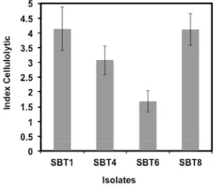
activity. We have successfully isolated 4
isolates showed potential cellulolytic activity from Odontotermes bhagwatii, and those were designated as SBT1, SBT4, SBT6 and SBT8, respectively. Molecular identifi cation based
on the 16SrRNA gene sequence showed that SBT1 and SBT8 belong to the group of
Paenibacillus. SBT1 was much closer to the
Paenibacillus cellulositrophicus, while the SBT8 close to the Paenibacillus sp. SBT4 and SBT6 were close to the Pseudomonas citronellolis
and Pseudoxantobacter soli, respectively.
Fig. 1. Index cellulolytic of cellulolytic bacteria isolated from Odontotermes bhagwatii.
Cellulolytic activity assay showed that SBT1 and SBT8 exhibited the highest cellulolytic activity compared to SBT4 and SBT6 (Fig. 1). Therefore, we have chosen the SBT1 and SBT8 as a model for the extracellular cellulase production.
Extracellular cellulase production.
To produce the extracellular cellulose,
Paenibacillus cellulsitrophicus SBT1 and
Paenibacillus sp. SBT8 were cultivated in
defined or limited medium containing 1% CMC as a carbon sources. Cultivation was carried out for 2 days at room temperature. From this batch fermentation system, Paenbacillus cellulositrophicus SBT1
and Paenibacillus sp. SBT8 could produce
extracellular cellulase roughly of 18.6 mg/l and 24.8 mg/l, respectively. This result indicated that those two isolates potentially could be used for large scaling production of the cellulase. For simplicity, hereafter, we designated the extracellular cellulase from
P. cellulositrophicus SBT1 and Paenibacillus
sp. SBT8 as SBT1 and SBT8 cellulase, respectively.
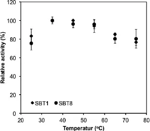
Effect of temperature on the cellulase activity. To test temperature dependencies of extracellular cellulase produced from SBT1 and SBT8, the SBT1 and SBT8 cellulase were test the their enzymatic activity on different temperature conditions. Both cellulases showed the temperature dependencies that were nearly identical (Fig. 2). Both celluases showed the highest activity at 35oC. At the temperature higher than 55oC, the enzymatic activity of both cellulases were roughly 20% reduced compared to the activity at the optimum temperatures. At the optimum temperature, the SBT1 and SBT8 cellulases exhibited the specifi c enzymatic activity of
0.17 and 0.12 U/mg, respectively. The result indicated that the SBT1 cellulase was more active than SBT1 cellulase. Based on the temperature dependencies assay, it clearly showed that both enzymes belong to the mesophilic enzymes at which optimum enzymatic activity were reached at moderate temperature.
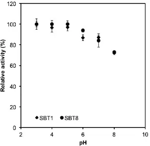
Effect of pH on the cellulase activity. The enzymatic activities as function of pH were
also examined in this work (Fig. 3). The results showed that both cellulase enzymes were exhibited the enzymatic activity at acidic pH, with the optimum pH was determined to be 3. The enzymatic activity of cellulose SBT1 and SBT8 were determined to be 0.19 and 0.13 U/mg, respectively. Lin et al
(2012) reported that the Bacillus thuringiensis
cellulase also exhibited enzymatic activity at the acidic pH (Fig.3).
Catalytic side of cellulase has two important catalytic residues, aspartate (D) and glutamate (E) (Davis and Henrissat, 1995). Aspartate and glutamate both have side chain confers with carboxyl group. The pK values of carboxyl group of asparatate (D) and glutamate (E) side chain are determined to be 3.65 and 4.25, respectively (Buxbaum, 2007). Those two residues function as acid and base during enzymatic catalysis. At the lower pH the carboxyl group of aspartate (D) residues undergoes deprotonated,
therefore it could be proton donor for nucleophilic activation of water molecules. The nucleophile activated water molecules attack the glycosidic bond of cellulose (Mosier et al., 1999).
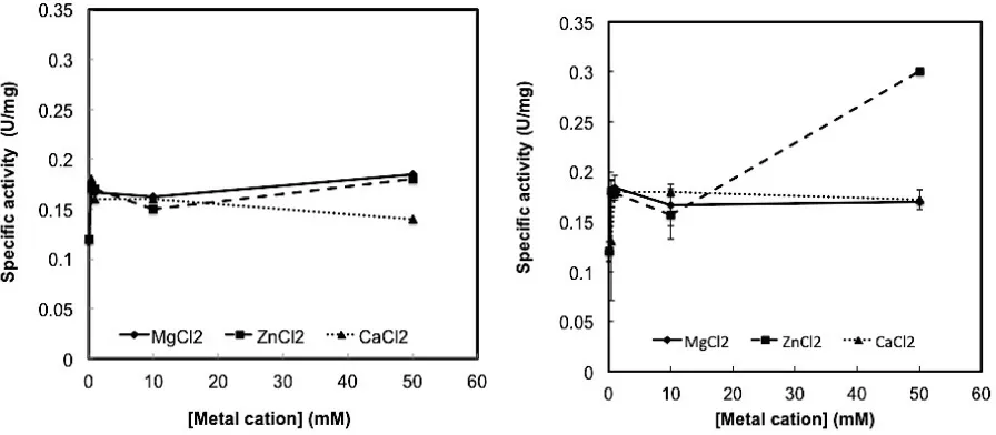
Effect of divalent metal cation on the cellulase activity. Several enzymes require the divalent metal cation for their enzymatic activity. Divalent metal cation plays important role for one third of the enzymes (Glusker et al., 1999). In order to know whether divalent metal cation affect the enzymatic activity of SBT1 and SBT8 cellulase, we have examined three divalent metal cations, Mg2+, Zn2+ and Ca2+ on the enzymatic activity of both cellulases. The concentration of divalent metal cations was varied from 0.5 to 50 mM.
The results showed that addition of divalent metal cations at concentration of 0.5 to 10 mM generally increases the enzymatic activity of both cellulases (Fig. 4). The addition of Mg2+ and Zn2+ at the concentration
above 10 mM, tend to increase further of SBT1 cellulase activity, while the Ca2+ tend to decrease enzymatic activity. However, in the case of SBT8, only Ca2+ at above 10 mM increases the enzymatic activity. It was surprisingly that both SBT1 and SBT8 showed similar dependencies on the present of Mg2+ and Zn2+ divalent metal cations. Both Mg2+ and Zn2+ have similar ionic radii, meanwhile Ca2+ has the largest ionic radii. The ionic radii of Mg2+ and Zn2+ are 0.65 and 0.72 Å, respectively, while ionic radius of Ca2+ is of 1.0 Å. In spite of the similar ionic radii, Mg2+ and Zn2+ both have dissimilar ligand binding. Mg2+ tend to form ligand with oxygen atom while Zn2+ readily to form ligand with nitrogen and sulfur. Although Ca2+ divalent cation has the largest ionic radii, this divalent cation has similar preference on the ligand binding to that of Mg2+ (Glusker et
al., 1999). To be note however, Zn2+ can bind to the oxygen atoms as ligand when it forms 6 coordination numbers. Therefore, it seems that the similarity or differences on the ionic radii cannot be ruled out for this discrepancies on the response of the enzymatic activity of SBT1 and SBT8 cellulases.
Fig. 4. Effect of divalent metal cations on the enzymatic acitivity of (a) SBT1 cellulase and (b) SBT8 cellulase. Error bars represent the standard deviation of the corresponding values.
Conclusion
Extracellular cellulase from SBT1 and SBT8 both exhibited enzymatic activity. Both were active at acidic pH and broad range of temperature. The addition of divalent metal cations at 1 -10 mM signifi cantly increased
both cellulases activities.
Acknowledgment
This work was supported by the grant from the Faculty of Agriculture, Universitas Gadjah Mada, under the program of Faculty of Agriculture Research Grant (Contract No.42/PN/KSP/2015).
Authors Contribution
MSR performed data analysis, data interpretation and writing the manuscript; EP performed enzyme isolation and data acquisitions for enzymatic activity, respectively; YK performed bacterial isolation and identifi cation; TY and EM contributed on
critical reading the manuscript.
References
Bignell, DE., Roisin, Y., and Lo. N. 2011. Biology of termites: a modern synthesis. Springer. 563pp
Buxbaum, E. 2007. Fundamentals of protein structure and function. Springer, NY.
367pp
Davis, G and Henrissat, B. 1995. Structure and mechanism of glycosyl hydrolase.
Struct. 3:853-859
Gao J, Weng H, Zhu D, Yuan M, Guan F, Xi Y. 2008. Production and characterization of cellulolytic enzymes from the thermoacidophilic fungal Aspergillus terreus M11 under solid-state cultivation of corn stover. Bioresour. Technol. 99:7623– 7629
Glusker, JP., Katz, AK and Bock, CW. 1999. Metal ions in biological systems. The Rigaku J., 16(2):8-16
Hongoh, Y., Deevong, P., Inoue, T., Moriya, S., Trakulnaleamsai, S., Ohkuma, M., Vongkaluang, C., Noparatnaraporn, N. and Kudo, T. 2005. Intra- and interspecifi c
comparisons of bacterial diversity and community structure support coevolution of gut microbiota and termite host. Appl. Environ. Microbiol. 71:6590 – 6599
Hongoh, Y., Sharma, VK., Prakash, T., Noda, S., Taylor, TD., Kudo, T., Sakaki, Y., Toyoda, A., Hattori, M. and Ohkuma. M. 2008. Complete genome of the uncultured Termite Group 1 bacteria in a single host protist cell. Proc. Nat. Acad. Sci. 105(14): 5555–5560
Horn, SJ., Vaaje-Kolstad, G., Westereng, B., Eijsink, VGH. 2012. Novel enzymes for the degradation of cellulose. Biotechnol. Biofuel. 45(5):1-12
Janda, JM and Abbott, SL. 2007. 16S rRNA gene sequencing for bacterial identifi cation in
the diagnostic laboratory: pluses, perils, and pitfalls. J. Clin. Microbiol. 45(9):2761-2764
Kumar, M. and Velayutham, TA. 2014. Screening of cellulase producing bacteria paper waste slurry, antibacterial activity, production of glucose and ethanol. Malay. J. Biosci. 1:31-36
Lin, L., Kan, X., Yan, H., and Wang, D. 2012. Characterization of extracellular cellulose-degrading enzymes from
Bacillus thuringiensis strains. Electron. J. Biotechnol. 15(3):1-7
Li, Y.H., Ding, M., Wang, J., Xu, GJ. and Zhao, F. 2006. A novel thermoacidophilic endoglucanase, Ba-EGA, from a new cellulose-degrading bacterium, Bacillus
sp. AC-1. Appl. Microbiol. Biotechnol.70(4): 430-436
Miller, 1959. Use of dinitrosaIicylic acid reagent for determination of reducing sugar. Anal. Chem. 31(3): 426–428
Mosier NS, Hall P, Ladisch CM, and Ladisch MR. 1999. Reaction kinetics, molecular action, and mechanisms of cellulolytic proteins. Adv. Biochem. Eng. Biotechnol. 65:23-40.
Ogawa A., Suzumatsu S., Takizawa, S., Kuboto H., Sawada, K., Hakamada, Y., Kawai, S., Kobayasi, T. and Ito, S. 2007. Endoglucanase from Paenibacillus spp. from new clan in glycoside hydrolase family 5. J. Biotechnol. 129:406-414.
Ohkuma, M. 2003. Termite symbiotic system: effi cient biorecycling of lignocelluloses. Appl. Microbiol. Biotecnol. 61:1-9
Rabinovic, ML., Melnick, MS., and Bolobova, AV., 2002. The structure and mechanism of action of cellulolytic enzymes. Biochem. 67:1026-1050
Rye, CS. and Withers. SG. 2000. Glycosidase mechanisms. Curr. Opin. Chem. Biol. 4:573–580
Shoseyov, O., Shani, Z., and Levy, L. 2006. Carbohydrate binding modules: biochemical properties and novel applications. Microbiol. Mol. Biol. Rev. 70:283-295