Serological Test for Surra Cases in Lombok Island
Ekawasti F1, Wardhana AH1, Sawitri DH1, Dewi DA1, Akbari RA2
1Indonesian Research Center for Veterinary Science Jl. RE Martadinata No. 30, Bogor 16114, West Java, Indonesia
2Faculty of Veterinary Medicine, Bogor Agriculture University, Jl. Agatis, Dramaga, Bogor fitrineek [email protected]
ABSTRACT
Surra or trypanosomosis caused by Trypanosoma evansi (T. evansi) is one of the most important livestock disease globally causing significant economic losses. Lombok Island possess high population of livestock but is endemic for surra. Diagnosis of surra by parasitological methods are considered easy, rapid and economic. However, the parasitological methods are insensitive to detect trypanosomes in all infested animals, particularly in case of low parasitaemia and chronic form of the disease. A chronic infestation of surra is often associated with severe production losses, but being subclinical the condition is often unrecognised. Accordingly, a rapid, sensitive and simple serological test such as the card agglutination Trypanosomiasis test / T. evansi (CATT/T. evansi) is required, especially in Lombok Island. The objective of this study was to determine serological prevalence of T. evansi based on CATT/T. evansi analysis. Samples were collected from cattle in three different districts in Lombok Island, i.e. West lombok, East Lombok and Central Lombok. Testing of samples with CATT/T. evansi was carried out following the instructions of the manufacturer. Of 114 cattle sera, 21.6% in West Lombok, 53.8% in East Lombok and 53.8% in Central Lombok were positive. This study indicated that the serological prevalence of Surra in Lombok was considered high, and a further confirmation by molecular methods is required.
Key Word: Surra, Prevalence, Serological Test, CATT, Lombok
INTRODUCTION
The government's target of self-sufficiency in beef and cattle meets some resistances. One of them is the spread of a disease affecting cattle. Animal health is one of the keys to success on efforts to increase the productivity of livestock. Incidence of the disease in cattle can potentially cause a decrease in the rate of productivity of livestock, causing economic losses in livestock (Patra 2007; Kertawirawan & Adijaya 2012). Diseases attacking cattles remains a major problem encountered in the livestock industry. In Indonesia, parasitic diseases have less attention from the farmers (Sudarisman 2006).
A blood parasite infesting most of mammals, especially livestock and causing high mortality and morbidity in cattle, horses and camels is Trypanosoma evansi (T. evansi). It generates Surra (trypanosomiasis) which is the most important single cause of economic losses in cattle production. Livestock agency of Sumba Timur (2012) stated that more than 4,268 cattle were vurnerable from Surra and 1,760 livestock died in the period of 2010-2012. The estimation of economic losses due to Surra attack in January to June 2012 was reported Rp. 1.4165 billion and if there is no treatment for the livestock, it will reach Rp. 167.224 billion.
Surra is most likely to affect horses, cattle, buffalo, pigs, and dogs. The prevalence of the disease in Buffalo in Sumatra, Java, South Kalimantan, Lombok Island, South Sulawesi, and North Sulawesi ranging was between 5.8 to 7%. Surra has been endemic in various regions in Indonesia, such as Banten, East Kalimantan, South Kalimantan, West Nusa Tenggara, East Nusa Tenggara, South Sulawesi, West Java, and West Sumatra. West and East Nusa Tenggara are central livestock regions in Indonesia, however some regions are endemic surra, so that it needs to pay attention to suppress the morbidity/mortality of livestock (Sutama 2008; Santosa et al. 2013).
Throughout the years 2012-2013 until the beginning of 2014 as many as 390 ponytail horse in Sumbawa died due to Surra, while there is no any report from the neighbour island, Lombok. Surra shows special problems with regard to diagnosis, the clinical signs are not typical (pathognomonis) and the standard techniques for the detection of trypanosomes are not sufficiently sensitive (Tran et al. 2009). Although significant improvement have been made recently on diagnosis, a high proportions of infections remains undetected as the chronic, more common form of the disease is often a parasitamic (Brown & Torres 2008).
Diagnosis is generally based on demonstration of the causative agent. Laboratory testing is necessary to confirm the causative agent by the standard trypanosome parasitological methods, serological tests have also been developed to detect antibodies (Radostits et al. 2007). Parasitological techniques such as Giemsa stained thin smears or thick drops and the microhaematocrit centrifugation technique (mHCT), are commonly used for the diagnosis of surra, but their sensitivity is low due to the fluctuating of parasitemia level, particularly during the chronic stage of the disease (Büscher 2014). Therefore, serological test based on the detection of T. evansi specific antibodies is recommended by the world organization for animal health (Organization Internationale des Epizooties, OIE) (OIE 2012). The CATT/T. evansi based on the RoTat 1.2 VAT is a quick and easy test which can be performed under field conditions for serological diagnosis of surra. Even though Ab-detection tests are sensitive, it cannot distinguish current from cured or past infections.
The serological test such as the CATT is used for the detection of antibodies circulating in the serum. The test could be used under both laboratory and field conditions. It is a quick, simple and easy to perform and sensitive method. However, serological techniques are not always distinguishing current from past infection due to the prolonged persistence of antibodies in the blood oftreated animals (Eyob & Matios 2013). As an alternative to parasitological tests, serological immunoassays can be applied for diagnosis of surra. Non-specific antibody tests like the formol gel, the mercuric chloride and the thymol turbidity test are routinely used for screening surra in poor-resource laboratories. More sophisticated antibody detection tests such as the card agglutination test (CATT/T. evansi) (Jahan et al. 2015).
Within the mammalian host, the cell membrane of thetrypanosomeis covered by a monolayer of variant surface glycoprotein (VSG). This VSG coat is highly immunogenic and induces a strong humoral immune response in the host. As a result, trypanosomes are recognised by VSG-specific antibodies are distroyed (Roge at al. 2014; Horn 2014).
higher sensitivity than the wet smears or buffy coat, MIT technique and MHCT (Magnus et al. 2002; Abdel-Rady 2008; Babeker & Elrasoul 2014).
The present work aimed to determine a serologycal prevalence of Surra in cattle raised in Lombok Island based on CATT/T.evansi test.
MATERIAL AND METHODS
Sample collection
A total of 114 blood sample was collected from cattle in West, East and Central Lombok regions. Samples collected using 10 ml plain vacutainer tubes were allowed to clot and then serum was harvested after 24 h. In the morning the blood samples were centrifuged at 1500 rpm for 10 minutes to separate serum. Each serum sample was collected in eppendorf tubes using sterile pasteur pipette. Serum samples were stored at cold temperature (4°C) in a sealed container and protected from light. These serum samples were preserved at -20°C until they were used for detection of trypanosome antibodies using CATT/T. evansi test (Reid 2002).
Serological test by card agglutination trypanosomiasis test (CATT)
CATT/T. evansi was used as one of the index tests. It is one of the OIE recommended tests for T. evansi-specific antibody detection CATT/T. evansi was carried out according to the instructions of the manufacturer with serum diluted 1:8 in CATT diluent.
Serum samples were tested using the card agglutination for trypanosomosis test CATT/T. evansi® following the instructions of the manufacturer (laboratory of serology,
institute of tropical medicine,Antwerp, Belgium). The CATT/T.evansi is a direct rapid card agglutination test, which uses formaldehyde fixed, freeze-dried trypanosomes expressing a predominant variable antigen type of T. evansi (RoTat 1.2) stained with coomassie blue (Bajyana Songa & Hamers 1988). Accordingly 20 μl of serum, diluted 1:8 in CATT-buffer
until 140 μl, was pipetted onto a reaction zone of a plastic coated test card and then added
with one drop (about 45 μl) of CATT reagent. The reaction mixture was spread out by a
clean stirring rod and allowed to react on a card test rotator for 5 min at 70 rpm. Blue granular deposits reveal a positive reaction visible to the naked eye (OIE 2012).
RESULTS AND DISCUSSION
Serum collection
These cattle serum samples from three districts in Lombok was found 114 samples to preparation for serological test (Tabel 1.).
Table 1. Serum samples from cattles in Lombok
District Total samples (cattles serums)
West Lombok 74
East Lombok 20
Central Lombok 20
Total 114
Serological test
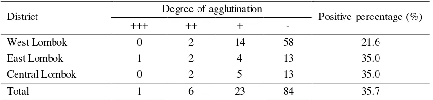
Serological tests have been developed and evaluated for diagnosis of trypanosomosis. The antibody-detecting tests such as the CATT/T. evansi so that, detection of infected cattles by serological test (CATT) revealed 35.7% (30/114) positive (Tabel 2.).
Table 2. The degree of intensity of aglutination of samples in CATT
District Degree of agglutination Positive percentage (%)
+++ ++ + -
West Lombok 0 2 14 58 21.6
East Lombok 1 2 4 13 35.0
Central Lombok 0 2 5 13 35.0
Total 1 6 23 84 35.7
-: Absence of agglutination; +: Moderate agglutination; ++: Strong agglutination; +++: Very strong agglutination
negative (Ngaira et al. 2005). Therefore, an emphasis is necessary to address the problem of diagnosis of T. evansi in the region. It is also important to note that serological tests need to be validated and standardized, if they are to be suitable for correct identification of infected animals, cross evaluation in different laboratories is thus required.
CONCLUSION
Serology prevalence to surra in Lombok Island was considerably high. Even though CATT/T. evansi technique is sensitive and suitable for mess screening, how ever is not always able to distinguish current from past infection. In addition, two or more technique should be used to diagnose reservoir host in Lombok island and for animal with sero negative should be re-tested after 30 days from the first examination due to serological latency.
ACKNOWLEDGEMENT
The author say thank to Eko Setyo Purwanto, Farlin Nefo, Edi Satria on technical assistance in the laboratory.
REFERENCES
Abdel-Rady A. 2008. Epidemiological studies (parasitological, serological and molecular techniques) of Trypanosoma evansi infection in camels (Camelus dromedarius) in Egypt. Vet World. 1:325-328.
Abule E, Snyman HA, Smit GN .2007. Rangeland evaluation in the middle Awash valley of Ethiopia: I. Herbaceous vegetation cover. J Arid Environ. 70:253-271.
Aquino LPCT, Rosangela ZM, Karen RL, Luiz CM, Garcia MV, Gustavo PB. 2010. Antiigenic characterization of Trypanosoma evansi using sera from experimentally and naturally infected bovines, equines, dogs and coatis. Rev Bras Parasitol Vet. 19:112-118.
Aregawi WG, Kassa ST, Tarekegn KD, Brehanu WT, Haile ST, Kiflewahid FZ. 2015. Parasitological and serological study of camel trypanosomosis (surra) and associated risk factors in Gabi Rasu Zone, Afar, Ethiopia. 7:234-240.
Babeker EA, Elrasoul YMH. 2014. Incidence and treatment of camel trypanosomosis (guffar) in West Omdurman in Sudan. J Anim Feed Res. 4:74-82.
Bajyana SE, Hamers R. 1988. A card agglutination test (CATT) for veterinary use based on an early VAT RoTat 1.2 of Trypanosoma evansi. Ann Soc Belg Med Trop. 68:233-240.
Bhutto B, Gadahi JA, Shah G, Dewani P, Arijo AG .2010. Field investigation on the prevalence of trypanosomiasis in camels in relation to sex, age, breeds and herd size. Pak Vet J. 30:175-177.
Birhanua H, Rogé S, Simon T, Baelmans R, Gebrehiwot T, Goddeeris BM, Büscher P. 2015. Surra SeroK-SeT, a new immunochromatographic test forserodiagnosis of Trypanosoma evansi
infection in domestic animals. Vet Parasitol. 211:153-157.
Brown C, Torres A. 2008. Foreign animal diseases. 7th Ed. St. Joseph (US): Animal Health Association.
Buscher P. 2014. Diagnosis of African trypanosomiasis. In: Magez S, Radwanska M, editors.
Dinas Peternakan Sumba Timur. 2012. Kejadian surra di Sumba Timur. Sumba Timur (Indonesia): Dinas Peternakan Sumba Timur.
Eyob E, Matios L. 2013. Review on camel trypanosomosis (surra) due to Trypanosoma evansi: Epidemiology and host response. J Vet Med Anim Health. 5:334-343.
Hagos A, Yilkal A, Esayass T, Alemu T, Fikru R, Feseha G, Goddeeris BM, Claes F. 2009. Parasitological and serological survey on trypanosomis (surra) in camels in dry and wet areas of Bale Zone, Oromyia Region, Ethiopia. Rev Méd Vét. 160:569-573.
Horn D. 2014. Antigenic variation in Africantrypanosomes. Mol Biochem Parasitol. 195:123-129.
Kertawirawan PA, Adijaya MR. 2012. Efektivitas penggunaan ivermectin untuk pengendalian parasit cacing pada usaha tani penggemukan sapi Bali. Seminar Nasional Teknologi Peternakan. hlm 48-55.
Luckins AG. 1988. Trypanosoma evansi in Asia. Parasitol Today. 4:137-142.
Mabey D, Peeling RW, Ustianowski A, Perkins MD. 2004. Diagnostics for the developing world. Nat Rev Microbiol. 2:231-240.
Magnus E, Lejon V, Bayon D, Buyse D, Simarro P, Verloo D, Vervoort T, Pansaerts R, Buscher P, Van Meirvenne N. 2002. Evaluation of an EDTA version of CATT/T. bruceigambiense for serological screening of human blood samples. Acta Trop. 81:7-12.
Ngaira JM, Olembo NK, Njagi ENM, Ngeranwa J. 2005. The detection of non-RoTat 1.2
Trypanosoma evansi. Exp Parasitol. 110:30-38.
OIE. 2012. Trypanosoma evansi infection (surra) by the World Assemblyof Delegates of the OIE in May 2012. Terrestrial Manual Chapter 2.1.17: Office International Des Epizooties. Rome (Italy): FAO.
Patra AK. 2007. Nutritional management in organic livestock farming for improved ruminant health and production an overview. Livest Res Rur Develop 19:1-21.
Radostits OM, Gay CC, Hinchcliff KW, Constable PD. 2007. Veterinary medicine. 10th Ed.
Philadelphia (US): Saunders Ltd.
Reid SA. 2002. Trypanosoma evansi control and containment in Australasia. Trends Parasitol. 18:219-24.
Roge S, Baelmans R, Claes F, Lejon V, Guisez Y, Jacquet D, Büscher P. 2014. Development of a latex agglutination test with recombinant variant surface glycoprotein for serodiagnosis of surra. Vet Parasitol. 205:460-465.
Santosa S, Agus S, Ratih W. 2013. Analisis potensi pengembangan usaha peternakan sapi perah dengan menggunakan paradigma agribisnis di kecamatan musuk Kabupaten Boyolali. Bul Peternakan. 37:125-135.
Sudarisman. 2006. Pencegahan penyakit virus pada hewan dengan vaksin mucosal. Wartazoa 16:181-189.
Sutama IK. 2008. Pemanfaatan sumberdaya ternak lokal sebagai ternak perah mendukung peningkatan produksi susu nasional. Wartazoa. 18:208-217.
Tadesse A, Omar A, Aragaw K, Mekbib B, Sheferaw DA. 2012. Study on camel trypanosomosis in Jijiga Zone, Eastern Ethiopia. J Vet Adv. 2:216-219.
DISCUSSION
Questions
1. The CATT useless for diagnostic tools what do you think?
2. How much the surra desease found in Indonesia?
3. How is the procedure of CATT, is the serum taken directly from the field?
4. What is an advantage of using CATT for surra test?
5. What type of medicine used for surra?
6. What is medicine for surra preventive and how much the dose for preventive?
Answers
1. The CATT can be used to test for screening only
2. Surra desease already found in some part of Indonesia, however there is no research undertaken about surra disease in Bali
3. In CATT method, blood from field is transported to Laboratorium by using ice box, then centrifuge to separate the serum. The serum then is used in CATT test.
4. The CATT test is faster and cheaper compared to other test. It is applicable in the field and do not need an expert to do the test
5. Many type of medicine are available for surra. However, due to the resistance cases to surra medicines in the field, the type of medicine will be used must be examined first