The treatment of anterior tooth crowded case of
upper jaw with dental transposition between
canine and lateral incisor tooth by fixed appliance
Tita Ratya Utari
Lecturer Dentistry Study Program, Faculty of Medical and Health Science University of Muhammadiyah Yogyakarta
ABSTRACT
Background: Dental transposition is an anomaly which the teeth position of the same jaw quadrant change position in the dental arch. The dental transposition of the teeth were classified into 2 types that are complete transposition and incomplete. A complete dental transposition describes a condition in which the dental crown and all structure of the dental root have a parallel position to the exchanged positions. Purpose: This paper aims to report the successful treatment of a canine complete transposition case with upper lateral incisor done at a patient. Case: There is a case based on a report from a boy aged 11 years who come with a complaint about irregular arrangement of front tooth of maxilla. Left central incisor (tooth 21) grew 90-degree rotation, there is mesiodens tooth in the part of palatal. There is still deciduous tooth that are tooth 63 and tooth 55. Result of radiography observation (OPG) seems that tooth 23 has not erupted and the tooth is located in between tooth 21 and 22, where it should be located in the distal side of tooth 22. Case Management: Orthodontic treatment has been carried out with fixed appliance using bracket straight wire slot 0.22. Tooth 23 is pulled toward the distal to place the correct position in the distal side of tooth 22 and extraction tooth 63 is also done. Conclusion: This dental transposition procedure requires a long time treatment, because this is done slowly and carefully. The treatment of transposition case between canines with upper lateral incisor that are done to the patient is successful enough. The root and crown are in good position, and class I occlusion.
Key words: Dental transposition, canine, lateral incisor, fixed appliance
INTRODUCTION
A dental transposition is a unique malocclusion and represents an extreme disturbance of the tooth arrangement and tooth eruption position (ectopic eruption) in which the position of two permanent teeth in one quadrant are exchanged.1,2 Dental transposition is anomalous of the teeth position where the teeth on the same jaw quadrant exchange position in the dental arch. Many studies show high prevalence of canine transposition with the first upper premolar transposition if it is compared to other types, besides that prevalence is higher among women and more unilateral. About two-thirds of the examples of this transposition are in the dental arch of left side.1
incisor (Mx.I2.I1) and the canines to the region of central incisor dental (Mx.C to I1).
Canines are the most often dental in experiencing transposition. 1 The dental of upper canine is probably dental who is the most varied position in the composition of human teeth and important for the orthodontist who often must find their location and move it. These teeth are often out of dental arch to the facial and palatal. It appears that from the canine transposition of first upper premolar sometimes show on the transposition of canine eruption in the side of facial between the first and second premolars, especially if there is still deciduous canine, so that it affects deficiency of arch length. Canine often rotates to the mesial, while the first premolar rotates to the distal and sometimes rotates to mesiopalatal. 5,6
Three studies provided sufficient data on the prevalence canine transposition - first premolar: 0.03% in a population sample of school-age children in Sweden, 0.13% in the population sample of dental patients in Saudi Arabia and 0.25% in a population sample of orthodontic patients in Scotland.5 The dental transposition has never been reported occurs of the deciduous teeth and never occurs in the upper and lower jaw at once. Often accompanied by other congenital anomalies such as hipodontia, peg shape, persistence canines eldest, severe rotation, malposition and root dilaserasi to be a malformation from the neighboring teeth. 2,3
Some theories state that tooth transposition is caused by multifactorial genetic factors. Hereditary, dental lamina or tooth seeds of the exchanged dental, the occurrence of trauma in the period of the oldest teeth, the occurrence of persistence of older canines, the teeth are excessive, or abnormalities in the bone and the presence of local factors such as tumors and cysts.7
Several case reports showed ectopic eruption with variations that are labeled as transposition, sometimes are qualified by additional modifications such as pseudo, incomplete, partial, simple or coronal. Ectopic eruption is a broad category that encompasses all abnormal tooth eruption path or deviate. Therefore, tooth transposition must be considered as subdivisions of ectopic eruption in which all the transposition are examples of ectopic eruption, but only a few ectopic eruption is transposisition.5
The treatment of tooth transposition case depends on the condition that occurred, such as leveling the arrangement tooth permanent of transposition position, the extraction of one or two teeth that transposes or autotransplantation.1,2,7
The purpose of this paper is to report the successful of cases treatment of canines with upper lateral incisor that are done to the patient.
CASE
A patient (male) was 9 years old, come with complaints of crowded upper teeth. The patient come from the tribe of Java with height 145 cm and weight 35 kg. The patient is still in a period of mixed teeth, seems that the oral hygiene is good enough; there are still deciduous in the upper jaw teeth that are tooth 55 and tooth 63. The teeth arrangement in the lower jaw is good and neat enough; there are still deciduous teeth that are tooth 75 and tooth 85. The patient feels that the dental of lower jaw has no problem and just want to be treated on the upper jaw only. Although he is already given the explanation that in the end of the treatment can occur difficulties in the interdigitation correction, but the patient has concluded to make treatment on the upper teeth only.
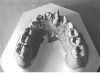
From the study analysis, seems that tooth 21 rotates 900 and there are mesiodens tooth between teeth 11 and teeth 21 in the side of palatine (Figure 1 and 2). Over jet 2 mm (on the teeth 11 and 41), overbite 2 mm (on the teeth 11 and 41). Molar and canine relationship both right and left is first class angle. A midline tooth 11 is aligned with teeth 41. From the analysis of mixed tooth, the space requirements for eruption of the canine tooth (tooth 23) sufficient with doing extraction of tooth 63.
From the analysis of panoramic photo, appears that teeth seed 23 located between tooth 21 and 22. Teeth seed 55, 75 and 85 are in a good position (Figure 3).
Figure 2. Intra oral photographs before treatment.
Figure 3. Pre-treatment panoramic radiograph.
CASE MANAGEMENT
Treatment began in July 2007. In the first stage, mesiodens is carried out which is located at the palatine part of teeth 21 and the opening of the buccal teeth 23 so that cleats bonding (button) can be mounted on the buccal of crown surface. Installation of braces is done on the upper jaw only (based to the patient wish) by fixed orthodontic appliance straight wire slot 0:22. In this stage, leveling is done mainly on teeth 21 that have rotated with using wire niti 0:12, then with 0:18 and niti rec 0.16x0.22. Teeth 63 were maintained and were ligated with ligature wire into a unity with
the teeth 24, 25 and 26 to pull teeth 23 steadily towards distal (Figure 4).
At month 3 (October 2007) treatment of tooth 21 that the rotation has been corrected, and the tooth 23 has moved to the distal in position collide with teeth 22 (Figure 5). Teeth 55 experienced luxation so that extraction will be done. Wire is replaced with ss wire, ties eight ligation of teeth 21 and teeth in the left upper region and the withdrawal of the teeth 22 is carried out towards the mesial and pull the tooth 23 to occlusal plane slowly.
The second stage, after the teeth 23 moves toward occlusal and touch the crown of tooth 63 so tooth 63 will be extracted, tooth 65 has extracted (on 18 November 2007). Then, shift tooth 22 towards the mesial and pull tooth 23 towards the distal and occlusal. On the month 5 (December) the tooth 22 has already coincided with tooth 21, the median line appears in a line, the button replacement of tooth 23 is done by canine braces (Figure 6). In the month 9 (April 2008), tooth 23 has reached occlusal plane on the position first class relation, but appear that the cervix part of gingival in the side of buccal still experienced inflammation (Figure 7).
The patient begins to not control routinely because inflammation appears gradually improved at month 15 (October 2008). Month 19 (February 2009), the gingival inflammation of canine dental had already improved, but the teeth 22 experienced hyperplasic gingival
Proceed to the third stage that is the median line and interdigitation correction. The correction of the
Figure 6. Intra oral photographs 5 months of treatment.
Figure 7. Intra oral photographs 9 months of treatment.
Figure 4. Intra oral photographs 3 weeks of treatment.
median line is by moving one by one the right upper anterior teeth towards to the mesial. Month 24 (July 2009) appears that the interdigitation is good enough with first class canine relation both right and left, the median line appears close to one line. The patient feels satisfied with treatment and in August 2009 and he wants to remove braces and continued to use retainer (Figure 8).
During the treatment of 24 months, the teeth 22 and 23 which experienced transposition have corrected in a good position. Teeth 21 which rotated have also corrected. The roots of the teeth 22 and 23 appear on the panoramic x ray in a good position. Teeth 55, 75 and 85 have erupted in a good position. Intra oral and extra oral photos are shown in the picture. Molar and canine relationship both right and left is in class I angle relationship with a fairly good interdigitasion; the median line of upper – bottom tooth closes to one line. However in the teeth 22, seems that gingival on the cervix became hyperplasic gingival so that the next stage needs to take gingivectomy treatment so better aesthetically.
DISCUSSION
The parents and patients want to do orthodontic treatment because of aesthetic considerations. The patient is still 11 years old. Teeth 23 and 22 experienced a complete transposition that is caused by the occurrence of the persistence of teeth 63. Rotation of teeth 21 is caused by the occurrence of the excess (mesiodens) teeth of the palate patients. Foster and Taylor states that a cone-shaped teeth are often associated with displacement of adjacent teeth.7
In this case, including the types of the canine-lateral incisor (Mx.C.I2) transposition and occurs in the left region which the case is often occur than the other tooth transposition.
The beginning diagnosis in the development of a transposition case based on a thorough intraoral diagnosis and is followed by complete radiographic analysis is better at the age of 6-8 years. This is especially important for early detection of malposition. If new transposition begins to occur can be detected early, the treatment should be started with interceptive to take deciduous teeth that are restrained and it guided ectopic teeth to its normal position in the arch. Interceptive procedure prevents the complete development of permanent canine eruption transposition.8
If the lateral incisor and canine has erupted in the position of transposition, it is usually not made an effort to correct the teeth into its normal position. The dental alignment are in a position of transposition and incisor reshape the surface of so that it will not damage the tooth or its supporting network and will obtain an aesthetic result that can be accepted. The dental rotation and alignment are corrected of transposition position of the position, and then used the retainer anterior mandibular fixed by debonding because the cut of supracrestal gingival fibers on the teeth that experience rotation.8
Transposition of maxillary offers several treatment options to be considered. Moving the dental transposition into its normal position on the arch in general would be better aesthetically and functionally. On the incomplete transposition, up righting and rotation is the most widely procedure done to put teeth into normal alignment, thus providing enough space on the arch.8,9
If the transposition is incomplete, with musty roots in transposition position, the teeth reposition into normal relation to the arch requires a complex procedure and likely to cause damage of teeth or supporting tissues. Alignment of the teeth are in the position of transposition must be done carefully because it is quite risky. 8 If one of tooth transposition or adjacent teeth experiences caries or severe trauma, should be considered to do the extraction procedure on the teeth. Extraction space is then used for alignment of canine and incisor crowding reduction.8 The treatment of this case of complete transposition was corrected by restoring the position of the teeth 23 and 22 to the right position with the consideration of aesthetics and function, the position of the root, and the availability of sufficient space Figure 8. Intra oral photographs after treatment.
to perform the extraction of tooth 63. The teeth 23 has not eruption yet so we can guide the eruption of 23 to the right position. Anterior teeth have an important function for arranging the continuing of dental arch, esthetic, and maintenance the form and function of the teeth.10 Canine is part of anterior teeth that give important support for esthetic and function, so it is very important to maintenance the teeth and guide the eruption in the right position.11
The more complex treatment options include efforts to move the teeth transposition into its normal position in the maxillary arch in certain cases, the treatment procedures can cause alignment of the transposition position that can be accepted aesthetically and functionally. To avoid disruption root or resorption during the treatment and to prevent Bony loss plat cortical of canines whose position into labial, firstly tooth transposition (premolars or lateral incisor) should be moved to a palatal, so that enough space to move the canine into its normal position. Moreover, the other (dental) transpositions can be moved into labial, back to its normal position in the arch.
In this case, we do not move tooth that is in transposition toward palatal but only pulling the canine toward distal, move lateral incisor to mesial, slowly and with light force. At the time of tooth 22 has occupied its proper position, coincide with tooth 21, so it seems that gingival of tooth 21 covers one-third of the tooth crown. Carranza et al.,12 states that, some causes are inflammation, the influence of drugs, systemic disorders, neoplastic or false enlargement. The cause of gingival enlargement in patients is probably false enlargement because of the gingival enlargement purely that is caused by the growth of connective tissue surrounding the teeth with the clinical picture, the gingival appears to cover part of teeth cervical, the occurrence of inflammation, and there is not a pathological condition. The enlargement therapy of this type is gingivectomy by a more competent experts periodontology.
It can be concluded that although this procedure requires a long time of treatment, but if it’s done slowly and carefully, the result will be very satisfactory aesthetically and functionally. The treatment of transposition case between canines with upper lateral incisor that are done to the patient is successful enough. The root and crown are in good position, and class I interdigitation. The enlargement therapy for the lateral incisor is gingival resection by a more competent experts in periodontology.
REFERENCES
1. Peck S, Peck L. Classification of maxillary tooth transposition. Am J Orthod Dentofac Orthop 1995; 107: 505-17.
2. Shapira Y, Kuftinec MM. Maxillary tooth transpositions: Characteristic feature and accompanying dental anomalies. Am J Orthod Dentofac Orthop2001; 119: 127-34.
3. Shapira Y, Kuftinec MM. Maxillary canine-lateral incisor transposition - orthodontic management. Am J Orthod Dentofac Orthop 1989; 95: 439-44.
4. Wasserstein A, Tzur B, Breznoak N, Incomplete canine transposition and maxillary central incisor impaction-a case report. Am J Orthod Dentofac Orthop 1997; 111: 635-9.
5. Peck L, Peck S, Attia Y. Maxillary canine-first premolar transposition, associated dental anomalies and genetic basis. 1993. 63(2): 99-109.
6. Shapira Y, Kuftinec MM, Stom D, A unique treatment approach for maxillary canine-lateral incisor transposition. 2001. 119: 540-5.
7. Suri L, Gagari E, Vastardis H. Delayed tooth eruptiopn: Pathogenesis, diagnosis, and treatment. A literature review. Am J Orthod Dentofac Orthop 2004; 126: 432-45.
8. Shapira Y, Kuftinec MM,Stom D. Tooth transpositions
-a review of the liter-ature -and tre-atment consider-ations. 1988. 59(4): 271-76.
9. Bocchieri A, Braga G. Correction of a bilateral maxillary canine-first premolar transposition in the late mixed dentition. 2002. 121: 120-8.
10. Bishara SE. Impacted maxillary canines: A review. Am J Orthod Dentofac Orthop 1992; 101(2): 159-17.
11. Steward JA, Heo G, Glover KE, Williamson PC, Lam EWN, Paul W. Factor that relate to treatment duration for patients with palatally impacted maxillary canine, Am. J Orthod Dentofac Orthop; 119(3): 216-25.


