How to cite:
Dewi DS, Chairinnisa ES, Sujuti H et al. (2018) Ƚ-SMA Expression Increased Over Cell Passages and Decreased by Exogenous TGF-Ⱦ1, In Vitro Stud-ies on Myofibroblast Derived from Orbital Socket Contracture J. Trop. Life. Science 8 (2): 200
– 205. *Corresponding author:
Debby Shintiya Dewi
Department of Ophthalmology, Faculty of Medicine, Brawijaya University
Jalan Veteran, Malang, Indonesia 65145 E-mail: [email protected]
JTLS | J. Trop. Life. Science 200 Volume 8 | Number 2 | April | 2018
Ƚ
-SMA Expression Increased Over Cell Passages and Decreased by Exogenous TGF-
Ⱦ1,
In Vitro
Studies on Myofibroblast Derived from Orbital Socket Contracture
Debby Shintiya Dewi *, Elsa Safira Chairinnisa, Hidayat Sujuti, Diana Lyrawati, Tinny Endang Hernowati Department of Ophthalmology, Faculty of Medicine, Brawijaya University, Malang, Indonesia
ABSTRACT
Ƚ-smooth muscle actin (Ƚ-SMA), a marker of myofibroblast, induces cytoskeleton reorganization, increases contrac-tility and stimulates cell migration in TGF-Ⱦ1 induced stress fibers. The aims of the present study were to determine the level of Ƚ-SMA expression and morphological cell changes in different passages of myofibroblasts with varied TGF-Ⱦ1 concentrations. Myofibroblast cell cultures were derived from fibrotic tissues of fourth degree socket con-tracture. The Ƚ-SMA expression level was measured in myofibroblast cultures passage I, II, and III with and without 10 ng/mL TGF-Ⱦ1, and in passage III with 2.5; 5; 10; and 20 ng/mL TGF-Ⱦ1. Results: The levels of Ƚ-SMA expres-sion level in passage I to III were I 31.42 ± 3.4; 40.34 ± 8.14 and 56.37 ± 7.57, respectively. Addition of 10 ng/mL TGF-Ⱦ1 into passage I-III myofibroblast cultures resulted in Ƚ-SMA expression level of 31.24 ± 2.93; 36.81 ± 6.09; and 14.29 ± 2.72, respectively. Myoblasts passage III showed the lowest Ƚ-SMA expression level following exposure to TGF-Ⱦ1 10 ng/mL (22.37 ± 12.86) and highest without TGF-Ⱦ1 (48.34 ± 13.36), however no morphological changes detected. Ƚ-SMA expression level increased with cell passages, decreases with addition of TGF-Ⱦ1 while not affecting morphology of myofibroblast derived from the orbital socket contracture.
Keywords: TGF-Ⱦ1, Ƚ-SMA, fibrotic, eye, socket, contracture INTRODUCTION
Orbital socket contracture is a wrinkled state of anophthalmic socket, thus, it cannot fit the prosthesis. The incidence of socket contracture was reported as 7.7% of the overall anophthalmic socket [1]. Our study, at dr. Saiful Anwar General Hospital Malang, Indonesia, in 2007, however, showed that the occurrence of socket contracture was 32.25% overall, mostly 5 years post-eye removal (60%) [2].
Our study showed that application of Mitomycin C during and after socket reconstruction surgery pre-vented eye socket contracture recurrences in 50% cases [3]. In one case report, the socket contracture did not recur on a patient undergoing 7 repair surgeries within one year with the use of 0.04% Mitomycin C intra- and post-operatively [4]. Prevention of recurrence of this case may be explained provided that the molecular mechanism of socket contracture has been known.
The socket contracture formed fibrotic tissue and granulation due to chronic irritation, one of which due to friction of the prosthesis with the socket, resulted in impairment of wound healing process [5, 6]. The for-mation of hypertrophic scars that dominates the socket contracture tissue is a deviation of wound healing in the form of hyperactivity of fibroblast, excessive formation of myofibroblasts and extracellular matrix (ECM) depo-sition [7, 8]
TGF-Ⱦ plays roles in all phases of eye wound heal-ing process by increasheal-ing growth factor secretion in-volved in cell migration, proliferation, ECM deposition and myofibroblast formation. Increased growth factor activity not only leads to an increase in wound healing rate, but also a higher risk for scar formation. Therefore, TGF-Ⱦ is one of the most potential targets for scarring intervention [8, 9, 10, 11].
tracture has not been clearly elucidated and may involve many factors. Studies on fibrosis were mostly performed using tissues other than socket contracture. Among few studies on socket contracture, one has demonstrated that myofibroblasts play an important role in modulating wound healing, tissue remodeling and organ defor-mation [12]. TGF- Ⱦ1 is a major profibrotic cytokine and increased expression plays an important role in the deposition of collagen and extracellular matrix, wound healing and scar formation in the socket contracture tis-sue [6]. TGF-Ⱦ1 is a key mediator that induce Ƚ- SMA [11, 12].
Alpha-smooth muscle actin (Ƚ- SMA) may serve as a reliable marker for myofibroblast cells [12], although others considered it as not a consistent marker for con-tractile fibroblasts and lots of collagen [13]. Alpha-SMA widely presents in stressed fibers induces cytoskeleton reorganization, increases contractility and stimulates cell migration. Stress fibers contracts myofibroblasts such as in muscle movements.
Myofibroblast cells morphologically resembling fi-broblasts and smooth muscle cells, rich in cytoplasmic microfilament (actin-rich stress fibers) and Ƚ-SMA ex-pression. Fibroblasts appear on day 2-3 following injury, while myofibroblasts appear on day 12 when wound contractions are nearly 80% complete [11, 12].
In the present study, we reported our in vitro study determining the level of Ƚ-SMA protein expression in myofibroblasts derived from orbital socket contracture
and whether addition of TGF-Ⱦ1 may affect Ƚ-SMA
ex-pression and morphology of such myofibroblasts. Since passage number of cells passaged several times may af-fect protein expression, we also determined whether pas-sage number alone, with or without addition of TGF-Ⱦ1, affect the level of Ƚ-SMA expression in myofibro-blast passages [14].
MATERIALS AND METHOD Sample of myofibroblast
This study was conducted in November to Decem-ber 2017 at Ophthalmology Department-dr. Saiful Anwar Hospital and Central Laboratory of Life Sciences
– Brawijaya University, Malang. The study design was approved by the Ethics Committee of dr. Saiful Anwar General Hospital and followed the guidelines of the Declaration of Helsinki.
The inclusion criteria for the patient was orbital socket contracture (degree 1 – 5) with medical indica-tion for surgical socket reconstrucindica-tion. The selected pa-tient was based on signed informed consent to partici-
pate in the study. The sample of this study was fibrotic tissue removed from a patient with fourth degree socket contracture.
Primary culture of myofibroblast was prepared from patient with socket contracture surgery [15]. Each tissue was cut into pieces approximately 1Å ~ 1 mm in size, placed in 12 well plastic culture plates in DMEM with 50 U/mL of penicillin, 50 g/mL streptomycin and 10% FCS, and incubated at 37°C in a humidified (95%) in-cubator under 5% CO2. Cell culture media were
changed thrice weekly. Upon reaching confluence, cells were trypsinized with 0.25% trypsin/EDTA solution in phosphate buffered saline (PBS) and passaged in 25 cm2 plastic cell culture flasks. Cells were cultured further for this study, or passaged 2-3 times and used directly for experiments, or harvested, resuspended in freezing me-dia and stored frozen in -80°C until used. For experi-ments, a series of cells passaged I-III were cultured for 3 weeks and incubated 24 hours with or without the addi-tion of 10 ng/mL TGF-Ⱦ1. Another series of experi-ments were performed using cells passage III, cultured for 3 weeks and incubated 24 hours with the addition of 2.5; 5; 10; or 20 ng/mL TGF-Ⱦ1.
Measurement of α-SMA level and myofibroblast mor-phology
The expression levels of Ƚ-SMA were measured in
passage I, II, III without and with the administration of
TGF-Ⱦ1 10 ng/mL as well as in passage III with
TGF-Ⱦ1 2.5, 5 ng/mL, 10 and 20 ng/mL.
The Ƚ-SMA expression level was measured with
anti Ƚ-SMA antibody [1A4, Cat. number ab7817,
Abcam]. Cells were fixed with paraformaldehyde, per-meabilized with 0.1% Triton X-100 and blocked with
3% BSA for 30 minutes at 25°C. Samples were
incu-bated with primary antibody (1/
400 in 1× TBS) for 3 hours at 25°C. An Alexa Fluor®633-conjugated Goat anti-mouse IgG polyclonal (1/
500) was used as the
sec-ondary antibody. Fluorescent images of Ƚ-SMA
myofi-broblasts were acquired in red channel using 400×
mag-nification, 0.5% power of excitation lines, and gain set at 850 of the laser scanning confocal microscope (CLSM FV1000, Olympus Corp., Waltham, MA). Confocal im-ages of the fluorophores were analyzed using Olympus Fluoview ver.4.2a software. Ƚ-SMA expression was measured in arbitrary unit (AU). Cells were observed also morphologically using 100× magnification with
in-verted microscope Olympus IX 71 and 400×
JTLS | J. Trop. Life. Science 202 Volume 8 | Number 2 | April | 2018 Data analysis
The results were analysed using IBM SPSS version 2.0 statistical analysis application. The data analysis in the passage group with TGF-Ⱦ1 exposure using Shapiro-Wilk normality test, followed by ANOVA or Kruskal-Wallis test, and post-hoc Tukey or Mann-Whitney test to know differences between each group. Results were considered statistically significant when p < 0.05.
RESULTS AND DISCUSSION
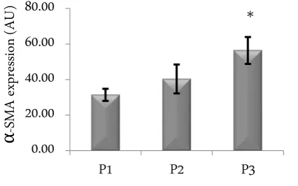
Effect of the number of passage on α-SMA level The level of Ƚ-SMA expression increased with in-creasing passage number, with values of 31.42 ± 3.4, 40.34 ±8.14 and 56.37±7.57 arbitrary unit (Figure 1).
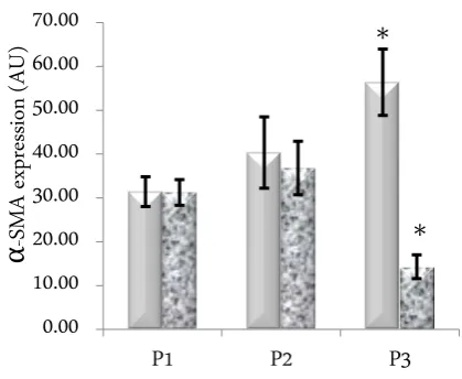
Addition of 10 ng/mL TGF-Ⱦ1 in cell culture de-creased Ƚ-SMA level only in passage III (p = 0.000). Val-ues of Ƚ-SMA level in each group was 31.24 ± 2.93; 36.81 ± 6.09; and 14.29 ± 2.72 arbitrary unit for passage I-III, respectively (Figure 2).
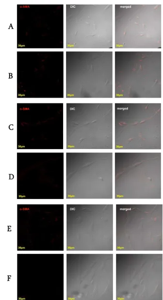
Microscopically, different passage with or without addition of TGF-Ⱦ1 did not change cell morphology (Figure 3).
When different passages were compared between with and without addition of TGF-Ⱦ1, our results showed that the most pronounced effects were seen on passage III and with addition of TGF-Ⱦ1 (Figure 4). On passage III, addition of TGF-Ⱦ1 decreased a-SMA ex-pression up to three folds.
Effect of TGF-Ⱦ1 concentration added on a-SMA expression was studied using passage 3 of myofibroblast primary culture from socket contracture. The results showed that compared to the myofibroblast without TGF-Ⱦ1, only 10 ng/mL TGF-Ⱦ1 decreased significantly
Ƚ-SMA expression level (p=0.000). The a-SMA
expres-sion level was 48.34 ± 13.36; 46.45 ± 47.04; 38.07 ± 9.54; 22.37 ± 12.86; and 29.80 ± 13.33 in cell culture with
TGF-Ⱦ1 0; 2.5; 5; 10; and 20 ng/mL, respectively (Figure 5).
Our study demonstrated that passaging influence Ƚ–SMA level on myofibroblast primary culture derived from orbital socket contracture tissue. Similar results were reported by Kinner et.al. (2001) that levels of Ƚ -SMA are increasing following the number of passages or passages performed. One of the main factors of physio-logical change from fibroblasts to myofibroblast is a me-chanical tension. [16] According to Wakatsuki et.al. (2000) there is progressive increase in matrix rigidity overtime if the tissue experienced a tension, including in our present study during passaging process. [17] Our study demonstrated that following passage and cultured
Figure 1. Level of Ƚ-SMA in cell culture with different passage (P1, P2, P3) without TGF-Ⱦ1
Figure 2. Level of Ƚ-SMA III in cell culture with different passage (P1, P2, P3) with addition of 10 ng/mL TGF-Ⱦ1. * de-notes significantly different compared to P1 (p = 0.000) without addition of TGF-Ⱦ1, a pro-fibrotic factor, myo-fibroblasts remained the same morphological while the Ƚ–SMA level increased. Probably, the passaging pro-cess itself induce stress response, which may increase the production of endogenous TGF-Ⱦ1, subsequently in-duced Ƚ–SMA expression, even without exogenous TGF-Ⱦ1. [18] Thus, the myofibroblast, a differentiated phenotype, did not convert into fibroblasts, an inactive phenotype of cells. [19] During physiological wound healing process, myofibroblasts undergo apoptosis. [20, 21, 22, 23].
On the contrary, the addition of exogenous TGF-Ⱦ1 on passage 3 resulted in significantly low Ƚ-SMA. In such case, others reported that TGF-Ⱦ1 may activate TGF-Ⱦ1 receptor, however, instead of inducing Ƚ-SMA expression, it activates focal adhesion kinase (FAK). Fur-ther, FAK binds integrins, fibronectin and collagen which all have roles as cell adhesion receptors in myofi-broblast differentiation. [24]. Another study also re-ported that TGF-Ⱦ1 did not always induce Ƚ-SMA ex-pression. In the presence of synthetic hyaluronan (HA)
Figure 3. Confocal microscopic observations. Bright field view (left), red filter immunofluorescent view for Ƚ-SMA (middle), and the merged view of both (right). Passage I (A), passage I with TGF-Ⱦ1 10ng/ml (B), passage II (C), passage II with
JTLS | J. Trop. Life. Science 204 Volume 8 | Number 2 | April | 2018
Figure 4. Effect of passage and addition of TGF-Ⱦ1. Significant effects were seen on passage 3 with and without 10 ng/mL TGF-Ⱦ1 addition.
myofibroblast differentiation was inhibited without de-creasing TGF-Ⱦ1 receptor expression. [25]. Further stud-ies are required to clarify whether FAK activation causes Ƚ-SMA degradation and apoptosis of myofibroblasts, or other pathways causing TGF-Ⱦ1 decreased Ƚ-SMA.
CONCLUSION
Passage number increased whereas TGF-Ⱦ1 could decrease Ƚ-SMA expression levels in dose-dependent fashion, however, both did not affect morphology of my-oblast culture derived from orbital socket contracture. Therefore, in vitro experiments to study myofibroblast from orbital socket contracture should consider passage number and level of TGF- Ⱦ1.
ACKNOWLEDGMENT
The authors wish to thank staff at Central Labora-tory of Life Science, Brawijaya University for technical assistance in confocal microscopy. This study receives no external funding.
REFERENCES
1. Adhikari RK, Khazai H, Usha KD (2007) Prospective evalu-ation of contracted sockets. Kathmandu University Medical Journal 5 (3): 391 – 395.
2. Lestari AT, Dewi DS, Sadono EG (2008) Pattern and man-agement of contracted socket in Saiful Anwar Hospital Ma-lang: A retrospective study. Makassar, Indonesian Ophthal-mology Meeting.
3. Suharyanto Y, Dewi DS (2012) Mitomycin-C Application in contracted socket reconstruction: A retrospective study. Ma-
lang, Department of Ophthalmology, dr. Saiful Anwar Gen-eral Hospital/Faculty of Medicine, Brawijaya University. 4. Dewi DS (2012) Successful of mitomycin C application in
the reconstruction of recurrent contracted socket: A case re-port. In the 27th Asia Pasific Academy of Ophthalmology Congress: 13 – 16 April 2012; Busan.
5. Stroncek JD, Reichert WM (2008) Overview of wound heal-ing in different tissue types. In: Reichert WM eds. Indsu-muraning neural implant: Strategies for contending with the in vivo environment. Boca Raton (FL), CRC Press. 6. Kurtul BE, Erdener U, Mocan MC et al. (2014) Clinical and
impression cytology findings of amniotic membrane and oral mucosal membrane transplantation for the management of socket contracture. International Journal of Ophthalmology. 7 (2): 340 – 344.
7. Su WH, Cheng MH, Lee WL et al. (2010) Nonsteroidal anti-inflammatory drugs for wound: pain relief or excessive scar formation? Mediators of Inflammation 2010: 413238. doi: 10.1155/2010/413238.
8. Li P, Liu P, Xiong R et al. (2011) Ski, a modulator of wound healing and scar formation in the rat skin and rabbit ear. Journal of Pathology 223 (5): 659 – 671. doi: 10.1002/path. 2831.
9. Ellis JS, Paull DJ, Dhingra S et al. (2009) Growth factors and ocular scarring. European Ophthalmic Review 3 (2): 58 – 63. doi: 10.17925/EOR.2009.03.02.58.
10. Falke LL, Gholizadeh S, Goldschmeding R et al. (2015) Di-verse origins of the myofibroblast—implications for kidney fibrosis. Nature Review Nephrology 11 (4): 233 – 244. doi: 10.1038/ nrneph.2014.246.
11. Stahnke T, Kowtharapu BS, Stachs O et al. (2017) Suppres-sion of TGF-Ⱦ pathway by pirfenidone decreases extracellu-lar matrix deposition in ocuextracellu-lar fibroblasts in vitro. PLoS ONE. 12 (2): e0172592. doi: 10.1371/journal.pone.0172592. 12. Tawfik HA, Abdulhafez MH, Osman WM (2016) Revisiting the role of the myofibroblast in socket surgery: An immuno-histochemical study. Ophthalmic Plastic and Reconstructive surgery 32 (4): 292 – 295. doi: 10.1097/IOP.0000000000000 510.
13. Sun K, Yongen C, Reed NI, Sheppard D (2016) Alpha-smooth muscle actin is an inconsistent marker of fibroblasts responsible for force-dependent TGF-beta activation or col-lagen production across multiple models of organ fibrosis. American Journal of Physiology Lung Cell Molecular Phys-iology 310: L824 – L836.
14. Pan GJ, Chang ZY, Schöoler HR, Pei DQ (2002) Stem cell pluripotency and transcription factor Oct4. Cell Research 12 (1): 321 – 329. doi: 10.2213/cl121321.
tic and reconstructive surgery. San Francisco, American Academy of Ophthalmology. pp. 351 – 352.
16. Kinner B, Spector M (2001) Smooth muscle actin expression by human articular chondrocytes and their contraction of a collagen glycosaminoglycan matrix in vitro. Journal of Or-thopaedic Research 19 (2): 233 – 241. doi: 10.1016/S0736-0266(00)00081-4.
17. Wakatsuki T, Kolodney MS, Zahalak GI, Elson EL (2000) Cell mechanics studied by a reconstituted model tissue. Bio-physical Journal 79 (5): 2353 – 2368. doi: 10.1016/S0006-3495(00)76481-2.
18. Hinz B (2015) The extracellular matrix and transforming growth factor-Ⱦ1: Tale of a strained relationship. Matrix Bi-ology 47: 54 – 65. doi: 10.1016/j.matbio.2015.05.006 19. Manapov F, Muller P, Rychly J (2005) Translocation of p21
Cip1/WAF1 from the nucleus to the cytoplasm correlates with pancreatic myofibroblast to fibroblast cell conversion. Gut 54 (6): 814 – 822. doi: 10.1136/gut.2003.036491. 20. Darby IA, Laverdet B, Bonté F, Desmoulière A (2014)
Fibro-blasts and myofibroFibro-blasts in wound healing. Clinical, Cos-metic and Investigational Dermatology 7: 301 – 311. doi: 10.2147/CCID.S50046.
21. Tomasek JJ, Gabbiani G, Hinz B et al. (2002) Myofibroblasts
and mechano-regulation of connective tissue remodelling. Nature reviews. Molecular Cell Biology 3 (5): 349 – 363. doi: 10.1038/nrm809.
22. O’Connor JW, Mistry K, Detweiler D et al. (2016) Cell-cell contact and matrix adhesion promote ȽSMA expression dur-ing TGFȾ1-induced epithelial-myofibroblast transition via Notch and MRTF-A. Scientific Reports 6: 26226. DOI: 10.1038/srep26226.
23. Negmadjanov U, Holmuhamedov A, Emelyanova L et al. (2016) TGF-Ⱦ1 increases resistance of NIH/3T3 fibroblasts toward apoptosis through activation of Smad2/3 and Erk1/2 pathways. Journal Patient-Centered Research and Reviews 3: 187 – 198. doi: 10.17294/2330-0698.1407.
24. Thannickal VJ, Lee DY, White ES et al. (2003) Myofibroblast differentiation by Transforming Growth Factor-1 is depend-ent on cell adhesion and integrin signaling via focal adhesion kinase. The Journal of Biological Chemistry 278 (14): 12384
– 12389. doi: 10.1074/jbc.M208544200.
25. Chen X, Thibeault SL (2012) Response of fibroblasts to transforming growth factor-b1 on two-dimensional and in three-dimensional hyaluronan hydrogels. Tissue Engineer-ing: Part A 18 (23 – 24): 2528 – 2538. doi: 10.1089/ten. tea.2012.0094.