The journal homepage www.jpacr.ub.ac.id
ISSN : 2302 ‐ 4690
27
Synthesis of Hematite Pigments (
α
-Fe
2O
3) by Thermal
Transformations of FeOOH
Lilik Miftahul Khoiroh1, Diah Mardiana1*, Akhmad Sabarudin1, Bambang Ismuyanto1
1
Departement of Chemistry, Faculty of Mathematic and Natural Science, Brawijaya University, Jl. Veteran Malang, East Java - Indonesia 65145
*Email :[email protected] phone : +62-341-575838 Fax: +62-341-575839
Received 15 January 2013; Revised 24 February 2012; Accepted 20 March 2013; Published online for 1 April 2013
ABSTRACT
Synthesis of iron oxide FeOOH from FeCl3, followed by thermal transformation to form
red hematite was conducted. The effect of pH and temperature calcinations was studied. The observed parameters were include crystal structure, grain size, color, particle size
distribution, and morphology. The result showed that α-Fe2O3 structure with relatively
pure and good crystallinity were found at all designated synthesis condition. The highest degrees of redness and chroma, uniformity of morphology and particle size distribution were achieved at pH 6, while the lowest degrees was formed at pH 8. The pigment at pH 6
has spherical-like shape with an average size of 0.16±0.057 μm. Instead, at pH 8 has
spherical-like and cubic-like shape with an average grain size of 0.17±0.051 μm. The
highest degrees of brightness, redness, yellowness and the highest chroma as well as
smallest grain size of 0.15±0.052 μm were achieved at 800 oC but at this temperature the
crystal orientation of 006 was not appear and the presence of 2θ angle (63.99o) were
supposed as crystal defects and impurity.
Key word: pigment, hematite, thermal transformations method, crystal, morphology
INTRODUCTION
Iron oxide is one of the metal oxide has been developed in various fields, for example as pigments [1]. These pigments have less toxicity, inert, and variety color ranging from black, yellow, brown, and red [2]. The highest demand of the iron oxide pigment is hematite which is usually provides red color [3]. Beside optical data, the geometrical important data of hematite are particle size, particle size distribution, and particle shape [4]. The particle size of hematite pigment is 0.1–0.5 μm. L* value of about 25-45, C* value of about 9-42, Ho value of about 21-57 [1]. Purity and properties of hematite pigments are influenced by some factors among other natural source iron [5], including method of synthesis, synthesis conditions such as pH and calcinations temperature [6].
The journal homepage www.jpacr.ub.ac.id
ISSN : 2302 ‐ 4690
28
thermal transformation which was able to produce hematite more selective. Thermal transformation of goethite was also produced hematite with particles size less than 1 μm [10], and with the same method, the resulting product was relatively pure hematite [11].
The condition of pH during synthesis significantly affect iron oxide resulted [6]. Thermal dehydration of iron oxide was undertaken in two stages. First, synthesis goethite at pH conditions between 5 and 7. Second, the reaction is followed by dehydration process at 750 oC. This process produced a relative pure hematite pigment, but the particle size is less than 0.05 μm [11], which is unqualified as red hematite pigments. The similar method has also been reported by Silva [12]. The first step synthesis was firstly done at pH 12 and then continued by dehydration of goethite at 350 oC. The product was reported contains a mixture of wustite, goethite, and hematite with particle size of 1.56 μm.
Calcinations temperature was reported affects on the particle size resulted and degree of color. Synthesis of hematite pigment at 800 oC for 2 hour produced a colored dark red hematite with L* (49.89) a* (8.14) b* (5.45) and particle size of less than 1 μm [10], while dehydration of goethite at 350 oC for 2 hour produced hematite with particle size 1.56 μm and L* (72.5) a* (5.1) b* (41.8) [12].
Based on those facts, this paper report an examination results of the effect of pH and calcinations temperature during synthesis hematite pigment through thermal transformation of FeOOH. Similar parameters was previously reported able to affect the quality of resulted hematite pigments, which include the grain sizes, particle size distribution, crystal structure, color, and morphology of the pigments.
EXPERIMENT Apparatus
Furnace (Heraeaus) was used for the dehydration of FeOOH, where pH during synthesis of FeOOH was measured by Orion model 420A pH meter. The color value of hematite were measured by color reader (Minolta CR 10), while morphology of hematite were assessed by scanning electron microscope (SEM, FEI type Inspect-S50), and structure crystal was determined by X-ray diffraction (PAN analytical type Expert Pro).
Reagent
All reagent used in this experiment had analytical grades except for HNO3 (Technical grade). FeCl3.6H2O, NaOH, NaHCO3, HCl 37%, were purchased from Merck. Distilled water prepared by Kottermann 1302 was used throughout this experiment.
Procedure Ferric Precursor
Solution FeCl3 0.16 M was adjusted to pH 3.94 by adding appropriate amount of NaOH 4 M. This mixture was heated at 40 oC for 30 minute, then temperature was raised to 70 oC before addition of concentrate HNO3 (30 mL) along with stirring at 750 rpm. Finally, solution was heated at 100 oC to form brown yellowness solid.
Synthesis hematite at various pH
The journal homepage www.jpacr.ub.ac.id
ISSN : 2302 ‐ 4690
29
HCl 1M. Solution was held to 70 oC for 1 hour and stirred at 750 rpm. The solution resulted was cooled to reach room temperature to afford precipitate, and further decantation, was filtered off, and was washed with 300 mL of distilled water and finally dried at room temperature. The iron oxide FeOOH obtained as powder continuously was heated in the furnace at 750 oC for 3 hour. The properties such as color, crystal structure, grain size, and shape were identified.
Synthesis of hematite at various calcinations temperatures
Ferric precursor was prepared in 250 mL of distilled water. A 25 ml NaHCO3 0.5 M was added and adjusted to pH 8 by addition of NaOH 1 M and/or HCl 1M. This solution was heated at 70 oC for 1 hour and stirred at 750 rpm. This was further cooled to reach room temperature, and the precipitate resulted was decanted, filtered off and washed with 300 mL of distilled water and finally dried to room temperature. Iron oxide FeOOH powder was continuously heated in a furnace at 750, 800, 850 oC for 3 hour. The properties such as color, crystal structure, grain size, and shape were further analysed for identification.
Characterization
The hematite pigments obtained were characterized through the determination of L*, a*, b* colorimetric by CIE Lab. Value of C* and Ho was calculated by formula [13]:
C* = [(a*)2 + (b*)2]1/2
H0 = tan-1 (b*/a*), degree 0o≤ Ho≤ 360o
The structure crystal of hematite pigment was analyzed by X-Ray Diffraction type Expert Pro (PAN analytical) with graphite monochromatic Cu Ka radiation (1.54060 A˚). Grain size was analyzed from XRD data using Schrerrer method [14]:
D =
In addition, morphology and particle sizes distribution was analyzed by scanning electron microscope.
RESULT AND DISCUSSION
Synthesis of α-Fe2O3 with pH variation
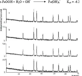
The phase and purity of the product were first examined by XRD. Figure 1 shows a typical XRD pattern of the synthesized sample from FeCl3. All the reflection peaks can be readily indexed to hexagonal phase of α-Fe2O3 with lattice constants a=b= 5.026oA and c= 13.735oA which are in agreement with the standard values (ICSD code 415251). No other phase was detected in Figure 1, which indicated the high purity of the product.
Figure 1 shows that all peaks at all pH variations have strong and sharp peaks, which indicates that the product has good crystallinity. The plane crystal was detected at all pH, which are 012, 104, 110, 006, 113, 202, 024, 116, 018, 214, 300, except direction crystal 006 at pH 9 does not appear. The product, which is formed from calcinations at pH 6,7, and 8, was α-Fe2O3. Transformation reaction of FeOOH to α-Fe2O3 by dehydration is [1]:
2 FeOOH(s) Fe2O3(s) + H2O(g)
The journal homepage www.jpacr.ub.ac.id
ISSN : 2302 ‐ 4690
30
Reaction of equilibrium stability constant of iron hydroxo complexes [1]:
α-FeOOH + H2O + OH- Fe(OH)4- Ks4 = -6.2
Figure 1. XRD pattern of hexagonal α-Fe2O3 a) pH 6 b) pH 7 c) pH 8 d) pH 9
Figure 2. The color of hematite pigment synthesized at pH 6 (a), 7 (b), 8 (c), and 9 (d)
The intensities of XRD at pH 8 is the highest. This is also recognized by the color. The color of change is represented by a* (degree of redness) and b* (degree of yellowness). The color of hematite pigments are found in terms value of degree of yellowness, which is the lowest and the degree of redness, which is the highest. At pH 8, degree of yellowness is the lowest compared to the others, which is the differences in lines a* and b* are significantly wide. Figure 2 shows the hematite pigment synthesized at different pH. The degree of lightness of pigment afforded from synthesis at pH 8 is lower than those afforded in other pH, meanwhile the highest degree of redness, the highest degree of lightness, and the highest was obtained at pH 6. Degree of lightness is affected by the high energy which was absorbed during synthesis. Della [10] reported that excited electron could drop to the ground state as photon was released in a specific wavelength, and this depends on the substance properties. The wavelength of the released energy gives the color of the object. Thus, the higher of the
The journal homepage www.jpacr.ub.ac.id
ISSN : 2302 ‐ 4690
31
material absorbed the energy, the higher color of the object and the lower brightness. The different of color was also influenced by grain size, shape, and particle size distribution.
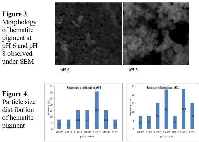
The grain size of hematite pigment determined with XRD by using Schrerrer method. The average grain size of pigment synthesized at pH 6, 7 was 16 μm, while for pH 8 was 17
μm and for pH 9 was 15 μm. The average size of the grain influenced the pigment color (Table 1). The pigment has spherical-like shape (Figure 3).
Figure 3.
Morphology of hematite pigment at pH 6 is spherical and the particle size is uniform than those obtained at pH 8. The particle of hematite at pH 8 has spherical-like and cubic-like shape, and the particle size distribution is less uniform (Figure 4). The morphology and particle size distribution are affecting the color of hematite pigment. The particle size distribution at pH 6 is better than that of at pH 8, therefore degree of lightness, degree of redness and chroma at pH 6 is better than other pH. The degree of lightness, degree of redness, degree of yellowness, chroma and hue (Table 1) indicated good result, because the value resulted within the range of standard. The value of hematite pigment standard are L* = 25-45, C* = 9-42, H0 = 21-57 [1].
Table 1. The result of characterization pigment at various pH
Value of Color Particle size distribution at pH 6
7.7 7.7 Particle size distribution at pH 8
The journal homepage www.jpacr.ub.ac.id
ISSN : 2302 ‐ 4690
32
Synthesis of α-Fe2O3 at various calcinations temperatures
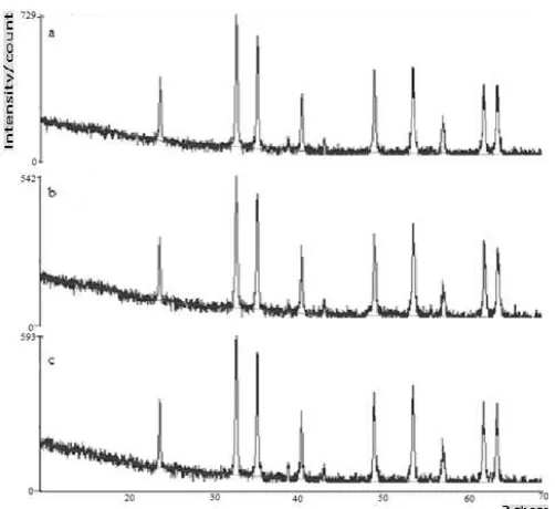
The temperature of calcinations and its effect on the formation of hematite pigment was also examined. The phase of products at various calcinations temperatures were also analyzed by XRD. Figure 5 shows the diffraction patterns of the resulted product at various calcinations temperatures, and shows a similar peak position with α-Fe2O3 standard. The plane of hematite crystal was detected are 012, 104, 110, 006, 113, 202, 024, 116, 018, 214, and 300, which indicates that the samples synthesized at each calcinations temperature was hematite, meanwhile at 800 oC, no appearance direction crystal of 006 and presence of 2θ angle (63.99o) were supposed as crystal defect and impurity.
Figure 5. XRD pattern of hexagonal α-Fe2O3 at a) 750 oC b) 800 oC and c) 850 oC
Figure 6. Hematite pigment synthesized at 750 (a), 800 (b), and 850 oC (c)
The highest intensities XRD of product is obtained at 750 oC. This tendency is also recognized by the color. The color of change is represented by a* (degree of redness) and b* (degree of yellowness). The colours of hematite pigment are found in terms of value degree of yellowness, which is the lowest and the degree of redness, which is the highest. At temperature 750 oC, the degree of yellowness is the lowest than others, the differences in lines a* and b* are significantly wide. Figure 6 shows color of synthesized hematite
The journal homepage www.jpacr.ub.ac.id
ISSN : 2302 ‐ 4690
33
synthesized. Based on data, the degree of lightness at temperature 750 oC is lower than those observed in other temperature. The different in color was influenced by grain size.
Table 2. The result of characterization hematite at various temperature
Value of color
The hematite pigment tends to reach the higher level of marron red color by increasing the calcinations temperature as shown at Figure 6. The highest degree of lightness, redness, and chroma were achieved at 800 oC, probably due to impurities and crystal defect which may occupy a ground state and various excited state similar to those in the transition metals. the movement of electron between these state gives rise both to color and the flouresence [16]. In addition, the highest degree of yellowness at 800 oC was caused by the smallest average grain size, in contrary the lowest degree of yellowness at 750 oC was caused the biggest average grain size than others.
CONCLUSION
Hematite pigment can be synthesized by thermal transformation of FeOOH method at pH 6 and 8, at 750 oC for 3 hour. Variation of pH and temperature gives effect to the degree of redness and degree of yellowness. The color of the product is red. The particle has spherical-like shape.
REFERENCES
[1] Cornell and Schwertmann, Iron Oxides: Structure,Properties, Reaction, Occurances, and Uses, Second edition, 2003, Wiley VCH , Weinheim.
[2] Potter., M.J, Iron Oxide Pigment, U.S. Geological Survey—Minerals Information, http://minerals.usgs.gov/minerals/pubs/commodity/iron_oxide/ ironpmyb 05.pdf, Accessed 27 October 2012.
[3] Tanner.,O, Iron Oxide Pigment [Advance release], U.S. Geological Survey Mineral Yearsbook, http://minerals.usgs.gov/ minerals/pubs/commodity/ iron_oxide/ myb1-2010-fepig. pdf, Accessed 27 October 2012.
[4] Buxbaum and Pfaff, Industrial Inorganic Pigment, Third edition, 2005, Wiley-VCH, Verlag GmbH & Co. KGaA, Weinheim.
[5] Neger, Parvin, Ahmed, and Alam, Bangladesh Journal of Scientific and Industrial Research, 2008, 43, 183-196.
[6] Mohapatra dan Anand, Int. J.Eng. Sci. Tech., 2010, 2, 127-146.
[7] Schwertmann., U and Murad, Clays and Clay Mineral, 1983,31 (4),277-284.
[8] R.M., Cornell and U., Schwertmann, Iron Oxides in the Laboratory Preparation and Characterization, Second edition, 2000, Wiley VCH , Weinheim.
The journal homepage www.jpacr.ub.ac.id
ISSN : 2302 ‐ 4690
34
[10] Della., V.P, Junkes., J.A, Montedo, O.R.K, Oliveira, A.P.N, Rambo., C.R, and Hotza., D, Am. Cer. Soc. Bull., 2007, 86 (5), 9101-9105.
[11] Legodi.,M.A and Wall., D, Dyes and Pigment, 2006, 74, 161-168.
[12] Silva., R.A, Castro, C.D, Petter, and C.A, Schneider, IMWA, 2011, 469-473.
[13] Blum, P., Reflectance Spectrophotometry and Colorimetry, PP Handbook, 1997, download from: http://www-odp.tamu.edu/publications/tnotes/tn26/CHAP7.PDF, Accessed 30 October 2012.
[14] F., Atay, V., Bilgin, I., Akyuz, S., Kose, J. Opto. Adv. Mat., 2007, 9 (11), 3604 – 3608. [15] Nagano, T, Nakashia, S, Nakayama, S, and Seno, M, Clays and Clay Minerals, 1994,
42 (2), 226-234.