EVALUATION OF CASPASE 3 AS AN INDICATOR FOR BREAST CANCER CHEMOTHERAPY RESPONSE
Kamal Basri Siregar1-3, TjakraWibawa Manuaba4, Muhammad NajibDahlan Lubis5, Rosita Juwita Sembiring6
1. Department of Surgery, Division of Surgical Oncology, Haji Adam Malik General Hospital Medan, Universitas Sumatera Utara, Indonesia.
2. Stem Cells Program, Universitas Sumatera Utara, Indonesia. 3. Nanomedicine Program, Universitas Sumatera Utara, Indonesia.
4. Departement of Surgery, Division of Surgical Oncology, Sanglah Hospital, Udayana University, Bali, Indonesia.
5. Department of AnatomicalPathology, University of Sumatera Utara, Indonesia. 6. Department of Clinical Pathology, University of Sumatera Utara, Indonesia. Email: [email protected]
ABSTRACT
Objectives:This study aimed to assess the difference of postchemotherapycaspase 3 level between TNBC subjects with and without clinical response.
Materials and Methods:A total of 48 subjects with intraductal and 12 subjects with intralobular triple negative breast cancer who were undergoing surgery at Adam Malik General Hospital were analyzed the response of neoadjuvant chemotherapy. Postsurgical breast tumour tissue was treated in Pathology Laboratory for caspase 3 analysis. The data was processed in SPSS 22 with significance limitation of 0.05.
Results:Median levels of caspase 3 postchemotherapy were higher both in intraductal and intralobular triple negative breast cancer subtype (6 vs 4.5 and 5 vs 3, respectively) the responsive group, while no changes detected in the group without clinical response. In statistical analysis, there were a significant difference of caspase 3 level postchemotherapy only in group with clinical response (p=0.005 in IDC and p=0.0031 in ILC).
Conclusion:Postchemotherapycaspase 3 level increased significantly in triple negative breast cancer, either intraductal or intralobular subtype, subjects with clinical response, but not in subjects without clinical response.
Keywords: triple negative breast cancer, caspase, neoadjuvant chemotherapy
INTRODUCTION
Breast cancer can be classified into 4 subclasses based on immunohistochemical examination: luminal A, luminal B, HER2/ERBB2, and triple negative breast cancer (TNBC). TNBC accounts for 17-21% of cases of breast cancer (3).Since this type of breast cancer did not have hormonal or targeted receptors, endocrine and target therapy had no benefit to TNBC. Therefore, chemotherapy plays an important role in the treatment of TNBC although with narrow therapeutic index (4).
Multicentre studies showed that 36% of patients receiving neoadjuvant chemotherapy with doxorubicin and cyclophosphamide showed complete clinical response and a good prognosis, especially for disease-free survival (p = 0.001) (5,6). In the EORTC study, it was reported that almost 23% of patients who did not suit for breast-conserving surgery could be benefited fromneoadjuvant chemotherapy.6 However, there were also many cases of chemoresistance yielded in causing harm to patients without beneficence (7).Therefore, there was an emerging need for a biomarker that can accurately identify the patient with chemosensitivityTNBC to avoid side effects in chemoresistance subjects (8). This study aimed to assess the difference of postchemotherapycaspase 3 level between TNBC subjects with and without clinical response.
MATERIALS AND METHODS
This is an analytic study with cohort prospective design to analyze the difference of caspase 3 level post-chemotherapy in the group with and without clinical response. In this study we also differentiate the analysis between intraductal (IDC) and intralobular (ILC) subtype. This study has been approved by the local ethical committee of Faculty of Medicine, University of Sumatera Utara.
All subjects with triple negative breast cancer who were undergoing surgery in Adam Malik General Hospital from 2015-2017 were included in this study. The subject must agree and sign an agreement to undergo neoadjuvant combination chemotherapy of 3 series, after first being given an explanation of chemotherapy. Subjects were excluded if they had other malignancies, congenital anomalies, chronic kidney disease, and chronic liver disease.
Subjects were given neoadjuvant chemotherapy, docetaxel and cisplatin before surgery.Chemotherapy clinical response was identified withResponse Evaluation Criteria in Solid Tumours(RECST), with or without clinical response (9). Postsurgical breast tumour tissue were treated in Pathology Laboratory for caspase 3 analysis. The tissues was fixated with buffer formalin and coloured to be analyzed with antibody kit. The intensity of caspase 3 were interpreted by two pathologists, scaling from 0 to 6 (10). We divided the IDC and ILC group for statistical analysis due to the difference features of each subtype. The median level of caspase 3 pre and post-chemotherapy was compared with the category of group with and without clinical response. The data was processed in SPSS 22 with significance limitation of 0.05 using Chi Square and Mann Whitney test.
RESULTS
had clinical response while 50% were not. In ILC, a majority of subjects (58.3%) had no clinical response, while only 41.7% subjects had a clinical response.
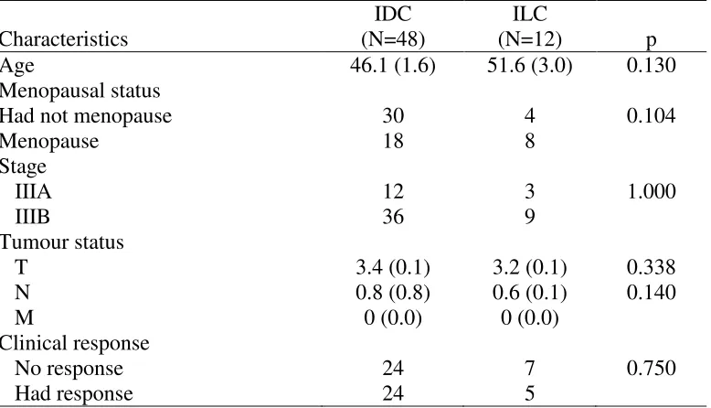
Table 1. Characteristics of subjects with triple negative breast cancer intraductal (IDC) and intralobular (ILC) subtype
Characteristics
The median level of caspase 3 prechemotherapy and postchemotherapy both in IDC and ILC were shown in table 2 and table 3. Compared to prechemotherapy level, both IDC and ILC group had higher median level descriptively (Table 2). There was a significant difference of caspase 3 level after chemotherapy in IDC (p=0.005) but there was no difference shown in ILC (p=0.250). Further analysis, the significant difference of caspase 3 level after chemotherapy only existed in the subgroup with clinical response (p=0.031), while no difference was found in the subgroup without clinical response (p=0.079).In ILC group, alhtough no significant difference of caspase 3 level was found in the whole group, a specific analysis in ILC subgroup with clinical response showed a significant difference of caspase 3 postchemotherapy (p=0.008) (Table 3).
Table 2. The difference of median level of prechemotherapypostchemotherapycaspase 3 betweenintraductal (IDC) and intralobular (ILC) subtype of triple negative breast cancer
Median level of caspase 3
IDC
Median level of
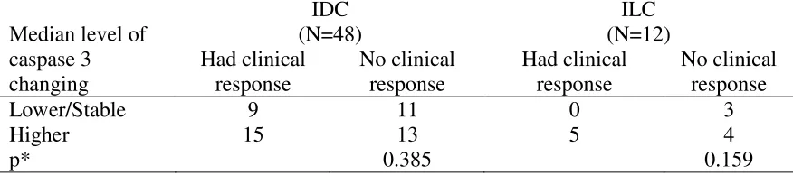
In IDC subgroup with clinical response, majority subjects (15 out of 24 subjects, 62.5%) showed higher caspase 3 level after chemotherapy. The similar description was showed in IDC subgroup without clinical response, in which majority subjects (13 out of 24 subjects, 54.2%) showed higher caspase 3 level after chemotherapy. Statistical analysis also showed no significant difference between both groups (p=0.762).In ILC subgroup with clinical response, all subjects had higher caspase 3 level after chemotherapy. In ILC subgroup without clinical response, a majority also had higher caspase 3 level after chemotherapy (4 out of 7 subjects, 57.1%). Statistical analysis showed no significant difference between both groups (p=0.240) (Table 4).
Table 4. Changing of caspase 3 level after chemotherapy in intraductal (IDC) and intralobular (ILC) subtype of triple negative breast cancer
Median level of
Not only IDC subgroup with or without clinical response showed no significant difference of caspase 3 after chemotherapy (p=0.762) but also ILC subgroup with or without clinical response showed no significant difference of caspase 3 after chemotherapy (p=0.240).
DISCUSSION
TNBC was a heterogenous group of cancer, with high grade, high proliferation rate, aggressive, and bad prognosis.Although TNBC sensitive to chemotherapy, there were reported many cases of chemoresistance of TNBC (11).Koyaet al. (2009) showed that neoadjuvant chemotherapy with doxorubicin, paclitaxel, andcyclophosphamide showed nearly 34% complete response overall and up to 57% in TNBC (12).Other study showed that platinum regiment even showed higher complete response in TNBC than non-TNBC type of breast cancer (80% VS 51%; P=0,005) (13).
(15). The incidence and prevalence of IDC has increased since 190s, in which 5.8 per 100,000 in 1975 increased to 32.5 per 100,000 in 2004 (16). It may be mammography, which increases the possibility of early detection and 60-70% reduction in mortality (17).In histologic examination, IDC was deficient with the presence of pleomorphic abnormality in epitheal ductal cells with irregular chromatin distribution which were bounded by the basement membrane of the breast ducts. Further, it could be divided into comedo, noncomedo, and the presence of necrosis or not (18).
On the other hand, the ILC develops from the epithelial tissue of the mammary glandular epithelium and often invades normal tissue without involving the desmopalstic response (19). In contrast to intraductal type breast cancer, ILC is characterized by mild thickening and induration of the breasts and may be clinically difficult to recognize and mammography (20). In ultrasound, ILC appears as a heterogeneous hyperioic mass, with unclear boundaries and posterior acoustic shadows (21). In molecular analysis, about 90% of ILCs have an E-cadherin protein expression deficiency, regulated by genomic alterations targeting the CDH1 gene on chromosome 16q22.1 (22).
Neoadjuvant chemotherapy response in TNBC patients was reported to vary in various studies. Gianni et al., (2010), studied the response to neoadjuvant chemotherapy in TNBC patients with 74% of the patients giving a positive response (23).Tewari et al., (2010), reported that neoadjuvan chemotherapy treatment in TNBC patients showed that 78% of patients responded positively with 64% of partial clinical responses and 14% complete clinical responses(24). Unresponsive to chemotherapy can be caused by chemoresistance or that the tumour size was too large (25).
As many as 50% subjects with IDC had clinical response while 50% were not. In ILC, majority of subjects (58.3%) had no clinical response, while only 41.7% subjected had clinical response. In this study, there are no differences between ICD and IDC. Thus, both groups were equal to be statistically tested. Based on these results, it is generally known that the probability of receiving a chemotherapy response in TNBC patients is only about 50%. With this result, consideration should be given to the cost and benefits of the chemotherapy treatment for TNBC patients later on.
Caspase-3 is in the cytoplasm and is transported into the nucleus during apoptosis (26). Caspase-3 functions in the cleavage of structural proteins, DNA repair enzymes and activates endonucleases that cause DNA fragmentation in apoptotic mechanisms (27). Caspase-3 as the executor or effector on the apoptotic process may play an important role in assessing the response or resistance of a cell to chemotherapy or cytotoxic substances (28).
These results indicated that caspase 3 that increases significantly after chemotherapy can predict clinical response in both IDC and ILC. Donovan's et al. (2003) conducted on 103 breast tissue samples, 25 fibroadenoma tissue and 5 normal tissues showed that caspase-3 precursor and caspase-3 active rates were higher in breast cancer tissue than in normal tissue (p = 0,0188; p = 0.0002) (29). Branham et al. (2012) showed that there was a significant incidence of caspase 3 postchemotherapy (30).dos Santos et al. (2017) also showed that caspase 3 could be a bioindicator of radiosensitivity.31 Research shows that the apoptotic forces caused by chemotherapy agents depend on the concentrations given. In MCF-7 cells administered doxorubicin for 18 h showed a linear increase of caspase 3 in accordance with drug levels (32).
The activation of apoptotic pathways is the primary mechanism of cytotoxic drugs killing tumour cells. The presence of defects in apoptosis signalling contributes to tumour resistance.32Salako (2007) has demonstrated that TNBC treated with chemotherapy activates caspase 3/7, disruption of the f-actin organization, leading to apoptosis and decreased cell motility. Thus, theoretically, the better the effectiveness of chemotherapy, the higher the mechanism of apoptosis produced.28
Several studies have reported the potential use of caspase-3 as a biomarker to predict tumour responses related to other prognostic variables (eg vascular invasion, lymph node metastasis, advanced stage clinical, and tumour size). This suggests that caspase-3 may be used as a potential factor for predicting tumour progression and poor prognosis in some cancers by controlling those factors (33).
In this study, it was shown that caspase 3 increased significantly in chemotherapy, but it has not been used as a marker to distinguish chemotherapy-sensitive and resistant groups as no exact level of increment was known to be a significant cut off point. Branham et al. (2012) shows that there is a higher probability of predicting chemoresistance or chemosensitivity before second cycles for showing less than 1 fold increase in caspase-3 (30).
This study used a prospective cohort design. A prospective cohort study was an analytic study that began observations since the patient had not been exposed to exposure, and followed until the exposure was completed. With the cohort design, it can be ensured that Caspase 3 is measured at the right time. However, this study is a preliminary study, no previous similar studies so that the determination of the sample size is done in accordance with the considerations of researchers.
CONCLUSION
Postchemotherapycaspase 3 level increased significantly in triple negative breast cancer, either intraductal or intralobulat subtype, subjects with clinical response.
CONFLICT OF INTEREST
This study has no conflict of interest to be declared.
ACKNOWLEDGEMENT
AUTHOR CONTRIBUTION
Kamal B. Siregar, as the first author, contributed to the idea, introduction, some part of results, and some part of the discussion. The second author, Manuaba TW, contributed to the methods and results part of this paper. The third and fourth authors, Lubis MND and Sembiring RJ, contributed to the analysis of caspase 3, and some parts of the discussion.
REFERENCES
1. Ponvinobala K, Kanchana G, Rubalakshmi G. In vitro anticancer acivity of hydro-alcohol extract of leaves of Andrographis Neesiana against PC-3 and MCF-7 cell lines. Int J Pharm Pharmaceutical Sci 2012;4(3):396-9.
2. PERABOI Cabang Medan. Laporan Kegiatan Dan Rencana Kerja Dua Tahunan Divisi Bedah Onkologi FK-USU /RSUP.H.ADAM MALIK, 2014.
3. Wu J, Li S, Jia W, Su F. Response and Prognosis of Taxanes and Anthracyclines Neoadjuvant Chemotherapy in Patients with Triple-Negative Breast Cancer.” J Cancer Res Clin Oncol 20011;137(10):1505-10.
4. Kirthi C, Adzal A, Reddy M, Ali SA, Yerramilli A, Sharma S. A study on the
adverse effects of anticancer drugs in an oncology center of a tertiary care hospital. Int J Pharm Pharmaceutical Sci 2014;6(2):580-3.
5. Fisher B, Brown A, Mamounas E, Wieand S, Robidoux A, Margolese R, et al. Effect of preoperative chemotherapy on local-regional disease in wome with operable breast cancer: findings from National Surgical Adjuvant Breast and Bowel Project B-18. J Clin. Oncol. 1997;15:2483-93.
6. Cleator S, Parton M, Dowsett M. The Biology of Neoadjuvant Chemotherapy for Breast Cancer.” Endocrine-Related Cancer 2002;9(3):183-95.
7. Indran R, Tufo G, Pervaiz S, Brenner C. Recent Advances in Apoptosis, Mitochondria and Drug Resistance in Cancer Cells. Biochimica et Biophysica Acta 2011;1807(6):735-45.
8. Rakha E, Ellis I, Reis-Filho J. Are triple-negative and basal-like breast cancer synonymous? Clin Cancer Res 2008;14:618-9.
9. Bai-lin Z, Tong S, Zhang B, Zheng S, Ning LU. Polymorphisms of GSTP1 Is Associated with Differences of Chemotherapy Response and Toxicity in Breast Cancer 2011;124(2):199-204.
10.Vakkala M, Pääkkö P, Soni Y. Expression of Caspases 3, 6 and 8 Is Increased in Parallel with Apoptosis and Histological Aggressiveness of the Breast Lesion. British Journal of Cancer 1999;81(4):592-99.
11.Dawood S, Broglio K, Esteva FJ,. Survival among women with triple receptornegative breast cancer and brain metastases. Ann Oncol 2009;20:621-7. 12.Koya S, Li Y, McDaniel S. Safety and effectiveness of dose dense neoadjuvant
chemotherapy in patients with stage II/III breast cancer. J ClinOncol 2009;27(15S):e11566.
14.West DC & McGrowder DA. Triple negative breast cancer: therapeutic and prognostic implications. Asian Pacific J Cancer Prev 2011; 12: 2129-33.
15.Lakhani S, Ellis I, Schnitt S, Tan P, van de Vijver M. WHO Classification of Tumours of the Breast. 4th ed. Lyon, France: International Agency for Research on Cancer; 2012.
16.Shampoo MA, Kyle RA. Pioneers of mammography-Warren and Egan. Mayo Clinic Proceedings. 1997;72(1):32-5.
17.Rosner D, Bedwani RN, Vana J. Noninvasive breast carcinoma. Results of a national survey by the American College of Surgeons. Annals of Surgery. 1980;192(3):139-47.
18.Sanders ME, Schuyler PA, Dupont WD, Page DL. The natural history of low-grade ductal carcinoma in situ of the breast in women treated by biopsy only revealed over 30 years of long-term follow-up. Cancer 2005; 103: 2481-4.
19.Hanagiri T, TadahiroNozoe, Makiko Mizukami, YoshinobuIchiki, Masakazu Sugaya, Manabu Yasuda, Mitsuhiro Takenoyama, Kenji Sugio and Kosei Yasumoto. Clinicopathological Characteristics of Invasive Lobular Carcinoma of the Breast. Asian J Surg 2009;32(2):76-80.
20.Iorfida M, Maiorano E, Orvieto E, Maisonneuve P, Bottiglieri L, Rotmensz N, et al. Invasive lobular breast cancer: subtypes and outcome. Breast Cancer Res Treat. 2012;133:713-23.
21.Butler RS, Venta LA, Wiley EL, et al. Sonographic evaluation of infiltrating lobular carcinoma. AJR Am J Roentgenol 1999; 172:325-30.
22.McCart RA, Kutasovic J, Lakhani S, Simpson P. Invasive lobular carcinoma of the breast: morphology, biomarkers and ‘omics. Breast Cancer Res 2015;17:12-5. 23.Gianni L, Eiermann W, Semiglazov V, Manikhas A, Lluch A, Tjulandin S, et al.
Neoadjuvant chemotherapy with trastuzumab followed by adjuvant trastuzumab versus neoadjuvant chemotherapy alone, in patients with HER2-positive locally advanced breast cancer (the NOAH trial): a randomised controlled superiority trial with a parallel HER2-negative cohort. Lancet 2010;375(9712:377-84.
24.Tewari M, Pradhan S, Singh U, Singh TB, Shukla HS. Assessment of predictive markers of response to neoadjuvant chemotherapy in breast cancer. Asian J Surg 2010;33(4):157-67.
25.Viale G. Characterization and clinical impact of residual disease after neoadjuvant chemotherapy. Breast 2013;22(2):88-91.
26.Blázquez S, Sirvent J, Olona M, Aguilar C, Pelegri A, Garcia JF, et al. Caspase-3 and Caspase-6 in Ductal Breast Carcinoma: A Descriptive Study. Histology and Histopathology 2006;21(12):1321-29.
27.Luo M, Lu Z, He S, Yuan K, Zhang Q, Sha M, et al. Nuclear Entry of Active Caspase-3 Is Facilitated by Its p3-Recognition-Based Specific Cleavage Activity.” Cell Research 20 (2). Nature Publishing Group 2010;1:211–22.
28.Salako O, Poku R, Nkembo A, Amissah F, Ntantie E &Lamango N. Treatment of Triple Negative Breast Cancer-Derived Cells with PolyisoprenylatedCysteinyl Amide Inhibitors Activates Caspase 3/7 and Disrupts F-actin Organization leading to Apoptosis and Diminished Cell Motility. Faseb 2016; 30(1): 1-3.
30.Branham KS, Krishna VM, Gopal SV, Raju AJ. Evaluation of caspase-3 as a possible indicator of apoptotic activity to assess the chemoresistance / sensitivity. J Pharm Res 2012; 5 (1): 229-231.
31.dos Santos N, Silva RF, Pinto MM, Silva EB, Tasat DR, Amaral A. Active caspase-3 expression levels as bioindicator of individual radiosensitivity. Anais da Academia Brasileira de Ciencias 2017; 89 (1): 649-59.
32.Yang XH, Sladek TL, Liu X. Reconstitution of Caspase-3 Sensitizes MCF-7 Breast Cancer Cells to Doxorubicin- and Etoposide-Induced Apoptosis. Cancer Res 2001;61:348–54.