Brain Research 881 (2000) 227–230
www.elsevier.com / locate / bres
Short communication
Interferon
b
-1a downregulates TNF
a
-induced intercellular adhesion
molecule 1 expression on brain microvascular endothelial cells through
a tyrosine kinase-dependent pathway
*
Giovanni Defazio , Paolo Livrea, Maurizio Giorelli, Davide Martino, Francesco Roselli,
Felicia Ricchiuti, Maria Trojano
Department of Neurologic and Psychiatric Sciences, University of Bari, I-70124 Bari, Italy
Accepted 8 August 2000
Abstract
TNFa(100 U / ml, 24 h) upregulated intercellular adhesion molecule 1 (ICAM1) expression on brain microvascular endothelial cell (BMEC) culture. The tyrosine kinase (TK) inhibitor genestein (100mg / ml), the protein kinase C (PKC) inhibitor staurosporin (1 nM), and interferon (IF)b-1a (1000 U / ml) antagonized TNFa effect. When an ineffective dose of IFb-1a (100 U / ml) was challenged with ineffective doses of either genestein (10 mg / ml) or staurosporin (0.1 nM), the combination IFb-1a–genestein significantly reduced TNFa-induced ICAM1 expression whereas IFb-1a–staurosporin did not. These findings indicate that a TK- rather than a PKC-dependent mechanism is involved in the modulation of TNFaresponse by IFb-1a on BMECs. 2000 Elsevier Science B.V. All rights reserved.
Theme: Cellular and molecular biology
Topic: Blood brain barrier
Keywords: TNFa; Tyrosin kinase; Interferonb-1a; Intercellular adhesion molecule 1; Brain microvascular endothelial cell
Disruption of the blood brain barrier (BBB) allowing It has recently been shown that prolonged IFb-1a / 1b inflammatory cells to infiltrate white matter tissue is a treatment downregulates TNFa-induced ICAM 1 expres-consistent initiating event in the development of de- sion on cultured BMECs [6,19]. The in vitro modulation of myelinating lesions in relapsing remitting multiple BMEC ICAM 1 expression by TNFa/ IFb-1a may be sclerosis (MS) [14]. TNFa released by activated T cells relevant to the results of recent clinical trials indicating probably contribute to BBB breakdown and transendotheli- that chronic treatment of RR MS with IFbs prevents both al migration of inflammatory cells [17] by upregulating the clinical exacerbation and the development on magnetic expression of adhesion molecules like intercellular adhe- resonance imaging of new lesions associated with BBB sion molecule 1 (ICAM 1) [21] on brain microvascular damage [11,18]. In the present study, we performed TK endothelial cells (BMECs). The signaling pathways uti- and PKC inhibition on BMEC culture to determine the role lized by TNFa for modifying BMEC properties involve of these kinases in the modulation of TNFa-induced both protein kinase C (PKC) and protein tyrosine kinase ICAM 1 expression by IFb-1a.
(TK). In fact, inhibition of PKC by staurosporin and of TK BMECs were isolated from male Wistar rats (100–120 by genestein reduced TNFa-stimulated lymphocyte adhe- g) and cultured according to Carson and Haudenschild [3] sion to BMECs [10]. In addition, staurosporin counteracted with some modifications [20]. Cells plated on collagen TNFa induced ICAM1 expression and fluid phase endo- type 1-coated 30 mm petri-dishes (Corning) were grown in cytosis on cultured BMEC [7]. M199 (Flow Laboratories, Irvine, CA) enriched with 20% heat inactivated fetal bovine serum (Hyclone, Logan, UT), 2.2 mg / ml sodium bicarbonate, 200 mg / ml sodium *Corresponding author. Tel.: 139-080-547-8511; fax: 1
39-80-547-heparin, 60 mg / ml endothelial cell growth supplement, 10 8532.
E-mail address: [email protected] (G. Defazio). mg / ml gentamicin, 0.5 U / ml penicillin G and 0.5 U / ml
228 G. Defazio et al. / Brain Research 881 (2000) 227 –230
glutamine (all from Sigma Chemical Co., St. Louis, MO). phosphate buffer saline containing 10% adult bovine serum Confluent monolayers were subcultured at 1:4 ratio by (to saturate non specific binding sites). Thereafter, cells 0.25% pancreatin (20 min, 378C) which removed endo- were sequentially incubated (2 h, room temperature) with thelial colonies from nearby and underlying cells [3]. 1:20 mouse anti-rat ICAM 1 (Seikagaku, Japan) and with Approximately 96% of cells in confluent passage 1 cul- 1:3000 peroxidase-conjugated rabbit anti mouse IgG (H1
tures had fibroblast-like appearance, and showed typical L) (Jackson Immunoresearch). Primary antibody controls granular cytoplasmic immunofluorescence with Factor VIII were cultures incubated in irrelevant isotype-matched mAb antiserum [6,12]. The other cells were identified as or normal mouse IgG. Finally, cultures were incubated for pericytes [3,20]. As reported earlier, our passage 1 BMEC 30 min with o-phenilendiamine dihydrocloride (0.5 mg / cultures maintained a lower level of endocytosis than ml) diluted in 0.5 M phosphate–citrate buffer containing non-barrier endothelial cells [5], and constitutively ex- 0.015 M H O . The reaction was stopped by 3 M H SO .2 2 2 4 pressed at low level basally ICAM1 [6]. Overall, passage 1 Absorbance was measured at 490 nm on a ELISA Microti-BMEC cultures used in this study indeed represent the ter Plate Reader (BIO-RAD). Background of the isotype brain capillary endothelium and preserved some of the control (ranging from 0.087 to 0.099 in different experi-characteristics in vivo. ments) was subtracted by data presented in figures. Similar Confluent BMECs were exposed to 100 U / ml human values were observed for background of normal mouse recombinant TNFa (Sigma, EC50 0.024 ng / ml), a dose IgG (0.089–0.099).
close to the range of concentration found in the cerebrospi- An overall test of significance using an F-ratio derived nal fluid (CSF) of MS patients with BBB damage. from 1-way ANOVA followed by a post hoc Newman– Incubation was continued up to 24 h, a time at which Keuls test was used to compare groups. P values ,0.05 upregulation of ICAM 1 elicited by TNFa reaches maxi- were considered significant.
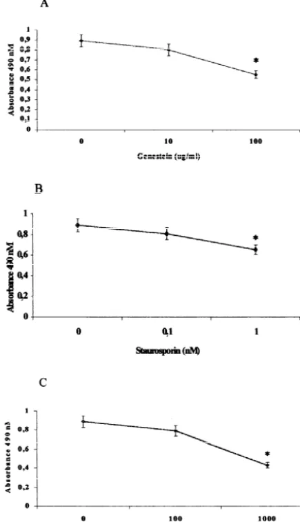
mal levels [20]. The TK inhibitor genestein was dissolved Twenty-four hours stimulation of confluent BMEC in growth medium to 10–100 mg / ml final concentrations. culture with 100 / U ml TNFa significantly upregulated The PKC inhibitor staurosporin was dissolved in dimethyl membrane-bound ICAM1 expression (mean sulfoxide (1:1000) thereafter in growth medium. As re- absborbance6SEM: untreated, 0.3460.04; TNFa 0.891
vealed by dual labeling with fluoresceine diacetate and 0.06 (F558.2, P50.002).
propidium iodide [13], dimethyl sulfoxide was not toxic to The exposure of TNFa-treated cultures to either 100 the monolayer at the concentrations used in the experimen- mg / ml genestein or 1 nM staurosporin 30 min before and tal wells. Staurosporin was used at concentrations (0.1–1 during TNFa treatment significantly reduced ICAM1 nM) lower than previously reported [7]. Kinase inhibitors expression due to TNFa(Fig. 1 A,B), whereas 10 mg / ml were added 30 min before TNFa, after which the incuba- genestein and 0.1 nM staurosporin were ineffective. As tion was continued for 24 h. At the doses and times used, reported earlier [7], 1000 U / ml IFb-1a 48 h before and kinase inhibitors did not affect BMECs viability. Human during 24 h TNFatreatment induced a significant suppres-recombinant IFb-1a (Serono-Pharma spa, Milan, Italy) was sion of ICAM1 elevation whereas 100 U / ml IFb-1a failed diluted into growth medium to the final concentration of to antagonize TNFa response (Fig. 1C).
100–1000 U / ml. The higher concentration was compar- In separate experiments, confluent BMECs were ex-able to that reached in serum following the systemic posed to the ineffective dose of IFb-1a (100 U / ml) for 48 administration of 8 MIU in MS patients [10]. Incubation of h, with ineffective doses of staurosporin (0.1 nM) or BMECs with IFb-1a alone for 48 h was followed by genestein (10 mg / ml) being added for the last 30 min. simultaneous treatment with IFb-1a and TNFa 24 h. As Thereafter, the incubation was continued for 24 h in the reported earlier [6], this treatment protocol reduced simultaneous presence of IFb-1a, staurosporin or genes-ICAM1 induction caused by 24 h TNFa in our culture tein, and TNFa (100 U / ml). Under this protocol, IFb-1a system. By contrast, we failed to observe ICAM1 dow- plus staurosporin failed to affect TNFa-induced ICAM1 nregulation under simultaneous administration of IFb-1a expression (Fig. 2A) whereas the combined IFb-1a / genes-and TNFa for 24 h, or when coincubation of IFb-1a and tein treatment significantly reduced ICAM1 expression due TNFafor 24 h was followed by 48 h of IFb-1a [6]. Thus, to TNFa (Fig. 2B).
to check possible interaction between IFb-1a and kinase To our knowledge, the antagonistic effect of the TK inhibitors, BMEC cultures underwent 48 h incubation with inhibitor genestein to TNFa-induced ICAM1 expression IFb-1a, with staurosporin or genestein being added for the reported herein has not been previously reported, whereas last 30 min. Thereafter, incubation was continued for 24 h downregulation of TNFa-induced ICAM1 expression by in the simultaneous presence of IFb-1a, staurosporin or the PKC inhibitor staurosporin and IFb-1a confirms
previ-genestein and TNFa. ous in vitro studies on BMECs [6,7]. Results from
G. Defazio et al. / Brain Research 881 (2000) 227 –230 229
Fig. 2. Effect of 100 U / ml interferonb-1a (IF) plus 0.1 nM staurosporin (STAURO) (A), or of 100 U / ml interferon b-1a (IF) plus 1 mg / ml genestein (Gene) (B) on the expression of ICAM 1 molecules on brain microvascular endothelial cell (BMEC) cultures stimulated with 100 U / ml TNFafor 24 h. Cultures were exposed to IF for 48 h with Stauro or Gene being added for the last 30 min. Thereafter, the incubation was continued for 24 h in the simultaneous presence of IF, Stauro or Gene, and TNFa. Background of the isotype antibody control was subtracted by presented data. Values are mean6SEM of triplicate wells from a representative experiment repeated twice. One-way ANOVA for (A):
F5NS. One-way ANOVA for (B): F514.2, P50.001; Newman–Keuls Fig. 1. Effect of the protein tyrosine kinase inhibitor genestein (A), the test: * TNF / Gene / IF different from the other groups, P,0.05. protein kinase C inhibitor staurosporin (B) and interferonb-1a (C) on the
expression of ICAM 1 molecules on brain microvascular endothelial cell cultures stimulated with 100 U / ml TNFafor 24 h. Both genestein and
ICAM1 expression by IFb-1a [7], an ineffective dose of staurosporin were added 30 min before TNFa, after which the incubation
was continued for 24 h. Background of the isotype antibody control was IFb-1a was challenged with ineffective doses of either subtracted by presented data. Values are mean6SEM of triplicate wells staurosporin or genestein. Under this schedule, the signifi-from a representative experiment repeated twice. One-way ANOVA for
cant combined effect of IFb-1a–genestein and the lack of Genestein: F514.6, P50.005; Newman–Keuls test: * 100mg / ml
differ-effect of IFb-1a–staurosporin provide evidence that a TK-ent from the other groups, P,0.05. One-way ANOVA for Staurosporin:
rather than a PKC-dependent intracellular signaling mecha-F57.43, P50.02; Newman–Keuls test: * 1 nM different from the other
groups, P,0.05. One-way ANOVA for Interferon beta-1a: F514.9, nism is involved in the modulation of TNFa response by
P,0.005; Newman–Keuls test: * 1000 U / ml different from the other IFb-1a on BMECs. groups, P,0.05.
230 G. Defazio et al. / Brain Research 881 (2000) 227 –230
expression and fluid phase endocytosis on brain microvascular observations are grounds for thinking that IFb may
endothelial cells, Brain Res 863 (2000) 245–248. differently affect TK and PKC signaling of distinct cell
[8] G. Eissner, W. Kolch, H. Mischak et al., Differential role of protein
types. kinase C in cytokine induced lymphocyte-endothelium interaction in
In conclusion, the information reported herein may have vitro, Scand. J. Immunol. 40 (1994) 395–402.
further elucidated some of the mechanism of action of [9] H. Fujisawa, Y. Naito, S. Horiuchi, T. baba, F. Otsuka, The effects of interferon beta on phorbol ester or calcium ionophore induced IFb-1a on activated BBB endothelium. Based on the
intercellular adhesion molecule 1 expression in epidermal carcinoma results of recent clinical trials indicating that activated
cells, J. Dermatol. 19 (1992) 78–81.
BBB endothelium may be an important target of IFb-1a in [10] S.J. Hudson, J.P. Cai, V. Thomas, Y.H. Chin, Intracellular signalling MS patients [11,18], a complete understanding of the of tumor necrosis factor-ain brain microvascular endothelial cells is mechanisms underlying IFb-1a effect on BMECs may lead mediated by a protein tyrosine kinase and protein kinase
C-depen-dent pathway, J. Neuroimmunol. 70 (1996) 199–206. to the identification of target pathways for the therapeutic
[11] L.D. Jacobs, D.L. Cookfair, R.A. Rudick et al., Intramuscular treatment of BBB damage in MS and other brain
in-interferon beta-1a for disease progression in relapsing multiple
flammatory disorders. sclerosis, Ann. Neurol. 39 (1996) 285–294.
[12] E.A. Jaffe, Endothelial cells and the biology of factor VIII, New Engl. J. Med. 296 (1984) 377.
[13] K.A. Jones, A. Senft, An improved method to determine cell Acknowledgements
viability by simultaneous staining with fluorescein diacetate-prop-idium iodide, J. Histochem. Cytochem. 33 (1985) 77–79. This work was supported by the Istituto Superiore di [14] A.G. Kermode, A.J. Thompson, P. Tofts et al., Breakdown of the
`
Sanita grants 93 / J / T60 and 93 / J / T20, Rome, Italy. blood brain barrier precedes symptoms and other MRI signs of new lesions in multiple sclerosis, Brain 113 (1990) 1477–1489. [15] M.J. May, C.P. Wheeler-Jones, J.D. Pearson, Effects of protein
tyrosine kinase inhibitors on cytokine-induced adhesion molecule References
expression by human umbilical vein endothelial cells, Br. J. Pharmacol. 118 (1996) 1761–1771.
[1] B.B. Aggarwall, R. Pandita, Both type I and type II interferons [16] I.L. Navarro, K. Mowen, S. Rodems, B. Weaver, N. Reich, D. downregulate human TNF receptors in human hepatocellular car- spector, M. David, Cytomegalovirus activates interferon immedi-cinoma cell line Hep G2. Role of protein kinase C, FEBS Lett. 337 ately-early response gene expression and an interferon regulatory (1994) 99–102. factor 3-containing interferon-stimulated response element-binding [2] G. Barbieri, L. Velazquez, M. Scrobogna, M. Fellous, S. Pellegrini, complex, Mol. Cell. Biol. 18 (1998) 3796–3802.
Activation of the protein tyrosine kinase tyk2 by interferon alpha / [17] C.S. Raine, Demyelinating diseases, in: R.L. Davis, D.M. Robertson beta, Eur. J. Biochem. 223 (1999) 427–435. (Eds.), Textbook of Neuropathology, 2nd Edition, William and [3] M.P. Carson, C. Haudenschild, Microvascular endothelium and Wilkins, Baltimore, MD, 1991, pp. 535–620.
pericytes: high yield, low passage cultures, In Vitro Cell. Dev. Biol. [18] L.A. Stone, J. Franck, P.S. Albert et al., The effct of interferon-bon 22 (1986) 344–354. blood–brain barrier disruptions demonstrated by contrast enhanced [4] C. Cernescu, S.N. Costantinescu, F. Balta, L.M. Popescu, N. Cajal, magnetic resonance imaging in relapsing-remitting multiple
Protein kinase C and the antiviral effect of human interferon, sclerosis, Ann. Neurol. 37 (1995) 611–619.
Virologie 40 (1989) 163–170. [19] M. Trojano, G. Defazio, C. Avolio, D. Paolicelli, F. Giuliani, M. [5] G. Defazio, D. Ribatti, B. Nico, F. Ricchiuti, R. De Salvia, L. Giorelli, P. Livrea, Effects of rIFN-beta 1b on serum circulating Roncali, P. Livrea, Endocytosis of horseradish peroxidase by brain ICAM1 in relapsing remitting multiple sclerosis and on the mem-microvascular and umbilical vein endothelial cells in culture: an brane-bound ICAM1 expression on brain microvascular endothelial ultrastructural and morphometric study, Brain Res. Bull. 43 (1997) cells, Neurovirology 6 (2) (2000) S47–S51.
467–472. [20] M. Trojano, G. Defazio, F. Ricchiuti, R. De Salvia, P. Livrea, Serum [6] G. Defazio, D. Ribatti, B. Nico, M. Giorelli, R. De Salvia, G. Russo, IgG to brain microvascular endothelial cells in multiple sclerosis, J.
L. Roncali, P. Livrea, ICAM1 expression and fluid phase endo- Neurol. Sci. 143 (1996) 107–113.
cytosis in brain microvascular endothelial cells following interferon [21] D. Wong, K. Dorovini-Zis, Upregulation of intercellular adhesion b-1a and TNFa, J. Neuroimmunol. 88 (1998) 13–20. molecule-1 (ICAM-1) expression in primary cultures of human [7] G. Defazio, B. Nico, M. Trojano, M. Ribatti, M. Giorelli, F. brain microvessel endothelial cells by cytokines and