Abstract. Aim: Inflammatory processes in nasal mucosa are reflected in various local mediators, found both in mucosal tissue and nasal discharge. In this prospective study, we assessed the effects of long-term low-dose oral administration of clarithromycin (CAM) on Th1 cytokines in nasal secretions and on clinical parameters of severity of nasal polyposis.
Methods: A total of forty nasal polyp patients (22 nonallergic and 18 allergic) received 500 mg/day single oral dose of CAM for eight weeks. We measured the levels of proinflammatory Th1 cytokines TNF-α, TNF-β, IL-1β, IL-2, IL-12, and IFN-γ in the nasal fluid samples, before and after treatment by CAM, using flow cytometric method. Before and after therapy, we scored each of the 40 patients according to nasal symptom score and endoscopic score. Results:
Following treatment, we found significantly reduced levels of TNF-α (p=0.006) in nasal secretions of nonallergic patients, and of IL-1β (p=0.008) in nasal fluid of allergic patients. Our results suggest an association between the reduction of nasal polyp size and reduction of TNF-α levels in nasal fluid of nonatopic patients and an association between the reduction of nasal polyp size and reduction of IL-12 levels in nasal discharge of atopic patients. Macrolide therapy decreased the size of polyps in 10/22 nonatopic and in 9/18 atopic patients. After macrolide therapy, we found 67.83% nonallergic subjects and 55.55% allergic subjects with improved nasal symptomatology. Conclusion: Long-term low-dose treatment with CAM is effective in the management of nasal polyposis, because of its antiinflammatory and immunomodulatory actions.
Key words: chronic inflammation, clarithromycin, nasal polyposis, nasal secretions, Th1 cy-tokines
Sažetak. Cilj: Upalni procesi u nosnoj sluznici očituju se u različitim lokalnim medijatorima, u tkivu sluznice i u nosnom sekretu. U ovoj prospektivnoj studiji procijenili smo učinke dugotraj-ne, niskodozirane terapije klaritromicinom (CAM) na Th1 citokine u nosnome sekretu, kao i na kliničke parametre očitovanja nosne polipoze. Metode: Četrdesetero (22 nealergičnih i 18 alergičnih) bolesnika s nosnom polipozom dobivalo je po jednu dnevnu dozu od 500 mg CAM-a tijekom osam tjedana. Mjerili smo koncentracije proupalnih Th1 citokina TNF-α, TNF-β, IL-1β, IL-2, IL-12 i IFN-γ u uzorcima nosnoga sekreta, prije i nakon terapije CAM-om, primjenom protočne citometrije. Prije i nakon liječenja klinički smo klasificirali svakoga od četrdesetero bolesnika prema nosnome simptom rezultatu i endoskopskom rezultatu.
Rezultati: Nakon liječenja detektirali smo značajno niže koncentracije TNF-α (p = 0,006) u nos-nome sekretu nealergičnih bolesnika i IL-1β (p = 0,008) u nosnome sekretu alergičnih boles-nika. Naši rezultati sugeriraju povezanost između smanjenja veličine nosnih polipa i snižavanja koncentracije TNF-α u nosnom sekretu u neatopičnih bolesnika, kao i povezanost između smanjenja veličine nosnih polipa i snižavanja koncentracije IL-12 u nosnome sekretu u atopičnih ispitanika. Terapija makrolidnim antibiotikom smanjila je veličinu polipa u 10/22 nealergičnih i u 9/18 alergičnih bolesnika. Nakon makrolidne terapije našli smo 67,83 % nealergičnih i 55,55 % alergičnih ispitanika s poboljšanim nosnim simptomima. Zaključak:
Zbog protuupalnih i imunomodulacijskih djelovanja, dugotrajna niskodozirana primjena CAM-a korisna je u liječenju nosne polipoze.
Ključne riječi: klaritromicin, kronična upala, nosna polipoza, nosni sekret, Th1 citokini
Corresponding author:
* Dr. Aleksandar Perić
Clinic of Otorhinolaryngology, Rhinology Unit
Military Medical Academy
Crnotravska 17, 11 040 Belgrade, Serbia
e-mail: [email protected]
1Department of Otorhinolaryngology,
Military Medical Academy, Belgrade, Serbia
2Institute of Medical Research,
Division of Clinical and Experimental Immunology, Military Medical Academy, Belgrade, Serbia
3Institute of Physiology,
Medical Faculty Kragujevac, Serbia
Primljeno: 12. 11. 2011. Prihvaćeno: 28. 2. 2012.
Effects of long-term low-dose treatment by
clarithromycin on Th1 cytokine levels in nasal
discharge of patients with nasal polyposis
Učinci dugotrajnog, niskodoziranog liječenja klaritromicinom na
koncentracije Th1 citokina u nosnome sekretu u bolesnika s nosnom
polipozom
INTRODUCTION
Macrolide antibiotics, such as erythromycin (EM), clarithromycin (CAM), and roxithromycin (RXM), are important chemotherapeutic agents in trea-tment of infections. Over the last decade, there has been a growing interest of various investiga-tors in the immunomodulatory and anti-inflam-matory action of 14-membered and 15-membe-red macrolides in the long-term low-dose treatment of chronic rhinosinusitis and nasal
pol-Macrolide antibiotics are known to have
anti-intlamma-tory and immunomodulaanti-intlamma-tory effects in treatment of
chronic rhinosinusitis and nasal polyposis. In this
pros-pective study, we investigated the effects of long-term
low-dose treatment by clarithromycin (CAM) on Th1
cytokine levels in nasal fluid of non-atopic and atopic
patients with nasal polyposis.
yposis. Nasal polyps develop usually in the ante-rior ethmoidal area and appear as grape-like structures, often in relation to inflammatory con-dition, but the exact etiology is still under debate. Oxidative stress and chronic persistent inflamma-tion are the main factors in the development of nasal polyps and inflammation triggers include bacterial, fungal and viral infection, allergy, and environmental pollution1,2.
Nasal polyps consist of loose connective tissue, oedema, inflammatory cells and some glands and capillaries, and are covered with various types of epithelium, but mostly pseudostratified epithelium with ciliary cells and goblet cells3,4.
Additionally, studies have found higher numbers of inflammatory cells, especially eosinophils, ne-utrophils and lymphocytes in nasal polyp lamina propria compared to healthy nasal mucosa3,4.
There have been many reports regarding the pharmacological actions of macrolides in trea-tment of chronic rhinosinusitis and its complica-ted form-nasal polyposis5-7. Those actions include
suppression of proliferation of nasal polyp fibro-blasts, shrinkage of nasal polyps, suppression of production of chemokines IL-8 and RANTES, etc 5-7. To our knowledge, there has been a little
studi-es about the role of T helper 1 (Th1) cytokinstudi-es in
etiology of nasal polyposis and there has been a little description of the influence of macrolide treatment on the production of these cytokines. Nasal secretion contains small amounts of cytoki-nes, potent biologic factors involved in the regu-lation of inflammation and immune defense, and other inflammatory mediators expressed by vari-ous epithelial and nonepithelial cells8. These
me-diators have a dominant role in the patophysiolo-gy of airway disease. Thus, the cytokine profile in nasal fluid may help to recognize mechanisms underlying nasal polyposis and the immunomo-dulatory effects of treatment by antibiotics. In contrast to biopsy, sampling of nasal discharge is easy, non-invasive and reproducibly accessible method.
In the present prospective, non-placebo contro-lled study, we analysed the effects of long-term low-dose administration of macrolide antibiotic clarithromycin on the clinical parameters of nasal polyposis and Th1 cytokine levels measured in nasal secretions. Our aim was also to investigate whether allergic and nonallergic patients with nasal polyposis have different outcome regarding the production of these cytokines during macroli-de treatment.
MATERIALS AND METHODS
Patients
The study population included forty (n = 40) pati-ents with nasal polyposis, 22 nonatopic and 18 atopic. Written informed consent was obtained from all subjects. This prospective study was per-formed according to the declaration of Helsinki and was approved by the Ethics Committee of the Military Medical Academy, Belgrade, Serbia. The diagnosis of nasal polyposis was based on a documented medical history and on the results of physical examination, nasal endoscopy and computerised tomography (CT) of the paranasal sinuses, according to the current European Gui-delines9. Nasal polyposis, which is considered to
be a complicated form of chronic rhinosinusitis, is defined as inflammation of the nose and the paranasal sinuses characterized by two or more symptoms, one of which should be either nasal blockage/obstruction/congestion or nasal
disc-harge (anterior, posterior nasal drip) ± facial pain/pressure, ± reduction or loss of sensation of smell and either endoscopic signs of polyps and/ or mucopurulent discharge primarily from midd-le meatus and/or oedema/mucosal obstruction primarily in middle meatus, and/or CT changes showing mucosal changes within the ostiomeatal complex and/or sinuses for more than 12 weeks9.
The exclusion criteria were diagnosis of cystic fi-brosis, primary ciliary dyskinesia, the presence of lower airways obstruction symptoms, bronchial asthma, aspirin sensitivity, antrochoanal and sp-henochoanal polyps. All subjects included in this investigation had no acute respiratory tract infec-tion. Glucocorticosteroid, antibiotic and antihi-stamine treatment was not allowed and any such treatment was withdrawn at least three weeks before study entry.
Allergy determination
The atopic status was evaluated in all subjects, on the basis of medical history of allergy and po-sitive skin-prick tests. Skin-prick tests were per-formed on the volar part of the forearm with a standard battery of common aeroallergens: bir-ch, timothy, mugwort (lat. Artemisia vulgaris), dog, cat, horse, mite (lat. Dermatophagoides
fari-nae, Dermatophagoides pteronyssinus), moulds
(lat. Alternaria alternata, Aspergillus fumigatus,
Cladosporium herbarum, Olea europaea,
Parieta-ria judaica, Plantago lanceolata, Platanus
aceri-folia). Negative (0.9% natrium-chloridum
soluti-on) and positive (1 mg/ml histamine dihydrochloride solution) controls were also in-cluded with each skin-prick tests. Reactions were read after 15 min and a test was considered posi-tive if the diameter of wheal was greater than 3 mm with respect to the negative control.
Drug treatment
Forty (n = 40) patients with nasal polyps, 22 no-nallergic and 18 allergic, received 500 mg/day (single oral dose) of the 14-membered ring ma-crolide antibiotic clarithromycin (CAM) for 8 weeks. There was no concomitant medication used during the macrolide therapy. The exclusion criteria for long-term low-dose macrolide trea-tment were: pregnancy, macrolide
hypersensiti-vity, age under 18 years, liver dysfunction or ga-strointestinal dysfunction.
Clinical score
To investigate the effect of CAM, the patients were asked to assess their symptoms associated with nasal polyposis (obstruction, anosmia, snee-zing, rhinorrhea, and itching) on the day of the enrollment in the study and after macrolide trea-tment and to score their symptoms from 0 to 3: 0 for no symptoms, 1 for mild symptoms, 2 for mo-derate symptoms, and 3 tor severe symptoms, so that the maximal nasal symptom score is 15. Nasal endoscopy was performed in a sitting posi-tion with a rigid endoscope 0° and 30° (Storz, Tut-tlingen, Germany). Neither topical anaesthesia nor decongestants were used. Before the CAM administration and within seven days after it, en-doscopic physical findings were scored according to Lildholtd et al.10. The degree of nasal polyps is
classified in relation to fixed anatomical landmar-ks in four steps: 0 = “no polyposis”, 1 = “mild pol-yposis (small polyps not reaching the upper edge of the inferior turbinate)”, 2 = “moderate polypo-sis (medium sized polyps reaching between the upper and lower edges of the inferior turbinate)”, 3 = “severe polyposis (large polyps reaching be-low the be-lower edge of the inferior turbinate)”. The maximal endoscopic score is 6, bilaterally. Treatment results were divided into the following two categories: improvement and no improve-ment. We have defined improvement as observa-tion of shrinkage of nasal polyps by more than one grade after the CAM administration.
Sampling of nasal fluid and Th1 cytokine determination
Nasal discharge samples were collected from na-sal cavities of all 40 subjects (22 nonatopic and 18 atopic nasal polyp patients) before and after treatment with CAM using modified absorption technique. This was done by placing cotton-wool sticks (Institute of Virrology, Vaccines and Sera, Torlak, Belgrade, Serbia) into the nasal cavity po-sterior to the muco-cutaneous junction, near by the middle nasal meatus for 60 seconds, as previ-ously described11-13. All samples were placed in a
medium (phosphate-buffered saline with genta-mycin 50 μg/ml, penicillin G 340 U/ml, fungizone 500 μg/ml) for 30 minutes to allow diffusion of cytokines into the medium and then stored at 4°C for a maximum of 2 h until processed. Nasal discharge was centrifuged at 1000 g for 10 minu-tes to separate the cellular components. After centrifugation, supernatants were portioned and stored at -70°C untill cytokine determination. The levels of Th1 cytokines (TNF-α, TNF-β, IL-1β, IL-2, IL-12 and IFN-γ) were measured in each of the 80 samples using commercial flow cytometric kit (Flow Cytomix, Bender MedSystems, USA) on the flow cytofluorimeter (Beckman Coulter XL-MCL, USA), which was connected with BMS Flow Cyto-mix Pro 2.2 Software, according to the manufacturer’s instruction. The sensitivity of de-tection was as follows: 22 pg/ml for TNF-α; 32 pg/ml for TNF-β; 17 pg/ml for IL-1β; 28 pg/ml for IL-2; 5 pg/ml for IL-12; 8 pg/ml for IFN-γ.
Statistical analysis
Data was presented as means ± standard deviati-on (SD), according to the normal distributideviati-on test. Differences in levels of Th1 cytokines, as well as differences in nasal symptom score and in nasal polyp size were examined using Wilcoxon’s signed rank test. Between-group comparisons were analyzed using the nonparametric Chi
squa-re-test. A p value of 0.05 or less was considered to be statistically significant. All statistical calcula-tions were performed with SPSS software (Stati-stical Package for the Social Sciences, version 11.0, SPSS Inc, Chicago, IL).
RESULTS
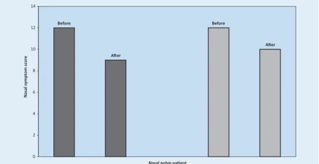
Six female and 16 male nonatopic patients with nasal polyps (mean age 42 (25-72) years) and 8 fe-male and 10 fe-male atopic patients (mean age 45 (19-65) years) received CAM. In nonallergic su-bjects, the average nasal symptom score improved from 12±2 before treatment with CAM to 9±4 af-ter treatment (p=0.041) (Figure 1). In allergic pati-ents, the average nasal symptom score decreased after therapy by CAM from 12±2 to 10±3 (p=0.046) (Figure 1). After macrolide treatment, we found 67.83% patients in the nonallergic group and 55,55% patients in the allergic group with impro-ved nasal symptoms. These differences were not statistically significant (chi square-test).
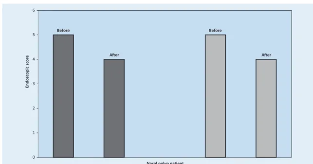
In nonallergic patients, we found a significant dif-ference in the endoscopic score before and after treatment (5±1 vs 4±2) (p=0.043) (Figure 2). The average size of allergic patients’ polyps was smaller after therapy (5±1 vs 4±1) (p=0.042) (Fi-gure 2). The size of nasal polyps decreased in 45.45% (10 of 22 cases) of patients without aller-gy and in 50% allergic patients (9 of 18 cases) but
After Before After Before 0 2 4 6 8 10 12 14
Nasal polyp patients
N as al sy m pt om s co re
Figure 1. Nasal symptom score of nonallergic patients (dark) and allergic patients (grey) with nasal polyposis, before and after CAM administration.
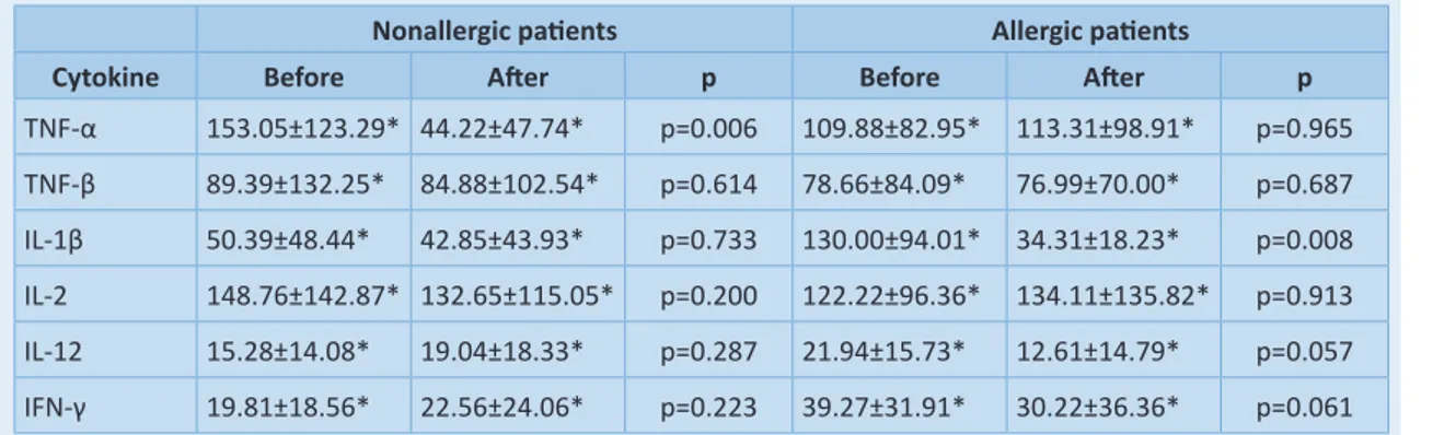
the difference was not statistically significant (chi square-test). In the improvement group, compa-ring the endoscopic findings before and after CAM therapy, there was a higher statistically si-gnificant difference in the nasal polyp size. In no-nallergic patients, the size of the polyps decrea-sed from 5±1 to 3±1 (p=0.007) (Figure 3). In allergic subjects, the average endoscopic score improved from 4±1 to 2±1 (p=0.006) (Figure 3). We found no significant differences in the levels of TNF-β, IL-2, IL-12 and IFN-γ in the nasal secre-tions before and after macrolide treatment (Ta-ble 1). Only the concentration of proinflamma-tory Th1 cytokine TNF-α in the nasal discharge of nonatopic patients was the highly statistically lower after CAM treatment (p=0.006) (Table 1). In the group of allergic patients with nasal poly-posis, we found significantly lower concentration of IL-1β (p=0.008) after therapy with CAM (Table 1). When we divided our patients into the
impro-vement and no improvement group, we found
new interesting details. In nonatopic patients, the levels of TNF-α significantly decreased in the
improvement group (p=0.003). However, in the
no improvement group, there was no difference
in the mean level of TNF-α before and after trea-tment (p=0.067) (Table 2). In subjects with aller-gic rhinitis, comparing the post macrolide trea-tment outcomes for levels of IL-1β, we found no differences between the improvement (p=0.007)
and no improvement group (p=0.006). However,
we found significantly lower levels of IL-12 in the
improvement group of atopic patients after CAM
administration (p=0.037) (Table 2).
DISCUSSION
Ichimura et al14 found that roxithromycin (RXM)
administered at 150 mg/day for at least 8 weeks shrank the nasal polyp size in 52% of twenty inve-stigated patients. They reported that the efficacy of macrolide therapy is not related to allergic symptoms. Our results also showed that there was no relationship between the presence of atopy and clinical efficacy of macrolide trea-tment. The results of the bacterial cultures sug-gest that the risk of selecting resistant bacteria is low15. In a small number of patients the cultures
were positive, but this was not always linked with
an increase in symptoms, which could be due to the fact that in addition to the direct bacteriosta-tic effect of macrolides, they may in some cases reduce the virulence of bacteria without eradica-ting them15.
However, the mechanisms of polyp shrinkage du-ring macrolide treatment are not well known. Nonaka et al16 demonstrated that in vivo RXM
treatment directly suppressed nasal polyp fibro-blasts (NPFs) proliferation, and that this effect of RXM on fibroblast growth was persistent, indica-ting that RXM may prevent the progression of na-sal polyps by inhibiting the development of fibro-sis.
Nasal polyposis is an example of an extreme im-mune dysregulation.Clinical, as well as experi-mental studies indicate that nasal polyp formati-on and growth are activated and perpetuated by an integrated process of mucosal epithelium, la-mina propria and inflammatory cells, which, in turn, may be initiated by both infectious and non-infectious inflammation4. Various toxic and
infectious agents, as well as allergens, encounte-red at the level of nasal/paranasal mucosa, may activate innate immune mechanisms and lead to induction of pro-inflammatory cytokines. Results presented by Fundová et al17 suggest that
dysre-gulations in innate immune mechanisms (for example signaling through toll-like receptors and induction of nuclear factor kappa B (NF-κB) and therefore cytokine production) and a defect in homeostasis of epithelial cells and prolonged cell survival, may play a role in pathogenesis and growth of nasal polyps. However, in the world li-terature, we can find small number of studies re-garding the role of Th1 cytokines in pathogenesis of this disease. The Th1 responses are dominated by phagocytic cell-mediated immune responses, with a marked increase in production of Th1 cytokines18.
IL-2 is an essential growth factor for T-cells and it acts in an autocrine fashion to stimulate T-cell proliferation and also serves to regulate immuno-globulin production and the growth of cytotoxic T-lymphocytes and natural-killer (NK)-cells18.
IL-12 is a heterodimeric cytokine consisting of p40 and p35 sub-units, that is secreted by macropha-ges/monocytes, B cells and other
antigen-pre-After Before After Before 0 1 2 3 4 5 6
Nasal polyp patients
En do sc op ic s co re
Figure 3. Endoscopic score of nonallergic patients (dark) and allergic patients (grey) in the improvement group, before and after CAM administration.
After Before After Before 0 1 2 3 4 5 6
Nasal polyp patients
En do sc op ic s co re
Figure 2. Endoscopic score of nonallergic patients (dark) and allergic patients (grey) with nasal polyposis, before and after treatment by CAM.
senting cells (APCs), and has multiple immunore-gulatory effects on various cell population in different inflammatory conditions19,20. IL-12 also
plays an important role in the proliferation, cytol-ytic activity and IFN-γ production by T cells and NK cells19,20. According to our results, long-term
low-dose CAM administration didn’t reduce the level of this cytokine in nasal discharge. However, when we divided our patients into the improve-ment and no improvement group, we found
redu-ced levels of IL-12 in the improvement group of allergic patients. This suggests a possible associa-tion between the reducassocia-tion of nasal polyp size and reduction of IL-12 levels in nasal secretions only in atopic subjects. IFN-γ is a Th1 cytokine which leads via macrophage activation to exten-sive inflammatory processes that also enable the killing of intracellular pathogens18. Dellacono et
al21 hypothesize that elevated levels of IFN-γ
na-Table 1. Th1 cytokine levels in nasal discharge of treated patients, before and after macrolide administration. Nonallergic patients Allergic patients
Cytokine Before After p Before After p
TNF-α 153.05±123.29* 44.22±47.74* p=0.006 109.88±82.95* 113.31±98.91* p=0.965 TNF-β 89.39±132.25* 84.88±102.54* p=0.614 78.66±84.09* 76.99±70.00* p=0.687 IL-1β 50.39±48.44* 42.85±43.93* p=0.733 130.00±94.01* 34.31±18.23* p=0.008 IL-2 148.76±142.87* 132.65±115.05* p=0.200 122.22±96.36* 134.11±135.82* p=0.913 IL-12 15.28±14.08* 19.04±18.33* p=0.287 21.94±15.73* 12.61±14.79* p=0.057 IFN-γ 19.81±18.56* 22.56±24.06* p=0.223 39.27±31.91* 30.22±36.36* p=0.061 *All cytokine levels are expressed in picograms/mililitres (pg/ml) and presented as means ± standard deviation (SD)
Table 2. The relationship between cytokine levels in nasal fluid and change of nasal polyp size: TNF-α levels in nasal secretions of nonallergic patients and IL-1β and IL-12 levels in nasal fluid of allergic patients in improvement
and no improvement group.
Improvement group No improvement group
Cytokine Before After p Before After p
TNF-α 224.38±174.68* 42.86±47.81* p=0.003 92.13±86.17* 78.86±81.62* p=0.067 IL-1β 143.28±128.37* 47.36±28.25* p=0.007 121.15±87.63* 32.26±19.87* p=0.006 IL-12 31.87±25.58* 11.73±12.06* p=0.037 26.71±19.22* 21.96±23.13* p=0.097 *All cytokine levels are expressed in pg/ml and presented as means ± standard deviation (SD).
sal polyp tissue. They found a positive correlation between the increased IFN-γ levels and presence of allergy and asthma in patients with nasal pol-yps21.
The results of our investigation showed that the TNF-α concentrations in nasal discharge of no-nallergic patients was significantly reduced after treatment with CAM. Therefore, the decreased TNF-α levels in nasal secretions were associated with reduction of polyp size only in nonatopic pati-ents. TNF-α is the main proinflammatory cytokine of Th1 immune response22. It is secreted by
ma-crophages, monocytes and natural killer (NK) cells and many other cell types, especially by activated eosinophils22,23. TNF-α, among other cytokines can
regulate fibroblast activity and collagen formation through modulation of collagenase activity23.
Rela-tionship between decreased levels of TNF-α in na-sal fluid and shrinkage of polyps can be explained by several recently published findings. Eosinophil infiltration is regulated by numerous chemokines and adhesion molecules such as eotaxin, regula-ted on activation of normal T cell expressed and secreted (RANTES), and vascular cell adhesion
mo-lecule (VCAM)-124. To infiltrate sites of
inflammati-on, eosinophils leave the bloodstream and pass through the endothelium in four steps, namely ro-lling, adhesion, transendothelial migration, and chemotaxis24. Adhesion molecules, such as
VCAM-1 play an important role during adhesion to endot-helial cells24. Experiments performed by Ohori et
al24 demonstrated that TNF-α stimulation induces
VCAM-1 protein production and mRNA expression in human nasal polyp fibroblasts. Epithelial and immunocompetent cells such as macrophages, mast cells and, especially, eosinophils produce TNF-α. These findings suggest that TNF-α increa-ses VCAM-1 production in nasal fibroblasts and ac-tivates the transmigration of eosinophils which in-duce further production of TNF-α and accelerate the accumulation of eosinophils in nasal polyps. Saji et al25 demonstrated that NPFs produced
RAN-TES by stimulation with TNF-α and IL-1β. Therefo-re, results published by Yoshifuku et al26 showed
that eotaxin secretion from fibroblasts was indu-ced by stimulation with IL-4 and synergistically en-hanced by simultaneous stimulation with TNF-α and IL-4. Iino et al27 demonstrated that long-term
Treatment by CAM decreased TNF-α levels in
non-aller-gic and IL-1β levels in allernon-aller-gic patients and showed a
strong anti-inflammatory and different
immunomodu-latory effects in non-atopic and atopic patients.
low-dose administration of erythromycin inhibits the production of TNF-α by human monocytes in
vitro. These results showed that treatment with
macrolide antibiotic could suppresse TNF-α pro-duction and the progression of nasal polyps due to inhibition of fibroblast and monocyte activity in nasal polyp tissue.
Our results also showed decreased levels of IL-1β in nasal secretions in atopic patients after trea-tment by CAM. IL-1β plays a crucial role in the pathogenesis of chronic rhinosinusitis and nasal
polyps. This strong Th1 proinflammatory cytoki-ne secreted by epithelial cells, monocytes, ma-crophages and fibroblasts upregulates the expre-ssion of E-selectin and intercellular adhesion molecule-1 (ICAM-1) in vascular endothelial cells, and thereby induces the extravascular transmi-gration of neutrophils28. The emigrated
neutrop-hils then secrete IL-1β, which amplifies the expre-ssion of E-selectin and ICAM-1, resulting in further neutrophil infiltration28. Miyanohara et
al29 revealed that clarithromycin suppressed IL-1β
gene expression in human nasal epithelial cells in
vitro. We demonstrated that macrolide
trea-tment of nasal polyposis have different immuno-modulatory and similar clinical effects in allergic and nonallergic patients. On the other hand, the-se results may be due to the fact that atopic and nonatopic patients have different mediator profi-les in their nasal secretions30, implying clear
diffe-rences in pathogenesis of their nasal polyps.
CONCLUSION
Evaluation of the cytokine levels in nasal dischar-ge could be an valuable path in monitoring nasal polyp patients, as well as sensitive way to assess new therapies for nasal polyposis and to study the pathogenesis of this disease. Low-dose ma-crolide treatment proved effective in the mana-gement of nasal polyposis in both atopics and noatopics. These results indicate the importance
of TNF-α and IL-1β regarding their influence on eosinophils and neutrophils in the pathogenesis of atopic and nonatopic form of nasal polyposis. Macrolide treatment may help minimize surgical treatment. We suggest that macrolides can be an alternative to topical and systemic glucocortico-ids in the management of nasal polyposis. AKNOWLEDGEMENTS
This study was supported by grants from the Mili-tary Medical Academy Research Fund. It was per-formed as a part of project of the Institute of Me-dical Research, Military MeMe-dical Academy, Belgrade, Serbia (VMA/11-14/B-6). The authors declare that they have no conflict of interest.
REFERENCES
1. Veyseller B, Aksoy F, Ertaş B, Keskin M, Özturan O, Yildi-rim YS. A new oxidative stress marker in patients with nasal polyposis: advanced oxidative protein products (AOPP). B-ENT 2010;6:105-9.
2. Bonfils P, Avan P, Malinvaud D. Influence of allergy on the symptoms and treatment of nasal polyposis. Acta Otolaryngol2006;126:839-44.
3. Bachert C, Robillard T. Management of nasal polyposis. B-ENT 2005;Suppl 1:77-86.
4. Norlander T, Brönnegård M, Stierna P. The relationship of nasal polyps, infection, and inflammation. Am J Rhinol 1999;13:349-55.
5. Cervin A, Wallwork B. Macrolide therapy of chronic rhinosinusitis. Rhinology 2007;45:259-67.
6. Nonaka M, Pawankar R, Saji F, Yagi T. Effect of roxithro-mycin on IL-8 synthesis and proliferation of nasal polyp fibroblasts. Acta Otolaryngol Suppl1998;539:71-5. 7. Suzaki H, Asano K, Yu M, Hisamitsu T. Influence of
roxit-hromycin on inflammatory cytokine production from nasal polyp fibroblasts in vitro. Acta Otolaryngol 2003;123:637-42.
8. Riechelmann H, Deutschle T, Friemel E, Gross HJ, Ba-chem M. Biological markers in nasal secretions. Eur Respir J 2003;21:600-5.
9. Fokkens W, Lund V, Mullol J. European Position Paper on Rhinosinusitis and Nasal Polyps group. European po-sistion paper on rhinosinusitis and nasal polyps 2007. Rhinol Suppl 2007;45(Suppl 20):1-136.
10. Lildholdt T, Rundcrantz H, Bende M, Larsen K. Glucocor-ticoid treatment for nasal polyps: the use of topical bu-desonide powder, intramuscular betamethasone, and surgical treatment. Arch Otolaryngol Head Neck Surg 1997;123:595-600.
11. Bachert C, Van Kempen M, Van Cauwenberge P. Regula-tion of proinflammatory cytokines in seasonal allergic rhinitis. Int Arch Allergy Immunol 1999;118:375-9. 12. Perić A, Vojvodić D, Radulović V, Vukomanović-Đurđević
B, Miljanović O. Correlation between cytokine levels in nasal fluid and eosinophil counts in nasal polyp tissue in asthmatic and nonasthmatic patients. Allergol Immu-nopathol (Madr) 2010;39:133-9.
13. Perić A, Vojvodić D, Radulović V, Vukomanović-Đurđević B, Perić AV, Miljanović O. Proinflammatory cytokine le-vels in nasal fluid as indicators of severity of nasal poly-posis. Acta Clin Croat 2010;49:395-403.
14. Ichimura K, Shimazaki Y, Ishibashi T, Higo R. Effect of new macrolide roxithromycin upon nasal polyps associ-ated with chronic sinusitis. Auris Nasus Larynx 1996;23:48-56.
15. Cervin A, Kalm O, Sandkull P, Lindberg S. One-year low-dose erythromycin treatment of persistent chronic si-nusitis after sinus surgery: clinical outcome and effects on mucoliliary parameters and nasal nitric oxide. Oto-laryngol Head Neck Surg 2002;126:481-9.
16. Nonaka M, Pawankar R, Tomiyama S, Yagi T. A macroli-de antibiotic, roxithromycin, inhibits the growth of na-sal polyp fibroblasts. Am J Rhinol 1999;13:267-72. 17. Fundová P, Filipovský T, Funda DP, Hovorka O, Holý R,
Navara M et al. Expression of IGF-1R and iNOS in nasal polyps; Epithelial cell homeostasis and innate immune mechanisms in pathogenesis of nasal polyposis. Folia Microbiol2008;53:558-62.
18. Král B, Krejsek J, Paráková Z, Kopecký O, Vokurková D, Derner V et al. Some serum activity markers of airways inflammation in difficult-to-control asthma patients. Acta Medica (Hradec Králové) 1997;40:61-70.
19. Davidsson Å, Danielsen A, Viale G, Olofsson J, Dell Orto P, Pellegrini C et al. Positive identification in situ of mRNA expression of IL-6, and IL-12, and the chemotactic cytoki-ne RANTES in patients with chronic sinusitis and polypo-id disease. Acta Otolaryngol 1996;116:604-10.
20. Shikano H, Kato Z, Kaneko H, Watanabe M, Inoue R, Ka-sahara K et al. IFN-γ production in response to IL-18 or IL-12 stimulation by peripheral blood mononuclear ce-lls of atopic patients. Clin Exp Allergy 2001;31:1263-70. 21. Dellacono FR, Eisma R, Lafreniere D, Leonard G,
Kreut-zer D. Interferon gamma expression in human nasal po-lyps. Laryngoscope 1997;107:626-30.
22. Jurišić V, Čolić S, Jurišić M. The inflammatory radicular cysts have higher concentration of TNF-α in comparison to odontogenic keratocysts (odontogenic tumour). Acta Medica (Hradec Králové) 2007;50:233-8.
23. Đorđević V, Zvezdanović L, Ćosić V, Vlahović P, Kundalić S, Jevtović-Stoimenov T et al. Serum levels and in vitro production of Th1- and Th2-type cytokines by periphe-ral blood mononuclear cells in patients suffering from systemic lupus erythematosus. Journal of Medical Bio-chemistry 2010;29:19-27.
24. Ohori J, Ushikai M, Sun D, Nishimoto K, Sagara Y, Fuku-iwa T et al. TNF-α upregulates VCAM-1 and NF-κB in fi-broblasts from nasal polyps. Auris Nasus Larynx 2007;34:177-83.
25. Saji F, Nonaka M, Pawankar R. Expression of RANTES by IL-1β and TNF-α stimulated nasal polyp fibroblasts. Au-ris Nasus Larynx 2000;27:247-52.
26. Yoshifuku K, Matsune S, Ohori J, Sagara Y, Fukuiwa T, Kurono Y. IL-4 and TNF-α increased the secretion of taxin from cultured fibroblasts of nasal polyps with eo-sinophil infiltration. Rhinology 2007;45:235-41. 27. Iino Y, Toriyama M, Kudo K, Natori Y, Yuo A.
Erythro-mycin inhibition of lipopolysaccharide-stimulatied tu-mor necrosis factor alpha production by human mono-cytes in vitro. Ann Otol Rhinol Laryngol Suppl 1992;101(suppl 157):16-20.
28. Tokushige E, Itoh K, Ushikai M, Katahira S, Fukuda K. Lo-calization of IL-1 beta mRNA and cell adhesion molecu-les in the maxillary sinus mucosa of patients with chro-nic sinusitis. Laryngoscope 1994;104:1245-50. 29. Miyanohara T, Ushikai M, Matsune S, Ueno K, Katahira
S, Kurono Y. Effects of clarithromycin on cultured hu-man nasal epithelial cells and fibroblasts. Laryngoscope 2000;110:126-31.
30. Perić A, Vojvodić D, Miljanović O. Influence of allergy on cytokine level in nasal discharge of patients with nasal polyps. Acta Medica Medianae 2010;49:40-4.