CHAPTER 5
RESULTS
5.1. Previous study: cell culture and organotypical slices
Initial experiments have been conducted to ensure that the tet-on system works. A neuronal cell culture from mice expressing rtTA was transfected with a construct containing tet promoter, CGG and GFP reporter. These cells showed expression of GFP after dox treatment (data not shown). It was concluded that this system worked in vitro and was ready to make the transgenic mice and cross them producing bigenic mice.
From the bigenic mice produced, this system was verified whether it also worked in the bigenic mice organs, especially the brains where the expression was expected. Organotypical cultures were conducted to prove it. The brains of bigenic mice Tet-on-99CGG-eGFP/GFA2-rtTA and Tet-on-99CGG-eGFP/PrP-rtTA at 6 days old were taken. Brain slices were cultured and treated with dox, and expression of GFP was observed under (confocal) microscope. The cultures showed strong GFP expression throughout the brain (data not shown). This result gives an insight that those bigenic systems work in vivo.
5.2. The workable of Tet-on-nCGG-eGFP: in vivo study using
Tet-on-nCGG-eGFP/hnRNP-rtTA
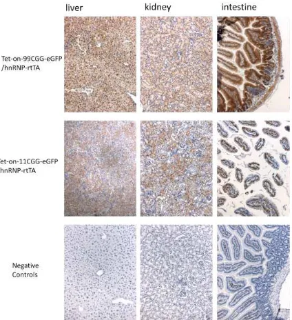
water. hnRNP-rtTA was used since these mice have previously been used and showed ubiquitous expression in all tissues (62).The bigenic mice with Tet-on-99CGG-eGFP/hnRNP-rtTA exhibited weight loss during 4 days of dox treatment, and died after 5 days of dox in drinking water treatment, while mice with Tet-on-11CGG-eGFP/hnRNP-rtTA and monogenic controls were normal, confirming expression of 99CGG as the cause of the death, neither the GFP nor the dox treatment.
Figure 3. Immunohistochemistry for GFP in liver, kidney, and intestine of bigenic
mice Tet-on-nCGG-eGFP/hnRNP-rtTA with dox treatment. Positive results are
5.3. The workable of Tet-on-99CGG-eGFP/GFA2-rtTA in vivo
The transgene expression of bigenic mice Tet-on-99CGG-eGFP/GFA2-rtTA were then tested in vivo by giving dox in drinking water. After 6 weeks of dox in drinking water, these bigenic mice were sacrificed and the brains were collected. Immunohistochemistry, Western blot and RT-Q-PCR for GFP were performed to check the expression. The result was disappointing. Immunohistochemistry did not show any signal of GFP in any brain regions. Western blot and RT-Q-PCR did not detect the expression either. To rule out the deficiency of the inducer, the dox treatment was lengthened until 9 weeks, of which a dose of 10 mg/ml of dox during the last 4 days was used, but still there was no expression detected. The rtTA expression in the brain of these bigenic mice was checked by rtTA immunohistochemistry. Apparently the absence of expression was due to the absence of rtTA expression, since the rtTA immunohistochemistry did not show any rtTA expression (data not shown).
5.4. The workable of Tet-on-99CGG-eGFP/PrP-rtTA in vivo
for GFP were performed to the RNA and protein extracted from the brains of these mice. Although GFP was expressed in only a small part of the brain, the GFP expression could still be detected by the RT-Q-PCR as well as the Western blot (Figure 14 and 15 sample A). This finding suggested that the transgene system for this bigenic line worked in vivo, and is ready to be used for FXTAS neuropathological studies. Before the main experiment and further studies using these bigenic lines were started, it was necessary to choose the best founders of the PrP-rtTA and the Tet-on-99CGG-eGFP, since there were several founders for both of the transgenic lines. The best founders were expected to be the transgenic lines which give the strongest and appropriate expression, and thus be able to produce enormous expression of expanded premutation CGG transcript. The abundant expanded premutation CGG expression is expected to provide the sufficiency to produce severe form of FXTAS outcomes.
Figure 4. Immunohistochemistry for GFP in brain slices of bigenic mice
Tet-on-99CGG-eGFP/PrP-rtTA after 2 weeks of dox in drinking treatment. Positive results
5.5. Transgene expression outside the brain in
Tet-on-99CGG-eGFP/PrP-rtTA bigenic mice
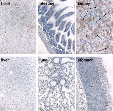
Figure 5. Immunohistochemistry for GFP in tissues outside the brain of bigenic mice
Tet-on-99CGG-eGFP/PrP-rtTA after dox intraperitoneal injection. GFP expression
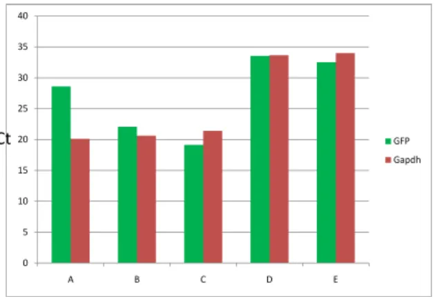
Figure 6. Western blot for GFP on kidney of tet-on-99CGG-eGFP/PrP-rtTA mouse
after dox IP treatment. The red bands are GFP; the green bands are Gapdh as the
internal control. We could see the kidney of this bigenic mouse (B) expressed the GFP, indicated by the presence of red band. Sample A was the negative control. Sample C was the positive control.
Figure 7.RT-Q-PCR for GFP RNA from kidney of tet-on-99CGG-eGFP/PrP-rtTA
mouse after dox IP treatment. Green bars represent the GFP. Red bars were Gapdh
5.6. Best founder of PrP-rtTA
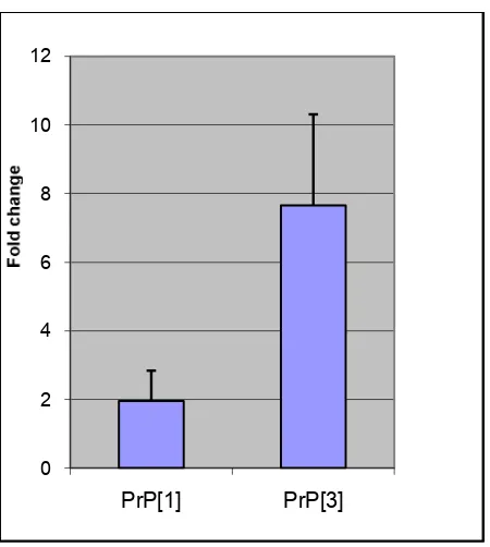
For the PrP-rtTA transgene, there were two founders available, founder 1 and founder 3. To choose the best founder to be used for further study, the rtTA expressions between these two founders were compared by PCR. RT-Q-PCR for rtTA was assayed to the mRNA isolated from the brain of both founders to compare the rtTA levels. The RT-Q-PCR showed more rtTA transcript in founder 3 than founder 1 (Figure 8). The rtTA mRNA levels of founder 3 were about 4 fold higher than founder 1. Based on this observation founder 3 gave more expression of rtTA than founder 1, and founder 3 of PrP-rtTA line was chosen for further studies instead of founder 1.
Figure 8. rtTA mRNA levels from the brains of different PrP-rtTA founder. rtTA
5.7. Best founder of Tet-on-99CGG-eGFP
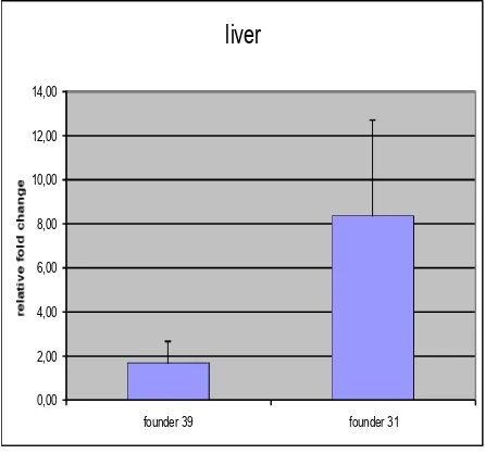
The best founder of Tet-on-99CGG-eGFP needed to be chosen. There were two founders available, founder 31 and founder 39. The strategy to come to the decision was by creating bigenic mice Tet-on-99CGG-eGFP/hnRNP-rtTA derived from both different founders of Tet-on-99CGG-eGFP. The bigenic mice were treated with dox water or food treatment for 4 days to induce the expression of the transgene. GFP expression in liver of the mice from those different founders, were compared by immunohistochemistry, Western blot and RT-Q-PCR. From the immunohistochemistry of the liver, the bigenic mice from founder 39 seemed to give the expression in mosaic pattern, with many cells not expressing the transgene. Expression of the transgene in founder 31 was found more homogenous throughout the cells, and gave stronger signal than founder 39 (Figure 9).
Tet-on-99CGG-eGFP founder. Based on the results, it was decided to use founder 31 for further studies, although it was still possible to use the founder 39.
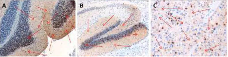
Figure 9. Immunohistochemistry for GFP in the liver slices comparing founder 31
and 39 of Tet-on-99CGG-eGFP. Positive results were indicated with the presence of
brown color staining (black arrow), which means the tissues expressed the transgene. The expression in founder 31 (B) was much more homogenous with more cells expressed the GFP than founder 39 (A). In founder 39 (A) there were many cells which did not express the transgene (pointed with red arrow). Sample C is negative control which was negative for GFP staining, which was indicated with the blue color staining. 200X magnification.
Figure 10. GFP RNA levels in the liver comparing founder 31 and 39 of
Tet-on-99CGG-eGFP. Founder 31 gave stronger expression than founder 39. It was more than 4
Figure 11.Western blot analysis for GFP in the liver comparing founder 31 and 39
of Tet-on-99CGG-eGFP. Figure X is the result of Western blot. A, B and C are mice
5.8. Formation of ubiquitin intranuclear inclusion in the
Tet-on-99CGG-eGFP/PrP-rtTA bigenic mice
The Tet-on-99CGG-eGFP/PrP-rtTA bigenic mice from a breeding between the best of each of the founders, then were subjected for ubiquitin-positive intanuclear inclusions assessment. These bigenic mice were treated with dox in drinking water for 4 weeks, and were sacrificed to collect the brains for the neuropathological studies. Immunohistochemistry was performed to check the GFP expression as well as the presence of ubiquitin-positive inclusions formation in the brain slices. Immunohistochemistry for GFP showed that the transgene was expressed by the presence of GFP staining in the brains (Figure 12). On the other hand, immunohistochemistry for ubiquitin did not detect any ubiquitin-positive inclusions (data not shown). It seemed that the expression of the expanded premutation CGG induced by dox in drinking for 4 weeks was not enough to produce the neuropathological hallmark of FXTAS, ubiquitin-positive intranuclear inclusion.
RT-Q-PCR and Western blot were gained. Although the expression of the transgene was limited to some parts of the brain, the GFP signal could still be detected through RT-Q-PCR and Western blot (Figure 13 & 14). The RT-Q-PCR showed increase levels of transgene transcript by the time of dox exposure. The 12 weeks of dox treatment gave the highest levels of RNA instead of the 16 weeks of treatment. Using the Western blot, the expression of GFP could be seen by the presence of the GFP bands. GFP levels tended to increase by the time of dox treatment except for the 12 weeks of treatment which gave lower levels than 4 weeks of treatment.
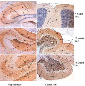
Figure 12. Immunohistochemistry for GFP in the brain slices of bigenic mice
Tet-on-99CGG-eGFP/PrP-rtTA after dox treatment. Positive results were indicated with
the brown color staining (arrow), which means the tissues expressed the transgene. These bigenic mice expressed the GFP in the brain mainly in hippocampus and cerebellum. 100X magnification.
Figure 13.GFP RNA levels in the brain of bigenic mice
Tet-on-99CGG-eGFP/PrP-rtTA after dox treatments. A is the bigenic mice with two weeks of dox treatment from
Figure 14. Western blot for GFP of bigenic mice Tet-on-99CGG-eGFP/PrP-rtTA
after different time points of dox treatment. The transgene expression could also be
Figure 15. Immunohistochemistry of ubiquitin in the brain slices of bigenic mice
Tet-on-99CGG-eGFP/PrP-rtTA with 12 and 16 weeks of dox treatment. The