L
Journal of Experimental Marine Biology and Ecology 244 (2000) 145–156
www.elsevier.nl / locate / jembe
Molecular cloning of a cDNA encoding a member of
CHH / MIH / GIH family from Penaeus monodon and analysis
of its gene structure
*
Apinunt Udomkit , Sunthorn Chooluck, Burachai Sonthayanon, Sakol Panyim
Institute of Molecular Biology and Genetics, Mahidol University, Salaya Campus, Nakhon Pathom 73170,
Thailand
Received 10 July 1999; accepted 28 August 1999
Abstract
We report the nucleotide and deduced amino acid sequences of Pem-CMG peptide, a member of crustacean CHH / MIH / GIH peptide family, in black tiger prawn (Penaeus monodon). The 59and 39 fragments of Pem-CMG cDNA were cloned by the method of rapid amplification of cDNA ends (RACE). The two fragments constitute a combined cDNA length of 593 bp with a 77 bp overlapping region. Sequence analysis reveals the presence of a 384 bp open reading frame which was subsequently cloned. The open reading frame encodes a precursor peptide that is comprised of 128 amino acids, with a putative processing site, KR. The mature peptide consists of 74 amino acid residues, the sequence of which is significantly homologous to those of the CHH / MIH / GIH family known from other crustaceans. Analysis of a genomic fragment of Pem-CMG reveals a single intron of 314 bp interrupting the coding sequence for the mature peptide. The presence of only one intron in Pem-CMG gene suggests that this gene is structurally different from the previously reported MIH gene of Charybdis feriatus and CHH-like gene of Metapenaeus ensis which possess two introns in their coding sequences. 2000 Elsevier Science B.V. All rights reserved.
Keywords: Eyestalk; Neuropeptide; Penaeid shrimp; Rapid amplification of cDNA ends
1. Introduction
A number of physiological processes in decapod crustaceans are known to be regulated by diverse neuropeptides synthesized by a neurosecretory system called
*Corresponding author. Tel.: 166-24-419-003 ext: 1237; fax: 166-24-419-906.
E-mail address: [email protected] (A. Udomkit)
X-organ-sinus gland complex (XOSG) located in the optic ganglia of the eyestalks (De Kleijn and Van Herp, 1995; Charmantier et al., 1997). Most of the peptide hormones isolated from the XOSG complex e.g. pigment dispersing hormone (PDH) and crustacean cardioactive peptide (CCAP) have also been identified in some other arthropods such as insects. Recently, a group of related peptides from XOSG complex that constitutes a novel hormone family has been described and thus far has been found only in crustaceans (Keller, 1992). This family includes crustacean hyperglycemic hormone (CHH), molt-inhibiting hormone (MIH) and gonad inhibiting hormone (GIH) that are involved in blood sugar regulation, inhibition of ecdysteroid synthesis, and regulation of reproduction, respectively (Cooke and Sullivan, 1982; Chang, 1993).
Direct amino acid sequencing of the hormones in this CHH / MIH / GIH family from several crustaceans has been conducted (Kegel et al., 1989; Chang et al., 1990; Webster, 1991; De Kleijn et al., 1994a,b). Comparison of the amino acid sequences of these hormones revealed significant degrees of similarity (Keller, 1992; Sun, 1994; Chang, 1997) suggesting that they constitute an authentic peptide family across species boundaries. In addition to several similarities in their structure, some hormones in the CHH / MIH / GIH family are multifunctional i.e. exhibit more than one biological activity (Chang et al., 1990; Tensen et al., 1991; Lee et al., 1995). However, the certainty concerning the primary physiological functions of different peptides still needs to be clarified.
In recent years, the cDNA coding for CHH, MIH and GIH in a number of crustaceans has been cloned based on the information of available amino acid sequences (Weideman et al., 1989; Tensen et al., 1991; Klein et al., 1993; De Kleijn et al., 1994a,b; Sun, 1994; Lee et al., 1995; Aguilar-Gaytan et al., 1997). Despite this increasing number of reports on cDNA cloning and nucleotide sequence analysis of neuropeptides in CHH / MIH / GIH family, the hormones in several crustaceans still await complete characterization, especially at the molecular level.
We report here the molecular cloning of a cDNA encoding Pem-CMG, a peptide in the family of CHH / MIH / GIH, from black tiger prawn, Penaeus monodon. An open reading frame encoding Pem-CMG peptide was cloned from a cDNA of a single pair of the eyestalks. A primary structure of the gene for Pem-CMG was also analyzed and
revealed that Pem-CMG gene of P. monodon has different structure from the genes
encoding MIH and CHH-like peptide that have been recently reported in Charybdis
feriatus and Metapenaeus ensis, respectively (Chan et al., 1998; Gu and Chan, 1998)
2. Material and methods
2.1. Animals
Black tiger prawns, P. monodon, were purchased from Shrimp Culture Research
2.2. Oligonucleotide primers
Abridged anchor primer (AAP) and abridged universal amplification primer (AUAP)
were provided with the 59 RACE System for Rapid Amplification of cDNA Ends
(GIBCO BRL). Other custom oligonucleotides were purchased from Bio-Synthesis, USA.
2.3. RNA isolation
A total of 100 eyestalks were collected from P. monodon immediately after
anesthetizing the live shrimp with crushed ice. After the cuticle and non-neural tissues were removed, the dissected eyestalks were ground to fine powder in liquid nitrogen by
means of mortar and pestle. Total RNA was extracted using TRIZOL Reagent (GIBCO
1
BRL). Poly (A) RNA was isolated using Dynabeads mRNA purification kit (DYNAL)
and was quantified by measuring the absorbency at 260 nm.
2.4. Amplification of the 39 ends of cDNA
The reaction was carried out according to the protocol of the 39 RACE System for
Rapid Amplification of cDNA Ends (GIBCO BRL). First strand cDNA was synthesized
1
from 75 ng of poly (A) RNA in a total volume of 20ml containing 20 mM Tris–HCl
(pH 8.4), 50 mM KCl, 2.5 mM MgCl , 10 mM DTT, 500 nM PRT primer, 5002 mM
each dATP, dCTP, dGTP, dTTP and 200 units of SuperScript II Reverse Transcriptase. The reaction was incubated at 508C for 50 minutes and then terminated at 708C for 15 minutes. The RNA template was degraded with RNaseH before proceeding to
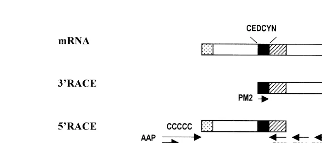
amplifica-tion step. A 2ml aliquot of cDNA was amplified with primers PM1 and PM2 (see Fig.
1) in a reaction containing 20 mM Tris–HCl (pH 8.4), 50 mM KCl, 2 mM MgCl , 2002
nM each primer, 200 mM each dATP, dCTP, dGTP, dTTP and 2.5 units of AmpliTaq
DNA polymerase (PE Applied Biosystems). Amplification was performed in a DNA Thermal Cycler (GeneAmp System 2400, PE Applied Biosystems) with 35 cycles of 948C for 1 minute, 508C for 1 minute and 728C for 2 minutes following with 7 minutes incubation at 728C as a final extension.
2.5. Amplification of the 59 ends of cDNA
The detailed protocol of the 59RACE System for Rapid Amplification of cDNA Ends
Version 2.0 (GIBCO BRL) was as follows. First strand cDNA synthesis was carried out as described in Section 2.4, except that 100 nM of primer PM3 was substituted for primer PRT. The cDNA synthesized was then purified using a GlassMax DNA isolation spin cartridge (GIBCO BRL).
A 10ml aliquot of purified cDNA was tailed with dCTP in 25ml of 10 mM Tris–HCl
(pH 8.4), 25 mM KCl, 1.5 mM MgCl , 2002 mM dCTP and 1 ml of terminal
deoxynucleotidyl transferase (TdT). The reaction was incubated at 378C for 10 minutes
and TdT was heat inactivated at 658C for 10 minutes. The PCR amplification of 5ml of
primer PM4 and abridged anchor primer (AAP). The second round PCR was performed using primer PM5 and abridged universal amplification primer (AUAP) to obtain specific amplified product.
2.6. Amplification of an open reading frame of Pem-CMG peptide
Total RNA extracted from a single pair of the eyestalks was used to synthesize a cDNA template for amplification of the open reading frame of Pem-CMG peptide. The condition for PCR was the same as described earlier in Sections 2.4 and 2.5. The primers used were CDF and CDR, the sequences of which are given in Fig. 1.
2.7. Amplification of a genomic fragment of Pem-CMG gene
Genomic DNA was prepared from abdominal muscle tissues of P. monodon by
phenol / chloroform extraction method (Sambrook et al., 1989). About 150 ng of genomic DNA was used as a template for PCR amplification of the Pem-CMG sequence
with the primers CMG-F and CMG-R (Fig. 2) designed from the most 59and 39ends of
the Pem-CMG cDNA. The PCR reaction mixture (150 ng of genomic DNA template, 20
mM Tris–HCl (pH 8.4), 50 mM KCl, 2 mM MgCl , 200 nM each primer, 2002 mM each
dATP, dCTP, dGTP, dTTP) was heated to 958C for 5 minutes, then 2.5 units of
AmpliTaq was added. Amplification was achieved by 35 successive cycles of denatura-tion at 948C for 1 minute, annealing at 508C for 1 minute and extension at 728C for 2
minutes, followed by a 10 minute final extension at 728C.
2.8. Cloning and sequencing
PCR Amplification products were purified using GeneClean II kit (Bio101). The
purified DNA fragments were digested with appropriate restriction before being ligated to pUC18 vector predigested with corresponding enzymes. The ligation products were used to transform E. coli JM109 and the recombinants were screened by restriction enzyme digestion. The nucleotide sequences of the recombinants were determined by the
method of ABI PRISME Dye Terminator Cycle using ABI PRISM Model 377
automated DNA Sequencer (PE Applied Biosystems).
3. Results
3.1. 39RACE cloning of the cDNA encoding Pem-CMG peptide
Nine individual recombinants, obtained by cloning of 39RACE products into pUC18,
that contain cDNA insert of different sizes ranging from 163 bp to 450 bp were selected for nucleotide sequence analysis. One of the clones harbors a 324 bp cDNA insert (including poly (A) tail), the nucleotide sequence of which exhibits more than 60% identity to those of mRNA encoding CHH and MIH from other crustaceans (data not shown).
3.2. 59 RACE cloning of the cDNA encoding Pem-CMG peptide
Using three specific primers generated from the nucleotide sequence of the 39RACE
fragment, a single band of amplification product of 370 bp (excluding G-rich tail) was
obtained from 59 RACE and was subsequently cloned into pUC18. The nucleotide
sequence analysis of three individual clones revealed that they contain cDNA insert homologous to the mRNA of CHH / MIH / GIH peptide family (data not shown). No variation in the nucleotide sequences was detected among the three clones.
3.3. Nucleotide sequence of Pem-CMG cDNA and its deduced amino acid sequence
The combination of nucleotide sequences of the 39and 59fragments of the Pem-CMG
cDNA revealed a 593 bp cDNA containing a 384 bp open reading frame, 65 bp 59
untranslated region and 141 bp 39untranslated region, with a potential polyadenylation signal AATAAA located 14 bp upstream from the poly (A) sequence (Fig. 3). The two fragments shared a 77 bp overlapping region (nucleotides 294 to 370) that exhibited identical nucleotide sequences. Using a cDNA that was synthesized from the total RNA of a single pair of the eyestalks, a fragment of 384 bp starting from a start codon to a stop codon of the open reading frame was cloned by PCR.
The nucleotide and deduced amino acid sequences of a 384 bp open reading frame of
Pem-CMG cDNA from nine individual recombinant clones are shown in Fig. 3. The
Fig. 3. Nucleotide sequence of Pem-CMG cDNA derived from the sequences of 59and 39RACE fragments. The 77 bp overlapping sequence is shaded. The 384 bp open reading frame is depicted by capital letters. The potential processing site, KR, is boxed. The deduced amino acids are written in one-letter symbols. The putative polyadenylation site is underlined.
contains several hydrophobic amino acid residues suggesting a possible role as a signal sequence.
3.4. Comparison of the amino acid sequence of Pem-CMG peptide with those of the
peptides in CHH /MIH /GIH family from other crustaceans
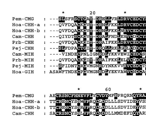
The deduced amino acid sequence of Pem-CMG peptide of P. monodon was
compared with those in the CHH / MIH / GIH family from several crustaceans. The result of this comparison (Fig. 4) revealed that Pem-CMG possessed a significant degree of homology to the peptides in this family (30%–50% identity). The Pem-CMG peptide contains six conserved cysteine residues which were found located at identical positions in all other members of CHH / MIH / GIH family. Most of the conserved amino acid residues were found in the N-terminal region whereas less homology was found in the C-terminal area.
3.5. Primary structure of Pem-CMG gene
A specific DNA fragment, about 900 bp, was amplified from genomic DNA of P.
monodon with primers CMG-F and CMG-R. This fragment, designated as g-CMG,
contained nucleotide sequence from the first start codon (ATG) to the stop codon (TAG) of Pem-CMG gene. Analysis of the nucleotide sequence of g-CMG fragment cloned in pUC18 revealed that this fragment contains one intron. An exon–intron boundary was detected using Gene Finder program of Baylor College of Medicine (BCM) on the WWW. The splice junction conformed to the splice donor and acceptor consensus sequence (Mounts, 1986).
Fig. 4. Comparison of the amino acid sequence of Pem-CMG peptide with those of H. americanus CHH-A and CHH-B (Hoa-CHH-A and Hoa-CHH-B, Tensen et al., 1991), C. maenas CHH (Cam-CHH, Kegel et al., 1989), P. bouvieri CHH (Prb-CHH, Huberman et al., 1993), P. japonicus CHH (Pej-CHH, Yang et al., 1995),
C. maenas MIH (Cam-MIH, Webster, 1991), P. bouvieri MIH (Prb-MIH, Aguilar-Gaytan et al., 1997), P.
japonicus MIH (Pej-MIH, Ohira et al., 1997) and H. americanus GIH (Hoa-GIH, De Kleijn et al., 1994b). The
amino acid residues that are identical to Pem-CMG peptide are highlighted.
intron is 314 bp long. This intron separates the two exons in the coding region for the
94
mature Pem-CMG peptide between the second and the third codons of Arg (Fig. 5).
4. Discussion
The 39and 59RACE strategy (Frohman et al., 1988) was employed in order to clone a
Pem-CMG cDNA due to the lack of information on amino acid sequences of the
peptides in CHH / MIH / GIH family in P. monodon. This technique requires only a single oligonucleotide primer which, in our case, was generated from the conserved amino acid sequence CEDCYN that is found conserved among the peptides in CHH / MIH / GIH family from a number of crustaceans (De Kleijn and Van Herp, 1995).
Fig. 5. Nucleotide sequence and predicted amino acid sequence of Pem-CMG gene. The gene consists of two exons that are separated by an intron of 314 bp (dashed line). Exon one harbors coding sequence for signal peptide and part of the coding sequence for the first 40 amino acids of Pem-CMG peptide. The second exon consists of the coding sequence for the rest 34 amino acids of the peptide. The deduced amino acids are written in one-letter symbols. The nucleotide G at position 90 is substituted for an A in the cDNA which changes the corresponding amino acid from threonine (in Fig. 3) to alanine in the genomic sequences (highlighted).
polymorphism can be excluded. Therefore it is likely that these differences represent gene isoforms or could be an error introduced during PCR amplification. The Pem-CMG peptide of P. monodon shared the highest degree of identity with CHH of P. japonicus (50%). Significant degrees of identity were also observed between Pem-CMG and CHH of Homarus americanus (45% for CHHa and 41% for CHHb), Carcinus maenas (42%) and Procambarus bouvieri (41%). These values were close to that between Pem-CMG and the MIH reported for P. bouvieri (39%), whereas lower degrees of identity are found between Pem-CMG sequence and MIH of C. maenas and P. japonicus (32% and 34%, respectively). The GIH of H. americanus also shows comparable degree of identity (30%) with Pem-CMG of P. monodon.
ecdysteroid secretion by the Y-organ in vitro, albeit at low level, (Webster and Keller, 1986) despite the low degree of identity (28%) it shares with MIH peptide form the same species. In addition, the MIH of H. americanus also seems to be involved in the regulation of methylfarnesoate synthesis by mandibular organ in this species (Chang et al., 1990). Methylfarnesoate has been proposed to play a role in controlling vit-ellogenesis in crustaceans (Laufer et al., 1987), therefore, H. americanus MIH is probably involved in the control of vitellogenesis, the function previously assigned to GIH, as well. Thus, it is unlikely to distinguish crustacean peptides in CHH / MIH / GIH family from each other by homology in their amino acid sequences.
The amino acid sequence deduced from the genomic copy of Pem-CMG differs from the sequence deduced from cDNA at one position in the signal peptide region (Fig. 5). This difference may reflect polymorphism as the genomic DNA and the cDNA used for amplification of Pem-CMG gene and cDNA were obtained from different individual shrimp. The gene for Pem-CMG of P. monodon contains only one intron that separates the coding sequence for the mature peptide. This is in contrast to the primary structure of the genes encoding MIH in Charybdis feriatus and CHH-like peptide in Metapenaeus
ensis that have recently been characterized (Chan et al., 1998; Gu and Chan, 1998). The MIH gene of C. feriatus contains two introns, intron 1 interrupts the sequence coding for
signal peptide and the second intron separates the mature peptide sequence. Two introns were also found in CHH-like gene of M. ensis. They interrupt homologous regions to those of C. feriatus MIH, one in the signal peptide sequence and the other in the mature peptide sequence. The differences in the structure of genes in the same family among different species is not uncommon and may reflect their evolutionary pathway. It is, however, too early to draw any conclusion concerning gene structure of the member of CHH / MIH / GIH family because only little information has been obtained from a few species so far. The structure of the genes coding for neuropeptides in CHH / MIH / GIH family from other crustaceans needs to be explored. This would give a clue to answer the question of how these structurally different genes have evolved from related gene families.
The sequence information from several members of CHH / MIH / GIH family together with their biological activities suggest that the neuroendocrine regulation of related physiological activities in crustaceans is more complicated than previously thought. A complete understanding of these regulatory processes requires further information from molecular studies as well as physiological studies to define more clearly the roles of individual peptides in this family.
Acknowledgements
References
Aguilar-Gaytan, R., Cerbon, M.A., Cevallos, M.A., Lizano, M., Huberman, A., 1997. Sequence of a cDNA encoding the molt-inhibiting hormone from the Mexican crayfish Procambarus bouvieri (Crustacean, Decapoda). As. Pac. J. Mol. Biol. Biotechnol. 5, 51–55.
Chan, S.-M., Chen, X.-G., Gu, P.L., 1998. PCR cloning and expression of the molt-inhibiting hormone gene for the crab (Charybdis feriatus). Gene 224, 23–33.
Chang, E.S., 1993. Comparative endocrinology of molting and reproduction: insects and crustaceans. Annu. Rev. Entomol. 38, 161–180.
Chang, E.S., 1997. Chemistry of crustacean hormones that regulate growth and reproduction. In: Fingerman, M., Nagabhushanam, R., Thompson, M.-F. (Eds.), Endocrinology and Reproduction, Recent Advances in Marine Biotechnology, Vol. 1, Science Publishers Inc, New Hampshire, USA, pp. 163–178.
Chang, E.S., Prestwich, G.D., Bruce, M.J., 1990. Amino acid sequence of a peptide with both molt-inhibiting and hyperglycemic activities in the lobster, Homarus americanus. Biochem. Biophys. Res. Commum. 171, 818–826.
Charmantier, G., Charmantier-Daures, M., Van Herp, F., 1997. Hormonal regulation of growth and reproduction in crustaceans. In: Fingerman, M., Nagabhushanam, R., Thompson, M.-F. (Eds.), Endocrinolo-gy and Reproduction, Recent Advances in Marine BiotechnoloEndocrinolo-gy, Vol. 1, Science Publishers Inc, New Hampshire, USA, pp. 109–161.
Cooke, J.M., Sullivan, R.E., 1982. Hormones and neurosecretion. In: Bliss, D.E. (Ed.), The Biology of Crustacea, Vol. 3, Academic Press, New York, USA, pp. 205–290.
De Kleijn, D.P.V., Van Herp, F., 1995. Molecular biology of neurohormonal precursors in the eyestalks of crustacean. Comp. Biochem. Physiol. 112B, 573–579.
De Kleijn, D.P.V., Janssen, K.P., Mortens, G.J., Van Herp, F., 1994a. Cloning and expression of two crustacean hyperglycemic hormone mRNAs in the eyestalk of the crayfish Orconectes limosus. Eur. J. Biochem. 224, 623–629.
De Kleijn, D.P.V., Sleutels, F.J., Martens, G.J., Van Herp, F., 1994b. Cloning and expression of mRNA encoding prepro-gonad-inhibiting hormone (GIH) in the lobster Homarus americanus. FEBS Lett. 353, 255–258.
Frohman, M.A., Dush, M.K., Martin, G.R., 1988. Rapid production of full-length cDNA from rare transcripts: amplification using a single specific oligonucleotide primer. Proc. Natl. Acad. Sci. 85, 8998–9002. Gu, P.-L., Chan, S.-M., 1998. The shrimp hyperglycemic hormone-like neuropeptide is encoded by multiple
copies of genes arranged in a cluster. FEBS Letts. 441, 397–403.
Huberman, A., Aguilar, M.B., Brew, K., Shabanowitz, J., Hunt, D.F., 1993. Primary structure of the major isomorph of the crustacean hyperglycemic hormone (CHH-I) from the sinus gland of the Mexican crayfish
Procambarus bouvieri (Ortman): interspecies comparison. Peptides 14, 7–16.
Kegel, G., Reichwein, B., Weese, S., Gaus, G., Peter-Katalinic, J., Keller, R., 1989. Amino acid sequence of the crustacean hyperglycemic hormone (CHH) from the shore crab Carcinus maenas. FEBS Lett. 255, 10–14.
Keller, R., 1992. Crustacean neuropeptides: structures, functions and comparative aspects. Experientia 48, 439–448.
Klein, J.M., Mangerich, S., De Kleijn, D.P.V., Keller, R., Wiedeman, W.M., 1993. Molecular cloning of crustacean putative molt-inhibiting hormone (MIH) precursor. FEBS Lett. 334, 139–142.
Laufer, H., Brost, D., Baker, F.C., Carrasco, C., Sinkus, M., Reuter, C.C., Tsai, L.W., Schooley, D.A., 1987. Identification of a juvenile-hormone like compound in a crustacean. Science 235, 202–205.
Lee, K.J., Elton, T.S., Bej, A.K., Watts, S.A., Watson, R.D., 1995. Molecular cloning of a cDNA encoding putative moult-inhibiting hormone from the blue crab, Callinectes sapidus. Biochem. Biophys. Res. Commun. 209, 1126–1131.
Mounts, S.M., 1986. A catalogue of splice junction sequence. Nucleic Acids Res. 10, 459–472.
Ohira, T., Watanabe, T., Nagasawa, H., Aida, K., 1997. Molecular cloning of a molt-inhibiting hormone cDNA from the kuruma prawn Penaeus japonicus. Zool. Sci. 14, 785–789.
Sun, P.S., 1994. Molecular cloning and sequence analysis of a cDNA encoding a molt-inhibiting hormone-like neuropeptide from the white shrimp Penaeus vannamei. Mol. Mar. Biol. Biotechnol. 3, 1–6.
Tensen, C.P., De Kleijn, D.P.V., Van Herp, F., 1991. Cloning and sequence analysis of cDNA encoding two crustacean hyperglycemic hormone from the lobster Homarus americanus. Eur. J. Biochem. 200, 103–106. Webster, S.G., 1991. Amino acid sequence of putative moult-inhibiting hormone from the crab Carcinus
maenas. Proc. R. Soc. Lond. B. 244, 247–252.
Webster, S.G., Keller, R., 1986. Purification, characterization and amino acid comparison of the putative moult-inhibiting hormone (MIH) of Carcinus maenas (Crustacean Decapoda). J. Comp. Physiol. B. 156, 617–624.
Weideman, W., Gromoll, J., Keller, R., 1989. Cloning and sequence analysis of cDNA for precursor of a crustacean hyperglycemic hormone. FEBS Lett. 257, 31–34.