The journal homepage www.jpacr.ub.ac.id p-ISSN : 2302 – 4690 | e-ISSN : 2541 – 0733
Synthesis and Characterization of Polyaniline Nanoparticle by
Inverse Micelle Microemulsion Method
Nuraini Uswatun Chasana, Siti Mariyah Ulfa, and Masruri MASRURI*
Department of Chemistry, Faculty of Mathematic and Natural Science, Brawijaya University Malang, Indonesia
*
Corresponding author: [email protected]
Received 8 May 2017; Revised 12 June 2017; Accepted 12 June 2017
ABSTRACT
Polyaniline (PANI) is a conductive polymer and potential for application in the electronic device manufacturing. This paper discloses a recent strategy in synthesis polyaniline nanoparticle (NP-PANI) using inverse micelle (IM) microemulsion method, and also characterization of the provided products. The synthesis step was conducted by mixing of aniline as a polymer precursor with PEG 400, n-hexane, n-butanol, ammonium persulfate and hydrochloric acid in three different reaction times, i.e. for 2, 6, and 12 hours at 26 oC. PANI was afforded as a brown powder in 2.87%, 1.97% and 2.30% yield for NP-PANI-IM-2, NP-PANI-IM-6 and NP-PANI-IM-12. The characteristic on ultra violet-visible and infrared spectra exhibited NP-PANI was formed as an emeraldine salt oligomer. Moreover, their morphology was observed through scanning electron microscopy (SEM) and showed a different range of nanoparticle size, i.e. 50-62, 68-72 and 58-62 nm for NP-PANI-IM-2, NP-PANI-IM-6 and NP-PANI-IM-12, respectively. These products open the potency application in material science.
Keywords: conducting, polyaniline, nanoparticle, microemulsion, emeraldine
INTRODUCTION
Organic polymer has played important rule for industrial manufacture such as paint, plastic, adhesive, electronic and energy. It is easily synthesized; modification and elasticity become an important consideration. Moreover, organic polymer with conductive properties also attracts attention. Complement to these physical properties, the organic conductive polymer also gain attention for certain purposes, such as for solar cell[1], light emitting diode[2,3], conductive ink, sensor[4–7], semiconductor[8], super-capacitor[9], energy storage[10], catalysis and catalyst supporting material[11–13]. In our ongoing research finding non-metallic material catalyst for alpha-pinene oxidation[14–16] into more valuable chemicals, it turned up on polyaniline nanoparticle (PANI-NP) as prospectus catalyst. Polyaniline (PANI) consists of a long chain of benzenoid ring series and quinoid ring[17,18] and gives moderate conductivity[19–21] about 200 s/cm with hydrochloric acid as dopant[22].
improve its catalytic reactivity[12]. The oxidation product of cyclohexane yielded in above 90% yield. However, it seems the preparation of polyaniline nanoparticle still hamper weakness with low yield. Microemulsion strategy by using polyethylene glycol 2000 in cyclohexane mixed by ammonium persulfate in hydrochloric acid provided a concentrate white emulsion. This is nanoparticle template, and experimentally slows down the polymerization process during addition of butanol and aniline. In addition, stirring into a longer time tend to increase nanoparticle size. This paper reports the strategy by modifying the nanoparticle template using less viscous polyethylene glycol 400 and n-hexane was also applied instead of cyclohexane to reverse the colloid system. Thus, the reaction undergoing much faster with similar nanoparticle size was obtained.
EXPERIMENT
Chemicals and instrumentation
The chemicals were purchased and used as given from manufacturer without any further preparation. Aniline (Merck), ammonium persulfate (Sigma), polyethylene glycol 400 (Bratachem), hydrochloric acid (Smartlab), acetone, n-butanol (Smartlab), demineralized and bi-distillated aqua (Smartlab).
The infrared spectra were recorded using Shimadzu FTIR 8400S using potassium bromide plate, a weigh of sample were measured by analytical balance PRECISION Advanced, UV-Vis absorption spectra were obtained from Shimadzu UV-VIS 1601 spectrophotometer, and morphology were observed using scanning electron microscopy (SEM) using SwiftED 3000 instrument, and the atomic composition were confirmed by X-Ray Microanalysis (EDX) HITACHI TM 3000.
Reaction procedure
Polyanilline nanoparticle was synthesized according to previous report with modification[13], and all solutions were prepared using double distillated of water (aquabidest). A 50 mL of poly ethylene glycol (PEG-400) 0.2 M in beaker glass was added 10 mL of n-hexane. This mixture was stirred, and was further added with ammonium persulfate (0.3 g) in 50 mL of hydrochloric acid 0.1 M. Then, n-butanol was added using syringe pump at flow rate 0.1mL/min for 30 min, and 50 mL of aniline was added. This reaction was stirred for 2 h at 26 oC, and 30 mL of acetone was added. Then, it was filtered and washed with acetone (10 mL) and aquabidest (10 mL x 3) until a colorless filtrate was afforded. Polyaniline nanoparticle (NP-PANI-IM-2) was obtained and further dried.
The other procedures were also undertaken by stirring the process for 6 and 12 h. This step afforded polyanilline nanoparticle (NP-PANI-IM-6) and (NP-PANI-IM-12). These were further analyzed for characterization.
UV-Vis analysis
A dried sample of NP-PANI (5 mg) was dissolved in ethanol (5 mL) and ultrasonicated for 15 min at room temperature. The mixture was analyzed on UV-Vis instrument at wavelength 190-800 nm.
FTIR analysis
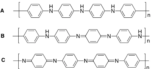
Figure 1. Three structures of polyaniline: (A) Leucoemeraldine base, LB, with fully reduced form. (B) Emeraldine base, EB, with half oxidized form, and (C) Pernigraniline base, PB, with fully oxidized form.
RESULT AND DISCUSSION
Structurally, PANI can be distinguished from its oxidation level[18] and can also be determined from the y value (0 ≤ y ≤ 1). The value (y = 1) indicates a fully reduced PANI and forms a leucoemeraldine base (LB) with a snuff color. PANI with purple appearance has a pernigraniline base (PB) with y value equal to 0, and its structure is fully oxidized. However, for PANI with y = 0.5 has an emeraldine base (EB) structure with a half oxidized structure and its color is blue. EB PANI has isolative properties, and the stability is in between PB and LB[12]. However, the doping of EB has attracted attention, due to its conductivity achieves to 100 S/cm. This dramatic changing, from isolative to conductive can be undertaken by doping process with proton acid such as hydrochloric acid. This conductive property provides emeraldine salt (ES) as a solid green powder and has conductivity level in semiconductor range. In addition, ES can also be turned back as EB by de-doping with reducing agent, such as ammonium hydroxide. However, PANI with PB and LB structure has no conductive properties.
The synthesized polyaniline nanoparticles using inverse micelle microemulsion strategy afforded a brown powder (Figure 2) in 2.87% (NP-PANI-IM-2), 1.97% (NP-PANI-IM-6), and 2.3% yield (NP-PANI-IM-12). This finding gives a better result than that previously reported[12].
Figure 2.
Polyaniline nanoparticle synthesized by inverse micelle microemulsion in different reaction time: IM-2 (2 h), IM-6 (6 h), and IM-12 (12 h).
Table 1. Yield of polyaniline nanoparticle Reaction
Time (h) Yield (%)
Color Particle size (nm) Reported Ref.28 Reported Ref.28
2 2.87 Brown Green 50-63 10-20
6 1.97 Brown Green 68-71 30-50
Previous paper reported a brown powder of polyaniline form as a leucoemeraldine base (LB) with a fully reduced state structure and contained without a quinoid ring. Similar color of its oligomer was also reported by synthesized using trehalose biosurfactant, and low yield was afforded[29].
Characterization by using ultra violet-visible spectrophotometer provided electronic
transition pattern of each polyaniline’s chromophore composed. The spectrum pattern and its
maximum wavelength in each maximum absorption were measured from the dispersing sample of polyaniline powder in ethanol. Sonication at room temperature for 5 minute was undertaken to assist the dispersion process (Figure 3).
Figure 3. Ultra violet-visible spectra of polyaniline nanoparticle synthesized in different reaction time: IM-2 (2 h), IM-6 (6 h), and IM-12 (12 h)
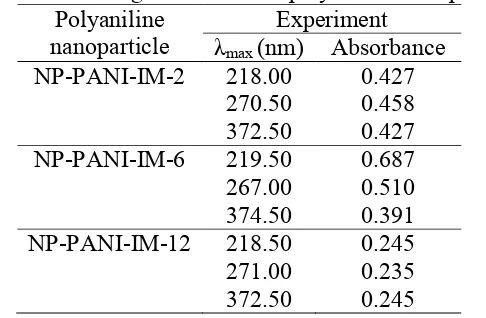
In general, the maximum wavelength of polyaniline nanoparticle recorded in three different peak absorptions. All these peaks similar to that previously reported for emeraldine salt with half and fully oxidized form[30]. The absorptions slightly shift to the lower wavelength (blue-shift). The effect of polar solvent such as ethanol commonly shifts the wavelength. The polyaniline nanoparticle synthesized from three different reaction time (PANI IM-2, IM-6, IM-12) give similar maximum peak adsorptions close to 218, 271, and 373 nm. The absorption in 218 nm correlate to * transition from benzenoid ring, adsorption in 271 nm due to * electronic transition of the doped the main chain, and absorption in 373 nm correlate to * electronic transition from the quinoid ring.
Table 2. Wavelength shift data of polyaniline nanoparticle Polyaniline
nanoparticle
Experiment
λmax (nm) Absorbance
NP-PANI-IM-2 218.00 0.427
270.50 0.458
372.50 0.427
NP-PANI-IM-6 219.50 0.687
267.00 0.510
374.50 0.391
NP-PANI-IM-12 218.50 0.245
271.00 0.235
372.50 0.245
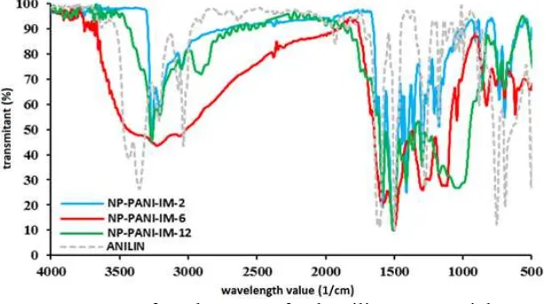
polyaniline nanoparticle (Figure 4). And the spectra agree to that previously reported[7,31]. Stretching vibration of the –N-H group is observed in 3220-3266 1/cm. As comparison, monomer aniline has amino group (-NH2) and observed as two absorption bands in 3350 and
3410 1/cm for vibration of N-H symmetry and asymmetric stretching. Conversely, two bands detected for direct coupling of nitrogen to carbon as C-N and C=N is observed in 1294-1298 cm/1 and 1582-1890 1/cm. These features indicate that polyaniline produced have structure with a half oxidized form, or as emeraldine salt (Table 3).
Figure 4. Infrared spectra of polyaniline nanoparticle
Table 3. Interpretation of NP-PANI-IM Infrared spectra
Functional Group Polyaniline Nanoparticle (NP PANI) Reference
IM-2 IM-6 IM-12
N–Hstretch 3265.26 (s) 3226.69 (s) 3262.26 (s) 3225a
=C-Hstretch 3053.11 (s) 3062.75 (w) 2906.53 (w) 3000a
C=C Quinoid 1583.45 (m) 1589.24 (m) 1589.24 (m) 1600a C=C Benzenoid 1510.16 (s) 1504,38 (s) 1504.38 (s) 1403 & 1500a
C-Nstretch 1298.00 (m) 1294.15 (m) 1298.00 (m) 1297b
N=Quinoid=N 1247.86 (w) 1247.86 (m) 1247.86 (w) 1119b C–C alkyl chain 1172.64 (w) 1149.50 (m) 1504.38 (m) 1100a
a
Ref. [31], bRef. [7]
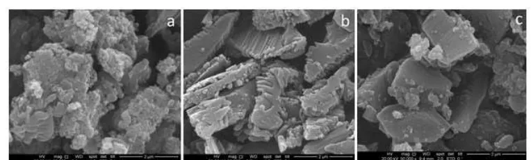
Figure 5. The morphology of NP-PANI-IM with 50.000 magnification using SEM (a) NP – PANI-IM 2 (b) NP-PANI-IM-6 (c) NP-PANI-IM-12
Table 4. The atomic composition percentage of polyaniline nanoparticle
Sample
Atomic Composition
C N
Wt% At% Wt% At%
NP-PANI-IM 2 45.63 52.26 08.04 07.89 NP-PANI-IM 6 64.92 69.28 22.93 20.98 NP-PANI-IM 12 66.33 70.78 19.75 18.07
Characterization of the polyaniline synthesized in different reaction time using SEM, confirms their surface morphology is nanoparticle (Figure 5). And also, the atomic composition analyzed using energy dispersion x-ray technique coupled to the SEM shows logical ratio of carbon and nitrogen atom composed the polyaniline (Table 4). At glance, the SEM image of polyaniline samples from three different reaction times; 2 h (NP-PANI-IM 2), 6 h (NP-PANI-IM 6), and 12 h (NP-PANI-IM 12) show uniformity structure. But, if the look into more detailed, the morphology structure of nanoparticle slightly different in size and shape. Some have a nanospheric and round shape with high rigidity. PANI-IM-6 and NP-PANI-IM-12 predominantly have a rectangle and rhomboidal shapes. This morphology has a low surface area. The other indicates a nanospheric and round shape. These shapes, then is measured and provide 50-63 nm for NP-PANI-IM-2, 68-71 nm for NP-PANI-IM-6, and 58-62 nm for NP-PANI-IM-12. This size is a slightly bigger than that previously reported[12]. In addition, the atomic compositions of polyaniline indicate proportional composition or ratio between carbon and nitrogen atom for three polyaniline samples. The product afforded in 2 h reaction has 52.26% of carbon and 7.89% nitrogen atom or equal to ratio 6 carbon atoms in benzenoid structure and one nitrogen atom attached to benzeneoid skeleton. However, the composition of carbon and nitrogen in polyaniline nanoparticle afforded in 6 and 12 h indicate increasing percentage (Table 4).
CONCLUSION
REFERENCES
[1] Hou, W., Xiao, Y., Han, G., Fu, D., Wu, R. J. Power Sources 2016, 322, 155–162. [2] Bera, A., Deb, K., Kathirvel, A., Bera, T., Thapa, R., Saha, B. Appl. Surf. Sci. 2016. [3] Ghushe, J. M., Giripunje, S. M., Kondawar, S. B. J. Inorg. Organomet. Polym. Mater.
2016, 26 (2), 370–375.
[4] Huang, J., Virji, S., Weiller, B. H., Kaner, R. B. J. Am. Chem. Soc. 2003, 125 (2), 314– 315.
[5] Nicolas-Debarnot, D., Poncin-Epaillard, F. Anal. Chim. Acta 2003, 475 (1), 1–15. [6] Xie, D., Jiang, Y., Pan, W., Li, D., Wu, Z., Li, Y. Sens. Actuators B Chem. 2002, 81
[14] Pradhita, M., Masruri, M., Rahman, M. F. Proc. 2015 IConSSE ISBN 978-602-1047-217 2016, 90.
[15] Masruri, M., Rahman, M. F., Ramadhan, B. N. IOP Publishing, 2016, Vol. 107, p 012060.
[16] Masruri, M., Amini, R. W., Rahman, M. F. Indones. J. Chem. 2016, 16 (1), 59–64. [17] Furukawa, Y., Ueda, F., Hyodo, Y., Harada, I., Nakajima, T., Kawagoe, T.
Macromolecules 1988, 21 (5), 1297–1305.
[18] Pouget, J., Jozefowicz, M., Epstein, A. e a1, Tang, X., MacDiarmid, A.
Macromolecules 1991, 24 (3), 779–789.
[19] Stejskal, J., Sapurina, I. Mater. Synth. 2008, 199–207.
[20] Das, T. K., Prusty, S. Polym.-Plast. Technol. Eng. 2012, 51 (14), 1487–1500. [21] Stejskal, J., Gilbert, R. Pure Appl. Chem. 2002, 74 (5), 857–867.
[22] Kumar, D., Sharma, R. Eur. Polym. J. 1998, 34 (8), 1053–1060. [23] Thiele, E. W. Ind. Eng. Chem. 1939, 31 (7), 916–920.
[24] Scirè, S., Fiorenza, R., Gulino, A., Cristaldi, A., Riccobene, P. M. Appl. Catal. Gen. 2016, 520, 82–91.