CHAPTER OUTLINE
“B
irds do it. Bees do it. Even educated
fleas do it. Let’s do it. Let’s fall in love.”
Songwriter Cole Porter got it right decades
ago, when he wrote this about sex. Okay, we’ll
admit that what he called “love” is actually
“sex-ual reproduction,” but you get the idea. There is
nothing new about sex, which plants have been
using as a means of ensuring reproductive
suc-cess for hundreds of millions of years. The need
to join gametes from two individuals traces
back to hundreds of millions of years ago in all
life forms, from fungi to flowering plants, from
bacteria to birds and bees and even humans.
Sexual reproduction has evolutionary
bene-fits: it speeds up the formation of new
geno-types (genetic configurations) that can be
tested against the environment. It also dilutes
or deletes harmful genes.
The urge to engage in sex is one of the
strongest human desires, ranking second only
to eating and breathing. Many biologists believe
this urge originates in evolution through natural
selection: without sex, we do not leave
descen-dants. The genes of people who have sex and
reproduce are found in the next generation,
and to the extent that reproduction is a genetic
urge, the mechanism is self-perpetuating.
Since reproduction is so critical to survival,
the urge needs to be managed; many of the
most common and critical human customs
con-cern reproduction: marriage, childbirth, and
family ties. In this chapter, we look at the
physi-ology and anatomy of reproduction, and
in-clude some scientifically based suggestions for
keeping the urge to reproduce in a healthy
framework.
510
The Reproductive
Systems:
Maintaining the Species
■ Survival of the Species Depends on Re-production and Gamete Formation p. 000
■ The Female Reproductive System Is Responsible for Housing and Nourishing the Developing Baby p. 000
Orgasm
Plateau
Excitement
1 2 3
R
esolu
tion R
esolu tio
n
Resolution Resolution
■ Structures of the Male Reproductive System Produce and Store Sperm p. 000
16
■ There Are Many Birth Control Choices, None of them Perfect p. 000
■ The Orgasm Is a Moment of Emotional and Physiological Epiphany p. 000
512 CHAPTER 16 The Reproductive Systems: Maintaining the Species Survival of the Species Depends on Reproduction and Gamete Formation 513 two to form a new individual. This mixes and blends
the allelesin the gene pool, creating new genetic
com-binations.
These new combinations are essential to the survival of the species. The genetic variation in popula-tions of sexually reproducing organisms is the basis for adaptation of organisms to their environment. Given enough variation, some individuals will always be better suited to the environment than others so that they can survive and pass on their genes. These “more fit” indi-viduals will produce more offspring, thereby increasing
Survival of the Species Depends on Reproduction
and
Gamete Formation
the percentage of their alleles in the gene pool. This line of reasoning is the underpinning for Charles Dar-win’s theor y of evolution through natural selection. The fittest organisms sur vive and pass their genes to the next generation (Figure 16.2).
In his now classic discussion, Darwin noted that the finches on the Galapagos Islands had beaks specifi-cally shaped to assist in eating the available food of that island. Some islands had large nuts and berries; those finches developed stronger, larger beaks. Other islands had grasses and thinner seeds; the finches on those is-lands developed delicate beaks able to pick the seeds from the grasses. In a recent press report, it has been shown that these finches’ beaks are still evolving. Just two decades after a competing finch with a large heavy beak arrived on one of the Galapagos Islands, the na-tive finch evolved a smaller, thinner beak to take advan-tage of a food source unavailable to the newcomer. As these species compete for food, they apparently can and do undergo descent with modification, or evolution.
Passing on your genes requires you to form haploid gametes. Gamete is a general term for the re-ender is an obvious structural and
func-tional difference between people. We are either male or female. We all know that the female produces eggs, and her anatomy is set up to house and nourish the developing baby. And we know that the male produces sperm, and his anatomy is designed to deliver that sperm to the egg. Because we rely on sexual reproduction, having two genders is necessar y to perpetuate the species (Figure 16.1).
Aside from the obvious anatomical differences, are there any homeostatic differences between men and women? Are we so different as to verify the flippant pronouncement “men are from Mars, women are from Venus?” Are we worlds apart just because of a difference in one chromosome? To answer these questions, we will start by looking at reproduction in general, and then at male and female anatomy. We will ex-plore hormonal differences, and finally, armed with this knowledge, we will explore birth control methods that help us to control when we re-produce.
The main purpose of the reproductive system is to produce haploid gametes (egg and sperm) and unite them to form a new individ-ual. Sexual reproduction in-volves choosing a mate based
on phenotype and mixing
and shuffling genes from the LEARNINGOBJECTIVES
Explainthe functions of the reproductive system. Placesexual reproduction in the context of the theory of evolution.
G
Haploid
Having half the number of chromo-somes of normal body cells, found in eggs and sperm.
Phenotype
An organism’s observable characteristics.
Alleles
Genes found on the same spot on the same chromosome in different individu-als, coding for sub-tle variations of the same protein.
Man and woman Figure 16.1
Sexual reproduction requires two sexes. Humans are dioecious, meaning the male and female reproductive organs are carried on different individuals. In contrast, earthworms and many plants are monoecious. One organism carries both male and female reproductive organs.
Natural selection: Darwin’s finches
Figure 16.2
In observing the finches living on the Galapagos Islands, Darwin noticed that there were subtle variations in beak shape. He then observed that these variations correlated to the type of food available on each island. His conclusion was that those birds better able to eat the available food dominated the bird population of each island. These successful birds were able to add their alleles to the population at a faster rate than those birds with less successful beak shapes.
productive cells that will form a new individual, the egg and sperm. These are produced via meiosis, a specialized type of cell division that ensures the equal and orderly division of chromosomes (Figure 16.3, p. 514). In order to
form gametes properly, the normally diploid chromo-some number must be cut in half, with the resulting ga-metes having exactly half the usual complement of alleles.
This way, when two haploid gametes unite to form a zy-gote, the original diploid number is restored. The divi-sion must be accomplished so that each gamete has a predictable and reliable half of the chromosomes. Rather than randomly splitting the chromosome,
ho-mologouschromosomes come together and are then
separated, one to each new gamete.
In the male, meiosis occurs exactly as depicted here, and four sperm are produced from two divisions
Diploid
Having the total number of chromo-somes of the body cells, twice that of the gametes.
Homologous
Similar in structure, function, or
of a primary spermatocyte. Females produce only one egg from each round of meiosis, investing almost all of the cytoplasm and organelles in one gamete. The extra genetic material that is split out at anaphase I and anaphase II is ejected from the developing egg with very little associated cytoplasm. These tiny capsules of DNA are called polar bodies. They are not viable, and they are quickly degraded in the female system.
Forming gametes is only one function of the re-productive system. The male and female gametes must be united in a protected environment, and the result-ing embryo needs to be nourished and protected as it
develops. In addition, the reproductive system must trigger puberty, maintain reproductive ability, stimulate secondary sex characteristics, and produce hormones involved in sexual maturation and general homeostasis. Both the male and female reproductive systems are composed of gonads, ducts, and accessory glands.
Gonadsare the organs that produce gametes. Ducts
transport the gametes and any fertilized egg that is pre-sent. Accessory glandsfacilitate gamete production and survival. Although all three components are found in both men and women, their structures and functions dif-fer with gender, so we’ll take up each gender separately.
514 CHAPTER 16 The Reproductive Systems: Maintaining the Species Structures of the Male Reproductive System Produce and Store Sperm 515 Chromosome
Sister chromatids
PROPHASE I Tetrads formed by synapsis of sister chromatids of homo-logous chromosomes
Crossing-over between nonsister chromatids
Kinetochore microtubule
Metaphase plate
Cleavage furrow Pairing of homologous chromosomes
METAPHASE I
Separation of homologous chromosomes ANAPHASE I
TELOPHASE I PROPHASE II
METAPHASE II
ANAPHASE II
TELOPHASE II MEIOSIS II MEIOSIS I
Stages of meiosis Synapsis of sister
chromatids
Crossing-over between nonsister chromatids
Genetic recombination Details of crossing-over during prophase I
A A
B B B B b B B b
a a b b
a a
G G g g G G
A a A a
G G g g g g
b b
A A
B A
Meiosis Figure 16.3
Meiosis is the orderly distribution of genetic material to newly formed haploid gametes. It includes steps very similar to those of mitosis, the main difference being the formation of tetrads in prophase I. These tetrads are pairs of homologous chromosomes that remain close to one another until they are pulled apart in anaphase I. Crossing over offers even more genetic variation, as the ends of these chromosomes are close enough to swap material. Telophase I then forms two “cells” that enclose doubled copies of half the chromosomes of the original diploid cell. The newly formed cell then immediately goes into prophase II , metaphase II, anaphase II and prophase II. These phases operate exactly the same way as those in mitosis, resulting this time in four haploid cells.
he function of the male reproductive system is to produce sperm and deliver it to the female reproductive system. This requires a gonad to produce sperm, some tubes to carry the sperm, and three types of accessor y glands to produce fluid to sustain the sperm (Figure 16.4, page 516).
STRUCTURES OF THE MALE
REPRODUCTIVE SYSTEM
The male reproductive system is essentially one long tube, with sperm generated in the gonads at one end, matured along the route, and released from the body at the other. Accessory glands add secretions to nourish and carry the sperm before it is released from the body. Sperm is produced in the testes. These paired organs are suspended in the scrotal sac, where their in-ternal temperature can be regulated with ease. Viable sperm can only be produced at temperatures 2 to 3 de-grees C below normal body temperature. The cremas-terand dartosmuscles of the scrotal sac move the testes
CONCEPT CHECK
How
does the production of haploid gametes help ensure survival of the species?How
does meiosis differ from mitosis? How does it produce haploid gametes?Structures of the Male Reproductive System
Produce and
Store Sperm
LEARNINGOBJECTIVES
Tracethe pathway of sperm through the male.
Describesperm production.
Outlinehormonal controls in the male.
T
Structures of the Male Reproductive System Produce and Store Sperm 517 516 CHAPTER 16 The Reproductive Systems: Maintaining the Species
to regulate their temperature. These muscles contract when the temperature drops. This elevates the testes, bringing them closer to the body and maintaining the required temperature by allowing the testes to absorb heat from the body. When the temperature within the testes rises, the muscles relax and the testes move away from the body, reducing their internal temperature.
The male reproductive organs usually begin de-velopment seven weeks after conception, forming from the embryonic mesonephros duct. By seven months af-ter conception, the testes migrate from their position in the abdominal cavity to the scrotal sac through the inguinal canal, dragging their associated vessels, nerves, lymph, and reproductive cords with them (Figure 16.5). Their path leaves a weak spot in the abdominal
wall, which can lead to a hernia later in life. A hernia is
a rupture of the abdominal wall accompanied by the protrusion of internal organs, usually the small intes-tine. Hernias often require surgery to reposition the protruding organs and close the hole.
This “descending” of the testes is vital to repro-ductive health. Recall that production of viable sperm requires a temperature three degrees C below body temperature. If the testes do not descend, the seminif-erous tubules of the testes will be too warm for sperm creation. Additionally, when the testes remain in the body cavity, they are far more prone to testicular cancer. In 3 percent of full-term male births and a full 30 percent of premature male births, the testes have yet to descend. The medical term for this condition is cryp-torchidism, literally “hidden orchid.” The male will be sterile if both testes remain in the body cavity, a
condi-Sagittal plane
Sacrum
Seminal vesicle
Ampulla of ductus (vas) deferens Coccyx
Ejaculatory duct Rectum
Urethra
Membranous urethra Anus
Bulbourethral (Cowper’s) gland
Bulb of penis
Epididymis
Testis
Scrotum
Sagittal section
Urinary bladder
Ductus (vas) deferens
Pubic symphysis
Corpora cavernosum penis Spongy (penile) urethra Penis
Corpus spongiosum penis
Glans penis Prepuce (foreskin) Prostate
External urethral orifice
The male reproductive system Figure 16.4
ulated to begin producing sperm. They first divide into spermatogonia. Spermatogonia in the walls of the semi-niferous tubules divide, forming primary spermato-cytes. As these cells continue to divide, they are pushed farther from the wall of the tubule into the lumen, where they become secondary spermatocytes and then
spermatids.
During this stage of development, the cells be-come progressively less like the cells of the male body and more like a separate entity. Eventually they become so different that these spermatids need protection from the immune system, which would otherwise destroy them as foreign cells. The Sertoli cells extend from the basement membrane of the seminiferous tubule all the way to the lumen. Their job is to isolate the developing sperm from the male blood supply, as protection against immune attack. The only cells of the seminiferous tubule
Spermatic cord
Cremaster muscle Inguinal canal
Ductus (vas) deferens Autonomic nerve
Testicular artery
Lymphatic vessel
Epididymis
Tunica albuginea of testis
Tunica vaginalis (peritoneum)
Anterior view of scrotum and testes and transverse section of penis Transverse section of penis:
Corpora cavernosa penis
Spongy (penile) urethra Corpus spongiosum penis
Scrotal septum
Cremaster muscle
Dartos muscle
Skin of scrotum
The scrotum, the supporting structure for the testes Figure 16.5
tion called bilateral cryptorchidism. Luckily, among ap-proximately 80 percent of cryptorchid males, the testes naturally descend within the first year. If they do not de-scend by 18 months, surgery is needed.
In normal development, each testis carries out
spermatogenesisindependently within the individual
pouches of the scrotal sac. The testes are actually a densely packed mass of seminiferous tubules, which are contained in 200 to 300 lobules within each testis. An individual lobule holds up to three tubules, providing a large number of seminiferous tubules per testis (
Fig-ure 16.6, page 518).
Within the seminifer-ous tubules are two types of cell: spermatogeniccells and
Sertolicells. At puberty, the spermatogenic cells are
stim-Spermatogenesis
Structures of the Male Reproductive System Produce and Store Sperm 519 518 CHAPTER 16 The Reproductive Systems: Maintaining the Species
Testes histology Figure 16.6
LM 270x
Spermatid (n)
Secondary spermatocyte (n)
Primary spermatocyte (2n)
Spermatogonium (2n) (stem cell)
Basement membrane
Leydig cell Sertoli cell
Transverse section of several seminiferous tubules
Transverse section of a portion of a seminiferous tubule
SPERMATOGENIC CELLS:
Sperm cell or spermatozoon (n) Late spermatid (n)
Blood–testis barrier (tight junction) Sertoli cell nucleus Basement membrane Blood capillary Leydig cell
Early spermatid (n) Secondary spermatocyte (n) Primary spermatocyte (2n) Spermatogonium (2n) (stem cell)
Lumen of seminiferous tubule
A
B Transverse plane
that do not require protection are the spermatogonia. These cells are identical to those of the male body, so they are in no danger of attack.
Beyond protecting the developing sperm, the Sertoli cells also assist in their survival. They provide nourishment for the developing sperm, assist in the fi-nal maturation of sperm by removing excess cytoplasm, control the release of sperm into the seminiferous tubule lumen, and mediate the effects of the hormones testosterone and inhibin.
One final type of cell in the testes occurs out-side the seminiferous tubules, between them in the lob-ules. The Leydigcells, or interstitial endocrinocytes, produce the hormone testosterone. Testosterone stim-ulates spermatogonia to produce sperm; stimstim-ulates bone growth; increases hair production on the arms, legs, underarms, chest, groin, and face; stimulates carti-lage growth of the larynx (thereby lowering the voice;, and increases libido. In short, testosterone from the Leydig cells turns the adolescent male into a fully re-productive man.
SPERMATOGENESIS IS THE PROCESS
OF SPERM FORMATION
The process of making and maturing a spermatozoon takes 65 to 75 days. It begins with the spermatogonia, which are stem cells. When the spermatogonia divide, one cell remains in contact with the basement mem-brane as a spermatogonium and the other moves to-ward the lumen to begin the process of spermatogene-sis. This second cell moves into a Sertoli cell and transforms into a primary spermatocyte. Both primary spermatocytes and spermatogonia are diploid cells. Once safely protected by the Sertoli cells, meiosis can occur. At the end of Meiosis I, two secondary spermato-cytes are formed. Each one has 23 chromosomes, but each chromosome is doubled. This results in a haploid number of alleles but a diploid number of actual chro-mosomes. Rather than 46 different chromosomes, there are two identical copies of 23 chromosomes held together by a centromere.
As meiosis proceeds, each secondary spermatocyte di-vides further to produce two
haploid spermatids. This yields a total of four haploid cells carry-ing the DNA of a sperm, but without the characteristic shape of the sperm cell. During the
process of spermiogenesis, these spermatids are slowly ejected from the Sertoli cell as they mature. When the sperm are free of the seminiferous tubule, they are called spermatozoa and are fully formed, if not yet
capacitated(Figure 16.7).
Some spermatogonia remain as precursor stem cells Basement membrane
of seminiferous tubule Superficial
Spermatogonium
Mitosis Some spermatogonia
pushed away from basement membrane
Differentiation
Primary spermatocyte
DNA replication, tetrad formation, and crossing-over
Secondary spermatocytes
Meiosis II
Spermatids
Spermatozoa Meiosis I
2n 2n
2n
n n
n n n n
n n n n
Lumen of seminiferous tubule Each chromosome has two chromatids
Deep MEIOSIS
SPERMIOGENESIS
2n
Sperm formation Figure 16.7
Spermatogonia undergo mitosis and produce two cells: one cell that migrates into the center of the seminiferous tubule becoming a primary spermatocyte, and a second one that remains on the periphery. The primary spermatocyte then undergoes meiosis I, producing two secondary spermatocytes. These spermatocytes go on to complete meiosis, producing a total of four spermatids. Once these spermatids undergo spermiogenesis, four functional sperm are produced.
Capacitated
Activated, i.e., capable of fertilizing an ovum.
Stem cell
A less differentiated cell that can give rise to a specialized cell.
Acrosome
Nucleus
Mitochondria Neck
Middle piece Centriole
Principal piece
HEAD
TAIL
End piece
Structures of the Male Reproductive System Produce and Store Sperm 521 520 CHAPTER 16 The Reproductive Systems: Maintaining the Species
Sperm Figure 16.8
A normal male produces about 300 million sperm (Figure 16.8) per day from puberty until
death. The sperm exist to reach and penetrate an egg, and each part of the sperm has a role in meeting this goal. The head of the sperm includes the acrosomeand the nucleus. The acrosome is a vesicle on the point of the sperm head that contains digestive enzymes. These will be useful when the sperm encounters the egg, as it can digest the oocytemembrane, allowing the nucleus of the sperm to penetrate. The mid-piece of the sperm contains many mitochondria, able to produce the ATP needed to reach the egg. The tail of the sperm consists of one long flagellum. The sperm is the only human body cell with a flagellum and is one of few human cells that must propel itself from its place of origin in order to perform its function.
THE EPIDIDYMIS STORES AND
MATURES NEWLY DEVELOPED SPERM
Once sperm is produced in the seminiferous tubules, it must be transported from the male to the female. This requires a series of ducts through which the sperm will pass. Semen, the fluid containing sperm and other ions, is formed as the sperm traverses these ducts.
The Sertoli cells create a fluid that fills the sem-iniferous tubule lumen and pushes the developing sper-matozoa along. The spersper-matozoa leave the seminifer-ous tubules in the lobules of the testes and travel through the straight tubules to the rete testes before leaving this organ. This network of testicular tubules ends at the epididymis.
The function of the epididymis is similar to that of the aging cellar at a winery. It serves as a storage area and final maturation center for the spermatozoa, just as the casks at a winery serve as a suitable environment for the young wine to age and mature before being sold. Spermatozoa reach final form
in the epididymis, losing that last bit of excess cytoplasm while gaining mobility and the ability to fertilize an ovum. This process takes about 10 to
14 days. Spermatozoa can remain quiescentin the epi-didymis for approximately one month. If an ejaculation event occurs, the walls of the epididymis aid in pro-pelling the sperm forward. A small peristaltic wave is generated in the smooth muscle of the wall, helping to push the spermatozoa into the next tube in the system, the ductus deferens(vas deferens).
The ductus deferens transports and stores
sperm
The ductus deferens runs approximately 50 centimeters from the scrotal sac through the inguinalcanal, looping over the ureter
and posterior to the urinar y bladder. It arises from the tail of the epididymis, where the epididymal tube expands in diameter. As the ductus defer-ens enters the abdominal wall, it sits at the anterior of the
spermatic cord, readily
acces-sible immediately beneath the skin of the scrotum. The length of the ductus deferens is used to store sperm for as long as several months. If there is no ejaculation dur-ing that time, the sperm are broken down and reab-sorbed. The placement of the vas deferens and its func-tion as a sperm transport vessel make it a prime candidate for sterilization surgery. You may have heard of a vasectomy; the term literally means “cutting the vas deferens.” (This procedure is explained in the section on birth control near the end of this chapter.)
Unlike the epididymis, the walls of the ductus deferens have a thick layer of smooth muscle to propel the sperm toward the urethra during ejaculation. The terminal end of the ductus deferens swells slightly as it runs down the back of the urinary bladder. This swelling is called the ampulla(FIGURE 16.9, page 522).
Seminal vesicles help nourish the sperm
After the ampulla, glands add fluid to the developing semen as it moves through the male system during ejac-ulation. The paired seminal vesicles, located on the posterior base of the urinary bladder, are the first of these glands. They secrete an alkaline, fructose-rich fluid that ser ves two purposes. The high pH helps to neutralize the potentially lethal, acidic environment of the male urethra and the female reproductive tract. The fructose serves as an energy source for the sperm as they become motile. Prostaglandinsare also released by the seminal vesicles. Prostaglandins have many physi-ological effects. They open airways, stimulate the sensa-tion of pain, reduce stomach acid producsensa-tion, and cause local irritation. Prostaglandins also seem to stim-ulate sperm motility. A final important component of seminal vesicle fluid is a clotting factor, which may be responsible for the coagulation of semen after ejacula-tion. In all, the seminal vesicles secrete approximately 60 percent of the total ejaculate volume.The ejaculatory duct runs through the
prostate gland
After the seminal vesicles, sperm travels along the short ejaculatory duct to the prostatic urethra. The union of the ampulla of the ductus defer-ens and the duct from the seminal vesicle marks the be-ginning of the ejaculatory duct. Semen does not reach this duct except during ejaculation. During an ejacula-tion, sperm and semen are forcefully pushed into theprostatic urethra, causing the prostate gland to add secretions.
The prostate gland is a golf-ball-sized gland ly-ing immediately at the base of the bladder. It com-pletely surrounds the uppermost portion of the ure-thra, secreting a milky fluid into the passing semen. This fluid includes citric acid for ATP production, pro-teolytic enzymes to break up the clot formed by the seminal vesicle secretions, and acid phosphatase, whose function is unclear. Another 25 percent of the semen volume comes from this gland.
Physiology of the male orgasm
Directed by the sympathetic nervous system, the male orgasm pro-pels sperm from the epididymis through the ductus def-erens, the ejaculatory duct and the urethra, releasing it from the male body. As sperm enter the ejaculator y duct from the ductus deferens, the prostate and bul-bourethral glands add their secretions, creating semen. Rhythmic, reflexive contractions of pelvic muscles cause the semen to be released from the penis in short bursts. During this reflex, the sphincter at the base of the urinary bladder closes, preventing urine from en-tering the urethra and sperm from enen-tering the uri-nary bladder.The total ejaculate released during orgasm is usually 2.5 to 5 ml. On average there are between 50 and 150 million sperm per ml, for a total of over 350 million sperm per ejaculate. If the sperm count drops below 20 million per ml, the male is said to be infertile. This number is usually too low to fertilize an egg be-cause so few of the ejaculated sperm reach the ovum.
Usually, a slight emission precedes ejaculation, as a peristaltic wave passes through the ejaculatory duct, ductus deferens, seminal vesicles, and prostate. The emission cleanses the urethra, removing potentially harmful cr ystals that might impair sperm function. While most ejaculations occur during the stimulation of sex, men can also experience “nocturnal emissions,” or ejaculations during sleep. These are normal, and may or may not be associated with sexually arousing dreams. We will return to orgasm later in this chapter.
The urethra travels the length of the penis
Once through the prostatic urethra, the semen trav-els the rest of the urethra. Immediately upon leavingQuiescent
Resting, quiet, inactive.
Spermatic cord
The artery, vein, nerve, lymphatics, and ductus defer-ens that lead from the abdominal cav-ity to the testes.
Oocyte
Structures of the Male Reproductive System Produce and Store Sperm 523 522 CHAPTER 16 The Reproductive Systems: Maintaining the Species
the prostate, the urethra dives through the urogenital
diaphragm (see F i g u r e 1 6 . 9). It then continues
the length of the penis, through the spongy (or pe-nile) urethra, and to the ex-ternal urethral orifice.
Where the urethra enters the spongy tissue of the penis, a final set of glands adds fluid to the ejacu-late. The bulbourethralglands lie on either side of the urethra, at the bulb, or base, of the penis. The glands are obviously named for their location. They secrete an
alkaline, mucous fluid into the urethra prior to sperm arrival that protects the sperm from the normally acidic urethra. The mucus of these glands lubricates the tip of the penis and the urethral lining, preventing damage to the sperm as they travel the final portion of the male reproductive system. Refer to figure 16.9for the
lo-cation of these structures.
The penis itself is a passageway for both urine and semen. The rootof the penis lies at the base of the prostate gland. The body of the penis contains three cylinders of erectile tissue. The two corpora cavernosa
lie on the dorsolateral surfaces, and the single corpus spongiosumencircles the urethra and expands at the
tip of the penis to form the glans. These three tissues contain numerous blood sinuses.
During arousal, the arteries that feed these tis-sues dilate under the influence of cyclic guanine monophosphate (cGMP). cGMP allows more blood to enter the erectile tissue, filling the sinuses and com-pressing the veins. This combi-nation results in an erection, an enlarged and stiffened pe-nis. This process is reversed by constriction of the arteries, in turn lessening the pressure on the veins and allowing blood to drain from the tissues. Via-gra® inhibits enzymes that naturally break down cGMP, thereby prolonging erections. cGMP is active in other processes in the body as well, for example in processing visual and olfactory information, memory, and learn-ing. The cGMP inhibitors in Viagra are extremely spe-cific; otherwise the drug would have negative side ef-fects on memory and vision.
When boys are born, the tip of the penis is cov-ered with a protective layer of skin, called the foreskin. Removal of this tissue does not seem to alter male func-tioning, nor does it have any clearly demonstrated posi-tive physiological effects, except that removal has been shown to reduce the rate of infection by the AIDS virus. Regardless, male circumcision continues to be prac-ticed in the United States and other countries around the world. The procedure involves the rapid removal of the entire foreskin of the penis, usually within the first few days of life.
The histor y of circumcision begins in East Africa long before biblical accounts. The practice was used to purify men and reduce sexuality and sexual pleasure. Jews, and later Muslims, adopted the practice, and it continues to this day. In the first century, Chris-tians strongly opposed circumcision. Not until 1870 did the medical practice of circumcision begin in the United States. Once medical professionals embraced the procedure, it became safer and more accepted. Al-though medical circumcisions were performed without question, no scientific studies of its benefits or con-traindications were undertaken. In 1949, Dr. Douglas Garnier wrote an article denouncing circumcision, ex-plaining that there seemed to be no medical reason for
the operation and that in fact it was causing unneces-sary deaths. As a result, the number of circumcisions dropped dramatically in England, but the United States was slower in questioning the procedure. In 1971, an-other article stated that there was no medical reason for circumcision, despite common opinion that a cir-cumcised penis is somehow “cleaner” or less likely to become infected. Since that time, the practice has slowly declined in the United States. Neonatal circum-cision was performed in only 60 percent of male births in 1996 and declined further to 55 percent in 2001.
In 2006, however, a number of studies showed that the foreskin may transmit AIDS during sex. A study in Uganda, for example, found a 30 percent reduction in disease transmission among circumcised men. Re-searchers reported that the reduction may be due to the fact that HIV binds strongly to the foreskin. Similarly, a 2004 study from India found that circumcised men were six times less likely to acquire HIV during sex.
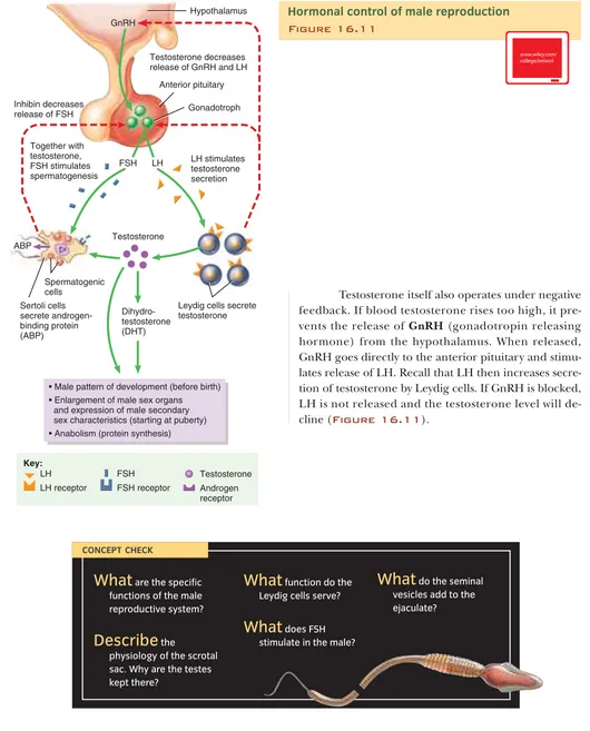
HORMONAL CONTROL OF THE MALE
Although it is true that males produce sperm endlessly from puberty until death, male hormones exert control over the rate of sperm production and the secretion of testosterone, which control male secondary sex charac-teristics. Lying deep in the brain, protected by the sphe-noid and attached to the brain by the hypothalamus, is the pituitar y gland. The anterior pituitar y gland se-cretes luteinizing hormoneand follicle-stimulating hor-mone, which are instrumental in governing the male reproductive system. The secretion of these hormones is governed by gonadotropin releasing factors pro-duced by the hypothalamus.
The names of these two hormones reflect their roles in the female, not male, system. When reproduc-tion was originally studied, it was assumed that only fe-males exhibited hormonal controls. Therefore, these anterior pituitar y hormones were first isolated and their functions were identified in females. Follicle stim-ulating hormone (FSH) stimulates immature oocyte (egg) follicles in the female ovar y. Luteinizing hor-mone stimulates the production of a yellow body; leutein roughly translates to yellow. After an oocyte is ovulated, a yellow body remains on the ovar y, hence
cGMP
Cyclic guanine monophosphate, an energy molecule.
Urogenital
Concerning both the urinary system and the reproduc-tive system.
ROOT OF PENIS:
BODY OF PENIS:
Transverse plane
Frontal plane
Urinary bladder Internal urethral orifice
Prostate
Orifice of ejaculatory duct Membranous urethra Bulbourethral (Cowper’s) gland
Bulb of penis Crus of penis
Corpus spongiosum penis
Corpora cavernosa penis Deep
dorsal vein
Superficial (subcutaneous) dorsal vein
Superficial fascia
Deep fascia Skin
Dorsal artery
Dorsal
Ventral
Transverse section Deep muscles of perineum
Corpora cavernosa penis
Tunica albuginea of corpora cavernosum Deep artery of penis
Corpus spongiosum penis
Spongy (penile) urethra Tunica albuginea of corpus spongiosum penis
Corona
GLANS PENIS
Prepuce (foreskin)
External urethral orifice
Frontal section Spongy (penile) urethra
Prostatic urethra
A B
Testosterone itself also operates under negative feedback. If blood testosterone rises too high, it pre-vents the release of GnRH(gonadotropin releasing hormone) from the hypothalamus. When released, GnRH goes directly to the anterior pituitary and stimu-lates release of LH. Recall that LH then increases secre-tion of testosterone by Leydig cells. If GnRH is blocked, LH is not released and the testosterone level will de-cline (Figure 16.11).
Structures of the Male Reproductive System Produce and Store Sperm 525
the name of the hormone responsible for ovulation. It came as a bit of a shock when scientists later discovered that the male pituitar y secretes the same hormones, with subtly different effects.
In the male, luteinizing hormone (LH) stimu-lates the Leydig cells, causing the release of testos-terone. For this reason, it is also called interstitial cell stimulating hormone (ICSH). The production of testos-terone is governed by a typical negative feedback loop. As more testosterone is produced, its levels increase, in-hibiting production of LH at the pituitary gland. In this way, the hormones testosterone and LH balance one another.
The functions of testosterone include stimula-tion of male patterns of development in utero, enlarge-ment of male sex organs dur-ing puberty, development of male secondary sex character-istics, development of sexual function, and stimulation of
anabolism.
Secondar y male sex characteristics are those asso-ciated with puberty: growth of skeleton and musculature; ap-pearance of body and facial hair; cartilaginous growth of the ears, nose, and lar ynx; thickening of the skin, and increased oil secretion in the skin.
Some tissues of the male convert testosterone to dihydrotestosterone, or DHT. You may have heard this compound being blamed for male pattern baldness on Web sites or television infomercials, which make it sound as if everybody’s hair will fall out if DHT concen-tration exceeds a certain level. In truth, male pattern baldness is due to varying sensitivity of hair follicles to circulating DHT. Hair follicles can produce the enzyme 5-alpha reductase, which converts circulating testos-terone into dihydrotestostestos-terone (DHT). This raises the concentration of DHT around these follicles, and DHT gets picked up by follicles that have many DHT recep-tors. A DHT-activated hair follicle has a shorter growing stage and a longer “resting” phase, and will produce a wispy hair. DHT also constricts blood vessels to the folli-cle, starving the hair of nutrition. All these factors even-tually cause the hair to fall out, and any replacement hair will be thin and slow-growing. This extreme
reac-tion to DHT is genetic, as are the patterns and num-bers of susceptible hair follicles on the head. Because factors that increase the likelihood of developing male pattern baldness are carried on the X (female) chromosome, it is considered a sex-related trait ( Fig-ure 16.10).
FSH is secreted by both the female and the male anterior pituitar y gland. In the male, where oocyte follicles are absent, FSH indirectly stimulates spermatogenesis. FSH and testosterone together cause the Sertoli cells to secrete androgen-binding protein
(ABP). ABP moves to the interstitial spaces of the testes, binding available testosterone and maintaining it in high concentration near the seminiferous tubules. Testosterone stimulates the final production of sper-matids. When the Sertoli cells are functioning to capac-ity to protect developing sperm, they secrete inhibin. This hormone inhibits FSH production from the ante-rior pituitary, slowing sperm production. In essence, the Sertoli cells are claiming that they are “full” and cannot protect any more developing sperm. In typical negative feedback, if sperm production slows too much the process reverses. The Sertoli cells no longer release inhibin, the anterior pituitary increases production of FSH, and sperm production rises.
524 CHAPTER 16 The Reproductive Systems: Maintaining the Species
[image:8.1440.804.1334.107.764.2]Male pattern baldness is hereditary
Figure 16.10
GnRH
Sertoli cells secrete androgen-binding protein (ABP)
Anterior pituitary
Inhibin decreases release of FSH
Testosterone decreases release of GnRH and LH Hypothalamus
LH stimulates testosterone secretion
Testosterone
Dihydro-testosterone (DHT)
• Male pattern of development (before birth) • Enlargement of male sex organs
and expression of male secondary sex characteristics (starting at puberty) • Anabolism (protein synthesis)
LH receptor FSH receptor Androgen receptor Gonadotroph
FSH LH
LH FSH Testosterone
Spermatogenic cells
ABP
Together with testosterone, FSH stimulates spermatogenesis
Key:
Leydig cells secrete testosterone
[image:8.1440.385.717.494.744.2]Hormonal control of male reproduction
Figure 16.11
Anabolism
The building up of larger molecules from smaller ones (contrast to catabolism).
CONCEPT CHECK
What
are the specific functions of the male reproductive system?Describe
thephysiology of the scrotal sac. Why are the testes kept there?
What
function do the Leydig cells serve?What
does FSH stimulate in the male?What
do the seminal vesicles add to the ejaculate?526 CHAPTER 16 The Reproductive Systems: Maintaining the Species The Female Reproductive System is Responsible for Housing and Nourishing the Developing Baby 527
The Female Reproductive System
is Responsible for Housing and Nourishing
the
Developing Baby
f the purpose of the male reproductive system is to deliver sperm, one purpose of the female reproductive system must be to receive sperm. But the female sys-tem must also produce eggs (or ova) for fertilization, provide an area for the fertilized egg to develop into a fully developed fetus, and give birth. Like the male
re-productive system, the female system also produces hor-mones that cause sexual maturity and stimulate the de-velopment of secondary sex characteristics.
The organs of the female reproductive system include the paired ovaries, the fallopian or uterine tubesleading from the ovaries to the uterus, the uterus
itself, and the vagina(Figure 16.12). Accessory
or-LEARNINGOBJECTIVES
Listthe functions of each female reproductive organ. Explainoogenesis. Describethe female hormonal cycles.
I
Sagittal section Sacrum
Coccyx
Rectum
Anus Vagina
Uterine (fallopian) tube
Fimbriae
Ovary
Urinary bladder
Pubic symphysis
Mons pubis
Clitoris
Urethra
Labium majus
Labium minus External urethral orifice Uterus
Round ligament of uterus
Cervix Sagittal
plane
The female reproductive system Figure 16.12
gans of the female system are fewer than the male, represented mainly by the mammaryglands and the external female genitalia.
While the anatomy of the fe-male reproductive system is simpler than that of the male, the hormonal control of the female system is far more complex. This is because two in-teracting hormonal cycles occur si-multaneously in the female. The ante-rior pituitary gland secretes FHS and LH, affecting the ovary, and the ovary then responds with the hormones es-trogenand progesteronethat affect the uterus. Ovarian hormones can in-hibit the anterior pituitary gland, pro-viding feedback control.
OVARIES ARE
RESPONSIBLE FOR
OOGENESIS—EGG
FORMATION
The ovaries are small, almond-shaped organs that lie in the pelvic cavity. They arise from the same embryonic tissue as the testes, making these
or-gans homologous. Similar to the testes, the ovaries pro-duce both gametes (ova) and hormones (estrogensand
progesterone). Oogenesisoccurs via meiosis but, un-like spermatogenesis, produces only one viable ovum per meiotic event (Figure 16.13).
Also unlike the production of sperm, oogenesis begins before the female is born, so that at birth the ovaries already contain all of the ova she will produce in her life (Figure 16.14, page 528). The ova are
found in the ovarian germinal epithelium, surrounded by the ovarian cortex. They wait there, suspended in early meiosis, until they receive hor-monal signals to continue de-velopment. At birth, each
ovary may contain from 200,000 to 2 million such cells. These primary oocytesundergo atresia, so that by pu-berty approximately 40,000 remain. Only 400 or so of these will actually mature to the point of ovulation dur-ing a woman’s reproductive lifetime.
Each primar y oocyte sits in the center of a group of follicular cells, which are stimulated to de-velop alongside the oocyte. A primary folliclehas one to seven layers of follicular cells surrounding the oocyte. These follicular cells produce the zona pellu-cida, a clear gel-like layer that surrounds the maturing oocyte. The innermost layer of follicular cells becomes attached to the zona pellucida, resembling a circular crown. These cells become the corona radiataof the oocyte.
Oogenesis
Figure 16.13Pr
ocess
Diagr
am
2n
2n
n n
n n
n
2n n n
n n
Primary oocyte
Secondary oocyte
Oogonium
First polar body
Second polar body
Zygote Fertilization
Ovulation
Secondary oocyte +
Sperm
cell Meiosis II Meiosis I
The nuclei of the sperm cell and the ovum unite, forming a diploid (2n) zygote. During fetal development meiosis I begins.
After puberty, primary oocytes complete meiosis I, which produces a secondary oocyte and a first polar body.
The secondary oocyte begins meiosis II.
A secondary oocyte (and first polar body) is ovulated.
After fertilization, meiosis II resumes. The oocyte splits into an ovum and a second polar body.
Ovum
Atresia
Reabsorption of immature ova prior to birth.
The Female Reproductive System is Responsible for Housing and Nourishing the Developing Baby 529
ovulation. The ends of these tubes fill with blood, dis-tend, and sway, creating small currents in the ab-dominopelvic fluid, in turn drawing the newly ovulated oocyte into the uterine tubes. Once collected in the uterine tube, ciliated epithelia lining the tube help wash the oocyte (or developing zygote if fertilization occurs) into the uterus. Smoking can inhibit the movement of the cilia of the uterine tube; this is one reason women who smoke have difficulty conceiving.
Because the oocyte is only viable for approxi-mately 24 hours, fertilization must occur within 24 hours of ovulation. Usually the egg can travel only the upper one-third of the uterine tubes during this time, meaning that if fertilization does occur, it will happen there. Sperm introduced into the female system travel up through the uterus and into the uterine tubes, while from the other direction, the oocyte is collected and swept into the uterine tube. The oocyte takes six to
seven days to reach the uterus itself, during which time it begins to degenerate unless fertilized.
The uterus is the site of development
The uterus is the womb where fetal development occurs. This organ has an outer covering, the perimetrium, a middle layer of smooth muscle, the myometrium, and an inner endometrium(Figure 16.15). Theen-dometrial lining thickens and sheds every 28 days or so in response to hormone levels, resulting in the men-strual flow. Implantationof the embryo occurs in the endometrial lining, which is
built up every month in antici-pation of receiving an embryo. If there is no successful fertiliza-tion, the endometrial lining is shed, resulting in most of the menstrual flow.
528 CHAPTER 16 The Reproductive Systems: Maintaining the Species Hormones released by the anterior pituitar y
gland affect these follicle cells, stimulating their matu-ration into a secondary follicle, and finally a mature, blister-like Graafian follicle. The Graafian follicle bursts during ovulation, releasing the secondary oocyte, along with its associated zona pellucida and corona radiata, into the pelvic cavity. Only if sperm are present and fer-tilization occurs will the secondar y oocyte complete meiosis II to form an ovum. The ovulated egg itself is short-lived, remaining viable for about 24 hours. There-fore, either the immature egg is fertilized by the sperm within 24 hours, resulting in a zygote, or it becomes nonviable and passes from the female body with the next menses.
THE UTERINE TUBES (FALLOPIAN
TUBES) CONDUCT THE OVA
Once the oocyte is ovulated, it must be swept into the uterine tubes because the ovary has no physical contact with the uterine tubes. The open ends of the uterine tubes are expanded into a funnel-shaped infundibulum
that ends in finger-like fimbriae. These tubes are ex-tremely close, but not physically connected, to the ovaries. The small gap between the two is open to the entire abdomino-pelvic cavity. The fimbriae must collect the ovulated oocyte and sweep it into the infundibulum. Successful pregnancy can occur only in the uterus, so the fimbriae must get the newly ovulated egg heading in the right direction. This is done by rhythmic swaying of the fimbriae in response to the hormonal controls on
Blood vessels in hilum of ovary
Primordial follicle
Primary follicle
Secondary follicle
Germinal epithelium
Corpus luteum Degenerating
corpus luteum
Blood clot
Frontal section
Ovarian cortex
Corpus albicans
Follicular fluid
Mature (graafian) follicle
Corpus hemorrhagicum (ruptured follicle)
Corona radiata
Ovulation discharges a secondary oocyte Ovarian medulla Frontal plane
Histology of the ovary Figure 16.14
The follicles on the ovary are shown here in clockwise order, with the least mature primordial follicles in the upper left of
the diagram. This arrangement of follicles maturing clockwise from left to right around the surface of the ovary is NOT how follicles appear in living ovaries! Follicles at various stages of maturity are randomly spread all over the ovarian germinal epithelium.
Infundibulum of uterine tube
Fimbriae of uterine tube Fundus of uterus
Uterine cavity Endometrium Myometrium Perimetrium
Internal os
Vagina External os Uterosacral ligament Isthmus Ureter Body of uterus Broad ligament
Ovarian ligament Ovary Uterine (fallopian) tube
Cervix of uterus
Cervical canal
Rugae
View
Suspensory ligament
Posterior view of uterus and associated structures
The anatomy of the uterus Figure 16.15
Implantation
The Female Reproductive System is Responsible for Housing and Nourishing the Developing Baby 531 530 CHAPTER 16 The Reproductive Systems: Maintaining the Species
The cells that line the cervixproduce a mucus that aids fertilization. During ovulation, the cer vical mucus is thin and water y, al-lowing sperm to enter the uterus. The mucus also be-comes more alkaline, improv-ing sperm survival in the usu-ally hostile acidic environment of the vagina. When no egg is present, the cervical mucus is thick and inhos-pitable to sperm, forming a cervical mucus plug.
Pregnancy is a phenomenally intricate process. Fertilization must occur within a specified window of time, and implantation must then precisely follow. To implant, the developing embryo must land on recep-tive endometrial tissue and then digest its way into the tissue and start to form the placental tissues.
In healthy females, endometrial tissue occurs only within the walls of the uterus. But in endometrio-sis, it also appears in the uterine tubes, on the external upper surface of the uterus, and even on the external surfaces of the urinary bladder and other pelvic organs. This causes trouble when the lining is shed, since the tissue is trapped inside the abdominal cavity. This mis-placed tissue can also cause abdominal cramps or pain as it grows.
Because the uterine tubes do not touch the ovaries, each ovulated egg floats in the abdominal cav-ity, hopefully swept into the uterine tubes by the fim-briae. Fertilization can occur outside the uterine tubes if sperm are present in the abdomino pelvic cavity when the ovum is released. If endometrial tissue is present, this developing embryo can implant on the superior surface of the uterus or bladder. Equally alarming, the embryo could be swept into the uterine tubes, and im-plant on endometrial tissue on the walls of the tube. Ec-topic pregnanciesoccur whenever implantation occurs outside the uterus (Figure 16.16). In all cases, the
embryo will not survive. If the implantation occurs in the uterine tubes, the life of the mother is also in jeop-ardy. The tubes cannot expand to accommodate the de-veloping embryo. As the embryo grows and the tube is stretched, the mother will feel pain, and if she does not get medical assistance, the tube will rupture causing in-ternal bleeding and perhaps death.
Some women past reproductive age develop uterine health problems, such as excessive bleeding re-lated to the uterus, or uterine cancer. One of the op-tions they are given is to undergo a hysterectomy. The suffix “ectomy” means to excise or remove a gland or organ. Hysterectomy means to remove the “hyster,”
which derives from the Greek for “womb.” What other words are rooted in “hyster”? Hysteria. Histrionics. All of these describe irrational behavior. Amazingly, it was once thought that the uterus was the root of this type of behavior, as it seemed that women suffered from more psychological disturbances than men. “Hyster” is still used to refer to the womb in medical terminology, even though the womb, or uterus, is not related to hysteria.
A hysterectomy, the removal of the uterus, is performed when uterine or ovarian cancer is detected, or as an emergency surger y to stop uterine hemor-rhage. An elective hysterectomy can be used to alleviate difficult menstrual cycles. Severe cramping, bleeding, or other menstrual discomfort are eliminated with re-moval of the uterus. Fibroids, or benign tumors of the uterus, can also cause severe discomfort and excessive bleeding each month. If fibroids become troublesome, a hysterectomy is often recommended. Other reasons for electing a hysterectomy in-clude endometriosis and
uter-ine prolapse, which
some-times occurs in older women, usually after they have had children. The entire uterus drops slightly in the pelvic
cav-ity, as the vaginal supporting ligaments sag. The blad-der and rectum may be drawn down, causing discom-fort and even displacement of these organs.
Uterine and ovarian cancers are common pathologies that often lead to the recommendation of a hysterectomy. In these cases, both the uterus and the ovaries are removed. The hormones produced by the ovaries may stimulate cancerous growth, so it is wise to remove them in either of these cancers, even if ovaries are healthy. If the patient suffers from endometriosis, the same principle holds. The ovaries are removed along with the uterus to prevent the misplaced en-dometrial tissue from responding to estrogens and progesterone. After the ovaries are removed, hormone replacement therapy is usually recommended to pre-vent postmenopausal symptoms such as night sweats, mood swings, and loss of bone density.
The vagina connects the uterus with the
ex-ternal environment
The vagina serves as the re-ceptacle for the penis during intercourse, an outlet for monthly menstrual flow, and the birth canal through which the developed fetus leaves the uterus. This 10-centimeter long muscular tube is lined with a mucous membrane. Because this tube must expand with the passage of the fetus, the walls feature trans-verse folds. The cells have a large store of glycogen, which breaks down to produce acids that retard microbial growth. Unfortu-nately, these acids are inhospitable to sperm as well and will kill them unless buffered. The aforementioned changes in cervical mucus during ovulation, together with the seminal vesicle fluids added to the semen, help the sperm to survive and reach the egg.The vulva
The external genitalia of the female are collectively called the vulva ( Fig-ure 16.17). The most sensitive area of thefemale external genitalia is the clitoris. This is a small tuft of erectile tissue homologous to the glans penis in males. It is extremely sensi-tive and plays a role in sexual stimulation.
Cervix
Base of the uterus.
Ectopic pregnancy Figure 16.16
Labia majora (spread) Labia minora (spread exposing vestibule)
Hymen
Anus
Prepuce of clitoris Clitoris
External urethral orifice
Vaginal orifice (dilated) Mons pubis
Inferior view Female external genitalia Figure 16.17
Prolapse
Sagittal plane Rib
Deep fascia
Suspensory ligament of the breast (Cooper’s ligament)
Lobule containing alveoli
Mammary duct
Lactiferous duct
Nipple
Nipple
Areola
Areola
Adipose tissue in superficial fascia Intercostal
muscles
Pectoralis major muscle
Sagittal section Anterior view, partially sectioned
A B
The Female Reproductive System is Responsible for Housing and Nourishing the Developing Baby 533 532 CHAPTER 16 The Reproductive Systems: Maintaining the Species
The mammary glands
The mammary glands are modified sweat glands located above the pectoralis ma-jormuscles (Figure 16.18). These glands aresup-ported by the Cooper’s ligaments and are protected by a layer of adipose tissue. They are composed of lactiferous
ducts, connected to lactiferous sinuses. Milk is produced in the lobules of the gland, stored in the lactiferous sinuses, and passed out of the breast via the lactiferous ducts. Naturally, this function is necessary only after childbirth. The breasts swell dur-ing the last weeks of pregnancy in response to the hor-mone prolactin(PRL) made by the anterior pituitary gland. Once milk is formed, it is released from the gland in response to oxytocin.
Oxytocin is released from the posterior pituitary gland when an infant suckles, in the “let-down” reflex. This response can also occur when the mother hears her baby cry, or even thinks about nursing her baby.
HORMONAL CONTROL OF THE FEMALE
REPRODUCTIVE SYSTEM
The female reproductive cycle is a study in feedback controls. Two separate cycles are occurring at once in the nonpregnant female: the ovarian cycleand the uter-ine cycle. Each affects the other, and together they cause the cyclic menstrual flow of the postpubescent female.
The ovarian cycle is a programmed series of events that occur in the ovary as eggs mature and ovu-late, governed by hormones from the anterior pituitary gland. Hormones released from the ovary, in turn, af-fect the endometrium of the uterus. Ovarian hormones are the cause of the uterine cycle, which in turn is re-sponsible for the appearance of the menstrual flow. The term female reproductive cycleusually includes both the ovarian and uterine cycles, as well as the hormones that regulate them and the associated cyclic changes in the breasts and cervix (Figure 16.19).
in the ovaries, maturing the follicles and associated ova, hence the name. Luteinizing hormone (LH) causes the most mature follicle to burst (ovulate), leaving a yellow body of spent follicular cells (corpus luteum) on the ovary.
Mammary glands Figure 16.18
The female reproductive cycle is ultimately reg-ulated by GnRH(gonadotropin releasing hormone) from the hypothalamus. Through its effects, FSH and LH are produced in the anterior pituitary. Follicle stim-ulating hormone (FSH) stimulates follicle cell growth
Uterine (menstrual) cycle
Stratum functionalis
Stratum basalis
M en
stru
ation
Mens
tru a
tion
1 2 3 4 5 6 7 8 9 10 11 12 13 14 15 16 17 18 19 20 21 22 23 24 25 26 27 28 1 2 Days
Menstrual phase
Preovulatory
phase Ovulation
Postovulatory phase Ovarian cycle
GnRH
Follicular phase Luteal phase
Primary follicles
Secondary follicle
Mature (graafian) follicle
Ovulation Corpus luteum Corpus albicans Hypothalamus
Anterior pituitary
Estrogens
Proliferative phase
Secretory phase
Primordial follicles
Progesterone and estrogens
Days
0 2 4 6 8 10 12 14 16 18 20 22 24 26 28
Hormone
concentration
LH
Estrogens
FSH
Progesterone Hormonal regulation of changes in the ovary and uterus
Changes in concentration of anterior pituitary and ovarian hormones LH
FSH
Corpus hemor-rhagicum
A
B
Female reproductive cycle Figure 16.19
Lactiferous
Milk-producing.
534 CHAPTER 16 The Reproductive Systems: Maintaining the Species The Female Reproductive System is Responsible for Housing and Nourishing the Developing Baby 535
I W
ONDER .
.
.
Can PMS really cause mood swings and emotional outbursts?
sion and anxiety. In stud-ies using mice, whose menstrual cycle is similar to humans, scientists have found that hor-mones related to proges-terone change a specific type of receptor in the brain, called GABA A re-ceptor. These changes oc-cur in the hippocampus, which is intimately in-volved in memory forma-tion. Changes in the re-ceptors affect the
behavior of the hippocam-pal neurons, which affects the mouse’s level of anxi-ety and therefore alters memory.
While any woman of reproductive age can suf-fer PMDD, the syndrome seems to be worse in women with a genetic predisposition or a family history of PMDD and of extreme physiological re-actions to normal hor-mone levels. Other risk factors for serious symptoms include high stress, multiple pregnancies, tubal ligation, use of oral contraceptives, ex-cessive change in weight, lack of exercise, and poor diet. Tobacco, alcohol, and caffeine all aggravate PMDD symptoms.
Medicine, including antianxiety medications, can be prescribed for severe symptoms, yet many women can moderate their symptoms through behavior or diet. Some studies show a benefit from calcium supplements, and it’s well established that stress reduction techniques such as exercise, yoga, and breathing exercises can help. If you or someone you love suffers from PMDD, it is important to be supportive and to seek medical assistance. A healthcare professional can work out a personal plan including med-ical intervention and behavioral changes that may reduce the intensity of this common syndrome.
The maturing follicle cells secrete estrogen into the bloodstream. Estrogen stimulates the development of the female secondary sex characteristics, including adipose deposition in the breasts, hips, and abdomen, and the development of groin and axillary hair. Estro-gen also increases protein buildup, working in har-mony with human growth hormone to increase body mass. In addition, estrogen lowers blood cholesterol. This hormone has been inplicated in PMS, the mood swings associated with the days immediately prior to be-ginning a new mentrual cycle. Investigate the truth of these accusations in the “I wonder . . .” box. In the blood, estrogen ser ves as a feedback mechanism in-hibiting the production of GnRH, FSH, and LH. As the estrogen level increases, GnRH, FSH, and LH levels all drop. Inhibin is also secreted by the cells of the grow-ing follicle as well as the corpus luteum. Inhibin pre-vents secretion of FSH and LH, adding another level of feedback to the system.
Once the corpus luteum has been formed, it begins to secrete progesterone, which stimulates the growth of, and glandular secretion in, the en-dometrium. As the uterine lining thickens, the uterine glands begin to function. The corpus luteum also se-cretes small quantities of relaxin, a hormone that quiets smooth muscle. It is thought that relaxin aids in im-plantation. Perhaps implantation occurs more success-fully in a quiescent uterus. Production of relaxin in-creases dramatically if implantation occurs, as the placenta begins secreting large quantities. A less irrita-ble uterus provides a better environment for the devel-oping embryo and permits placental development.
FEMALE REPRODUCTIVE CYCLE
OVERVIEW
The physiological changes in the ovaries and uterus, and the hormonal changes during the female repro-ductive cycle, are part of an integrated system (see Fig-ure 16.19). The uterine cycle is the regular growth
and loss of the endometrial lining. At the beginning of the cycle, the month-old lining is shed. This usually takes from three to seven days to complete, allowing the female to know precisely when her “period,” or menstrual flow, began. The low levels of all female
hor-mones in the blood impair blood flow to the stratum
functionalis of the
en-dometrium, causing the lining to slough off. The volume of a typical menstrual flow is ap-proximately 50 to 150 ml, made up of tissue fluid, mu-cus, blood, and epithelial cells.
The next 6 to 13 days make up the preovulatory phase. The variable length accounts for the individual differences in menstrual cycles. FSH secretion in-creases, stimulating follicles in the ovary, and causing maturation of approximately 20 follicles. By day 6, one follicle in one ovary has grown faster than the others, becoming the dominant follicle. This follicle secretes estrogen and inhibin, preventing further release of FSH and therefore quieting the development of the remain-ing follicles in both ovaries.
The dominant follicle will enlarge until it ap-pears as a swollen area on the surface of the ovary. This Graafian follicle increases estrogen production under the influence of LH from the anterior pituitary. This stage of ovarian activity is called the follicular phase ow-ing to the involvement of the follicle cells.
An increased estrogen level in the blood repairs the blood vessels damaged during the previous men-strual flow and stimulates mitosis of the endometrial cells. Glands develop in the stratum functionalis of the endometrium, but they do not yet function. Because the endometrium is growing (proliferating), this is the
proliferative phase.
Increasing levels of estrogen stimulate in-creased production of GnRH, which in turn stimulates surges in LH. The Graafian follicle reacts to this LH spike by popping, extruding fluid and the ovum into the abdominopelvic cavity. This violent, often painful action is ovulation. A slight temperature increase indi-cates that ovulation has taken place. This normal re-sponse to trauma is the basis of some natural birth con-trol methods, such as the sympto-thermal method, that involve charting body temperature every morning. A slight spike in recorded temperature indicates ovula-tion, when an ovum is released and made available for fertilization.
Stratum
functionalis
Outer layer of endometrium that grows and sheds in response to hormone levels in the blood.
W
hat is the truth to thescare stories? Can PMS cause mood swings and emotional outbursts? Pre-menstrual syndrome (PMS) is a cyclical disor-der of severe physical and emotional distress that appears during the post-ovulatory phase of the fe-male reproductive cycle and disappears when menstruation begins.
A severe form of PMS called premenstrual dys-phoric disorder (PMDD), describes as many as 150 physical and emotional symptoms that are linked to the menstrual cycle. Common symptoms in-clude nausea and acne. Breast tenderness and swelling are linked to fluid retention. But many symptoms are psycholog-ical, including severe mood swings, anxiety, and depression. While as many as 80 percent of
American women may have some of these symptoms dur-ing their reproductive years, PMDD itself affects only 8 to 20 percent.
Women with severe cases of PMDD often have high blood levels of two stress hormones, cortisol and norepi-nephrine. That’s significant because scientists link many of the symptoms of PMDD to the interaction of hormones and the brain. Just as the brain (acting through its control of the hypothalamus gland) can regulate hormones, hormones can affect the brain, as we see in the way that estrogen and testosterone can stimulate sexual arousal.
In PMDD, one example of how hormones and neural tissue interact concerns progesterone. Progesterone level varies with the menstrual cycle. Progesterone can be me-tabolized into a related compound called allopregnanolone, which has been linked to such PMDD symptoms as
depres-F
F
F
F
P
P
P
P
O
O
O
O
ll
536 CHAPTER 16 The Reproductive Systems: Maintaining the Species The Orgasm Is a Moment of Emotional and Physiological Epiphany 537 Disordered Eating
Osteoporosis
Amenorrhea
Female athlete triad Figure 16.20
This syndrome is more common in women who are
perfectionists, highly competitive, and have low self-esteem.
LEARNINGOBJECTIVES
Understandthe physiological role of orgasm in males and females. Describethe four phases of the human sexual response.
After ovulation, the follicle cells are dormant and the corpus luteum cells begin to function. This phase, the postovulatory phase, has the most uniform duration, taking 14 days in almost every woman. The corpus luteum formed during ovulation will survive for exactly 14 days. If no fertilization occurs, the corpus lu-teum degenerates into the corpus albicans. During the lifespan of the corpus luteum, the progesterone level increases. As it degenerates, progesterone declines.
In the uterus, the endometrial lining is main-tained by progesterone. The endometrial glands begin to function, and the lining is prepared for a possible implantation. This phase is often called the secretory phasein reference to these glandular activities. Assum-ing there is no implantation and no pregnancy, proges-terone, estrogen, and inhibin levels all drop by the end of the postovulatory phase. As the progesterone levels decline, the endometrial lining loosens. With such low hormone levels in the blood, the endometrial lining cannot be maintained and is lost from the underlying tissues, and menstruation begins again.
Correct functioning of the female reproductive cycle depends on many variables. Lifestyle has a pro-found effect, as can be seen in postpubescent elite fe-male athletes. True, girls who participate in sports are healthier and get better grades, and they are less likely to suffer depression or use illegal substances. But in-tense involvement in sports can be risky. The female ath-lete triadis a condition in which health deteriorates due to overemphasis on sports (Figure 16.20). Three
related problems can arise: disordered eating, amenor-rhea(lack of a menstrual cycle), and osteoporosis.
Often coaches or others involved in girls’ sport inadvertently feed into this triad by emphasizing
in-The Orgasm Is a Moment of Emotional
and
Physiological Epiphany
CONCEPT CHECK
Compare
oogenesis tospermatogenesis. What are the main differences?
Describe
theconnection between the uterine tubes and the ovaries.
Describe
thestructure of a mammary gland, including the function of the Cooper’s ligaments.
How
, specifically, does the ovarian cycle direct the uterine cycle?ale and female sexual responses share some similarities (Figure 16.21). In both sexes, blood
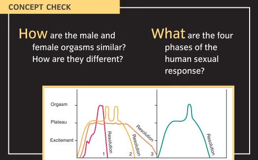
flow to the genitals is altered, gland secretion increases, and orgasm results in rhythmic contractions of pelvic muscles. In the mid-1950s, sex researchers Masters and Johnson began research on the human sexual response that spawned the study of human sexuality. They identified four phases of the human sexual re-sponse: arousal, plateau, orgasm, and resolution, which appear in both males and females.
During arousal, or excitement, blood flow is altered to the penis or clitoris, glands be-gin to secrete lubricating fluids, and heart rate and blood pressure increase. Arousal is governed by the parasympathetic nervous system. This phase is highly responsive to sensory stimulation, such as touch-ing of the genitals, breasts, lips, or earlobes. Other sen-sory stimulation, including visual, auditory, or even ol-factory stimuli, can increase or dampen the arousal.
As excitement builds, plateauis reached. This can last from a few seconds to many minutes. During this phase many females, and some males, experience a rash-like flush to the skin of the upper neck and face.
Orgasm, a series of wave-like muscular contrac-tions, and an intense pleasurable sensation, marks the end of the plateau. Orgasm and resolution are con-trolled by the sympathetic nervous system. In the male, orgasm accompanies ejaculation. In the female, receiv-ing the ejaculate does not provide much stimulation. Simultaneous orgasm is not automatic, nor should it be expected. Once males reach orgasm, they experience a refractory period of a few minutes to a few hours. Dur-ing this time, a second ejaculation is physiologically im-possible. Females do not require a refractor y period
and can experience two or more orgasms in rapid succession.
The last phase, resolution, begins with a sense of intense relaxation. Heart rate, blood pressure, and blood flow all return to pre-arousal levels. Resolution time is variable, taking longer to arrive when no orgasm occurred.
<