Ch. Tri Nuryana, Department of Anatomy, Embryology & Anthropology, Faculty of Medicine Gadjah Mada University, Yogyakarta
The effect of cyperus rotundus root ethanol
extract on the epithelization in the healing
process on skin excision wound
Ch. Tri Nuryana1, E. Suryadi1, Harijadi2
1 Department of Anatomy, Embryology & Anthropology,
Faculty of Medicine Gadjah Mada University
2 Department of Anatomy Pathology
Faculty of Medicine Gadjah Mada University
ABSTRACT
Ch. Tri Nuryana, E. Suryadi & Harijadi - The Influence of Ethanol Extract of Purple Nutsedge (Cyperus rotundus) on Epithelization of Skin Wound Healing Process
Background: If skin is wounded, epithelial cells will migrate to the wound area in order to keep homeostatic. Epithelization consists of mitotic and proliferation of epithelial cells. Rhizome of purple nutsedge (Cyperus rotundus) contains cyperene, flavonoid, ²-sitosterol dan ascorbic acid that can accelerate epithelization in wound healing process and has been used in traditional medicine.
Objective: To investigate the influence of ethanol extract of Purple Nutsedge (Cyperus rotundus) in epithelization of wound healing process.
Methods: Fourty five Balb/c male mice 8-10 weeks old, 25-35 g weight, were excised punch biopsy on back 0.5 cm right and left of columna vertebralis, 2.5 cm in cutis from the ear area. The mice were divided into 5 groups. Negative control group was treated with vehicle of extract, positive control group was treated by gel containing placenta extract 1% & neomycin sulfate 0.5% and the others were treated with C. rotundus 1%, 2%, and 4% in concentration. Each of the groups was divided into 3 subgroups. Each of the subgroups composed of 3 mice based on the period of termination, i.e.: 3rd, 7th and 12th day
after wounded. Histological evaluation was done to investigate the thickness of epithelial layer.
Results: The data were analyzed by Two-way Anova and the results showed that there was a significant difference (p<0.05) in every decapitation period, groups, and interaction between decapitation period and groups. The results of LSD test showed that there was a significant difference (p<0.05) in the thickness of epithelial layer between negative control and C. rotundus groups.
Conclusion: Cyperus rotundus extract accelerates epithelization process of wound healing in mice.
Key words: Wound healing - Cyperus rotundus - epithelization - mice
ABSTRAK
Ch. Tri Nuryana, E. Suryadi & Harijadi - Pengaruh ekstrak etanol umbi teki (Cyperus rotundus) terhadap epitelisasi pada proses penyembuhan luka eksisi kulit
Latar belakang: Jika kulit terluka, sel epitel akan bermigrasi ke daerah luka untuk mempertahankan hmoeostasis. Epithelisasi terdiri atas mitosis dan proliferasi sel epitel. Umbi teki (Cyperus rotundus) mengandung cyperene, flavonoid, ²-sitosterol dan ascorbic acid yang dapat meningkatkan proses epitelisasi luka dan telah digunakan dalam pengobatan tradisonal.
Bahan dan cara : Empat puluh lima tikus Balb/c umur 8-10 mingggu, berat 25-35 g, dibiopsi tusuk pada punggung 0.5 cm kanan dan kiri columna vertebralis, di kulit 2.5 cm dari daeraha telinga. Tikus dibagi menjadi 5 kelompok yaitu : kontrol negatif diberi perlakuan dengan bahan pelarut ekstrak, kontrol positif diberi perlakuan dengan gel yang mengandung ekstrak plasenta 1% & neomisin sulfat 0,5%, sedangkan yang lain diberi perlakuan dengan C. rotundus kadar 1%, 2%, and 4%. Masig-masing kelompok dibagi selanjutnya menjadi 3 subkelompok berdasarkan terminasi percobaan, yaitu hari III, VII, dan XII setelah perlukaan. Evaluasi histologis dilakukan utuk menilai ketebalan lapisan epitel yang timbul.
Hasil: Dengan analisis Anova-dua jalan ditemukan ada perbedaan bermakna (p<0.05) pada tiap periode dekapitasi, dalam kelompok, dan interaksi antara periode dekapitasi dan kelompok Hasil uji LSD menunjukkan bahwa terdapat perbedaan bermakna (p<0.05) dalam ketebalan lapisan epitel antara kontrol negatif dan kelompok perlakuan dengan C. rotundus groups.
Simpulan : Ekstrak Cyperus rotundus mempercpat proses epitelisasi dalam penyembuhan luka pada tikus.
INTRODUCTION
All of the body surface is lined with epithelial cells whose integrity is needed to maintain homeostasis. If the surface of the body is injured, the epithelial cells will migrate towards the wound to cover the injured skin1. The epithal cells of the skin are of keratinized
stratified squamous type. Keratinocytes will move towards the area of the wound in 24-48 hours after the injury, and will stop when they meet the keratinocytes from the other side of the wound. Suprabasal keratinocytes are the first cells that migrate to the wound. Epithelization is started by the increase in epithelial cell mitosis activity towards the margin of the wound, followed by their proliferation. Epithelial cells slide from the margin of the wound are in amoeboid movement. They use fibrin bands and extracellular matrix (ECM) compo-nents such as fibronectin as their guide. Epithelial cells from the remains of the epithelial structure separate their hemidesmosome bonds and reattach on the basal membrane and move rapidly over the wound. The proliferation of epithelial cells is affected by several growth factors and an enzyme that responible for the migration of keratinocytes and epithelization, i.e. matrix metalloproteinase-1 (MMP-1)2,3.
The high cost of treatment has caused the community to choose traditional alternative treatment. One of traditional drugs used to cure wounds is purple nutsedge (Cyperus rotundus) root. C. rotundus is a plant that grows in tropical and subtropical areas, especially in open and shaded areas, and is an ineradicable weed. Several studies have been done to find out the contents and the effects of C.
rotundus4.
C. rotundus contains essential oils, terpenes, flavonoids, b-sitosterol, and ascorbic acid. The main
terpenes in C. rotundus are cyperenes, which include sesquiterpene hydrocarbones5,6,7. Cyperene has a
role in protein synthesis and an inducer of the growth factors needed in epithelization8,9. Flavonoids can
restrict the quantity of free radicals and maintain the stability of collagen fibers6,9. b-sitosterol is a natural
steroid with estrogenic effect and able to maintain the humidity of the wound area to enable the cell growth10. Ascorbic acid has a role in stimulating the
synthesis of collagen, fibroplasia (?) and epithelizat-ion11,12.
Previous studies on the activity of C. rotundus
on the wound healing showed that 2% w/w cream of C. rotundus root alcohol extract had higher activity in wound healing compared to nitrofurazone ointment8.
The aim of this study was to find out the effect of C. rotundus root ethanol extract given topically on the epithelization in wound healing process.
MATERIALS AND METHODS
sulphate, and three treatment groups with topical C.
rotundus root ethanol extract in the concentration of 1%, 2%, and 4%, respectively. Each group was put into individual cages. Each group was devided further into 3 subgroups based on the decapitation period, that is, day 3, 7, and 12 after the wound. Histological slides were made with HE staining for evaluation of the epithelial layer growing over the wound..
C. rotundus root were obtained from Yogjakarta Province, ±1000 m from sealevel, in January 2007.
C. rotundus root ethanol extract in this study was 96% ethanol extract dissolved in simple cream base. The extraction was conducted in the Department of
Pharmacy, Faculty of Medicine, Gadjah Mada University.
RESULT AND DISCUSSION
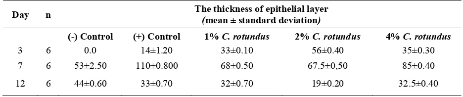
Epithelization process was assessed by measuring the thickness of epithelial layers. TABLE 1 shows that on day 3, the negative control group had no epithelization, while the other groups showed some epithelization process. The layer of epithelial cells in negative control group was the thinnest, followed by 1%, 4%, and 2% C. rotundus groups.
TABLE 1. Mean and standard deviation of the thickness of epithelial layers (micrometers) in the skin excision wound healing process after the application of C. rotundus
root ethanol extract (400x magnification, 10 visual fields).
Day n The thickness of epithelial layer
(mean ± standard deviation)
(-) Control (+) Control 1% C. rotundus 2% C. rotundus 4% C. rotundus
3 6 0.0 14±1.20 33±0.10 56±0.40 35±0.30
7 6 53±2.50 110±0.800 68±0.50 67.5±0,50 85±0.40
12 6 44±0.60 33±0.70 32±0.70 19±0.20 32.5±0.40
Note: n: sample size
On day 7, positive control, 4% and 1% C.
rotundus groups had the thickest epithelial layers, the 2% C. rotundus group started showing a thinning of epithelial layer, while negative control group still showed hyperplasia or a thickening of epthelial layer. On day 12.2% C. rotundus group had the thinnest epithelial layer, almost similar to
the thickness of normal epithelial layer (10 micrometer), while negative control group still showed hyperplasia. A graph illustrated the thickness of C. rotundus groups and control groups epithelial layers in each decapitation periods showed in FIGURE 1.
FIGURE 1. The graph of the thickness of C.
rotundus groups and control group epithelial layers in excision wound healing process on Balb/c mice back skin in each decapitation
periods
The thickness of epithel layer (
µ
)
FIGURE 1 showed that each group had a specific graphic pattern. The epithelial layers of C.
rotundus groups were thicker than positive and negative control groups on day 3. On day 7, positive control, 4%, and 1% C. rotundus groups had the thickest epithelial layers, and then they were thinning back to the normal epthelial layer thickness on day
12. On day 7, 2% C. rotundus group started showing a thinning in epithelial layer, while negative control group still showed hyperplasia. Two-way Anova analysis was used to find out the difference between decapitation periods and groups (TABLE 2).
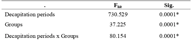
TABLE 2. Two-way Anova analysis on the thickness of epithelial layers (micrometer) between decapitation periods and groups in excision wound healing process on the back skin of Balb/c mice
. Fhit Sig.
Decapitation periods 730.529 0.0001*
Groups 37.225 0.0001*
Decapitation periods x Groups 80.154 0.0001*
Note: * : significant (p<0.05)
TABLE 2 showed a significant difference (p<0.05) in decapitation periods (F=730.529, p=0.0001), in groups (F=37.225, p=0.0001), and between decapitation periods and groups (F=80.154, p=0.0001).
The difference between groups was analyzed with LSD to find out the dominant effect between groups. The analysis showed that there was a significant difference (p<0.05) between negative control, positive control, and 1% C. rotundus
groups. The difference between positive control and 4% C. rotundus groups was not significant (p>0.05). The 1% C. rotundus group was not significantly different (p>0.05) from 2% C.
rotundus group, but significantly different (p<0.05) from 4% C. rotundus group. The 2% C. rotundus
group was significantly different (p<0.05) from positive control group, but not significantly different (p>0.05) from 4% C. rotundus group.
On day 3, the epithelial layer in 2% C. rotundus
group was the thickest and significantly different from positive control, negative control, 1%, and 4%
C. rotundus groups; followed by 4%, 1% C.
rotundus, positive control and negative control
groups. It showed that the epithelization process in
C. rotundus groups occurred earlier than in control groups. This was caused by cyperene, the active compound in C. rotundus, which induced the growth factors, such as EGF and FGF. Positive control group showed thinner epithelial layer compared to C. rotundus groups, probably caused by the negative effect of growth factor TGF-b18,9,13.
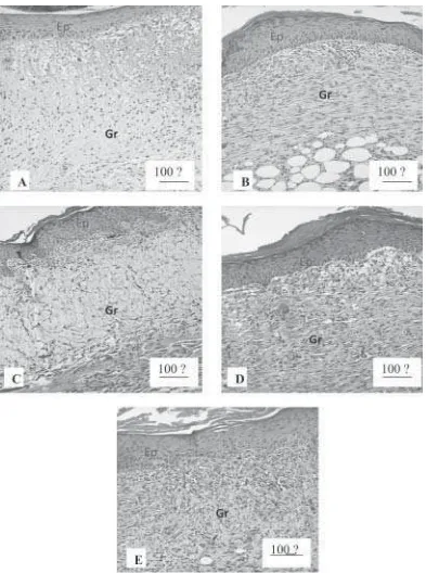
Based on histological observations (FIGURE 2), on day 7, the thickness of epithelial layer in 2% C. rotundus group had decreased, with higher granulation tissues. The peak of hyperplasia of epithelial layer in 2% C. rotundus group was assumed to be occured on day 5, and after that, it was thinning back to its normal thickness. Positive control group still showed process towards hyperplasia, similar with the process in 1% C.
rotundus group. It was assumed that keratinocytes in 2% C. rotundus group migrated and had mitosis more optimal than other groups. The epithelization in this group is facilitated by ascorbic acid12,14.
FIGURE 2. The microscopic pictures on day 7, with HE staining (100x magnification), of C. rotundus and control groups. (A) Negative control, (B) Positive control, (C) 1% C. rotundus, (D) 2% C. rotundus, (E) 4% C. rotundus groups.
Previous studies used Wistar male rats as subjects, and the study showed that 2% w/w C.
rotundus root alcohol extract cream had a higher activity wound healing (100% wound closure on day 18) compared to nitrofurazone ointment. The study used three kinds of wounds, i.e. excision to observe the wound closure and contraction, incision to observe the inflammatory cells, and dead space to observe the granulation8. The result
of that study was consistent with the result of this study, which concluded that C. rotundus could accelerate the wound healing.
Meanwhile, Zhu et al.15 reported that C. rotundus had cytoprotective effect on gastric ulcer. This study used rats as subjects, who were given
C. rotundus root ethanol extract with the dose of 0.25 gr/kgBW orally, after induced with alcohol per oral.
C. rotundus contains cyperene, an inducer of the growth factors such as EGF that stimulates the epithelization. EGF acts as a mitogen on epithelial cells, keratinocytes, and fibroblasts, and stimulates the migration of keratinocytes and the formation of granulation tissues14,16. In the wound
healing process, EGF is produced by keratinocytes, macrophages, and other inflammatory cells migrating to the wound area. FGF has functions in angiogenesis, fibroblast proliferation, and epithelial migration to compose new epidermis. TGF-b in placental extract given in positive control is an inhibitor in epithelial cell growth13,17.
Epithelization is also influenced by matrix metalloproteinase-1 (MMP-1) produced by epithelial cells, fibroblasts, myofibroblasts, chondrocytes, osteoclasts, endothelial cells, and leukocytes. Overproduction of MMP causes pathological conditions, so that an inhibitor is needed, that is, tissue inhibitor of metallo-proteinase (TIMP) to control the activity of MMP18,19,20.
CONCLUSION
Topical C. rotundus root ethanol extract could accelerate the epithelization in wound healing process in mice.
ACKNOWLEDGEMENT
Most of the research in this article was funded by DIKTI through BPPS research grant.
REFERENCES
1. Torre J. & Sholar A. Wound Healing, Chronic Wounds. eMedicine Web MD. http://www.emedicine/ woundhealing/chronicwound.html, 2006.
2. Takeda H, Katagata Y, Hozumi Y & Kondo S. Effects of Angiotensin II Receptor Signaling during Skin Wound Healing. Am J Pathol. 2004; 165:1653-62
3. Wagener FADTG, Beurden HE, Hoff JWV, Adema GJ & Figdor CG. The Heme-heme Oxigenase System: Molecular Switch in Wound Healing. Blood Paper. 2003; 1182(10): 2248-52.
4. Hall DW, Vandiver VV & Ferrell JA. Purple Nutsedge, Cyperus rotundus L. IFAS, University of Florida. EDIS Website at http://edis.ifas.ufl.edu/purplenutsedge.html, 2004.
5. Kilani S, Abdelwahed A, Amma RB, Hayder N. Chemical Composition, Antibacterial and Antimutagenic Activities of Essential Oil from (Tunisian) Cyperus rotundus. J Ess Oil Res. 4091(11). http://www.findarticle.com/p/articles/ mi_qa4091/is_200511/ai_n15935895/19_2. 2005. 6. Pal DK & Dutta S. Evaluation of the Antioxidant activity
of the roots and Rhizomes of Cyperus rotundus L. Indian J Pharmaceut Sce. 2006; 68(2):256-58. 7. Parekh J & Chandra S. In-vitro Antimicrobial Activities
of Extracts of Launaea procumbens Roxb. (Labiateae), Vitis vinifera L. (Vitaceae) and Cyperus rotundus L. (Cyperaceae). African J Biomed Res. 2006; 9(2): 89-93. 8. Puratchikody A, Nithya C & Nagalakhsmi G. Wound healing activity of cyperus rotundus linn. Indian J Pharmaceut Sci. 2006; 68(1): 97-101.
9. Fraga BM. Natural sesquiterpenoids. The royal society and chemistry journal. 2002; 19:650-72.
10. Radenahmad N, Vongvatcharanon U,
Withya-chum-narnkul B & Connor JR. Serum levels of 17²-estradiol in ovariectomized rats fed young-coconut-juice and its effect on wound healing. Songklanakarin J Sci Technol. 2006; 28(5):898-910.
11. Jagetia GC, Rajanikant GK, Baliga MS, Rao KV & Kumar P. Augmentation of wound healing by ascorbic acid treatment in mice exposed to gamma-radiation. Int J Radiat Biol. 2004; 80(5): 347-54.
12. Percival M. Nutritional Support for Connective Tissue Repair and Wound Healing. Clinical Nutrition Insights. NUT026. Rev.6/98, 1997.
13. Kenyon NJ, Ward R, McGrew G & Last JA. TGF-ß1 causes airway fibrosis and increased collagen I and III mRNA in mice. Biomed J. 2003; 58:772-77.
15. Zhu M, Luk HH, Fung S & Luk, CT. Cytoprotective Effects of Cyperus Rotundus Against Etanol-Induced Gastric Ulceration in Rats. Phytother Res. 1997; 11(5): 392-94.
16. Jonkman MF, Hoeksma EA & Nieuwenhuis P. Accelerat-ed Epithelization Under a Highly Vapor-Permeable Wound Dressing Is Associated with Increased Precipitat-ion of Fibrin(ogen) and Fibronectin. J Invest Dermatoly. 1990; 94: 477–84
17. Ashcroft GS, Mills SJ, Lei KJ, Gibbons L, Jeong MJ, et al. Estrogen modulates cutaneous wound healing by downregulating macrophage migration inhibitory factor. J Clin Invest. 2003; 111: 1309-18
18. Atiyeh BS, Al-Amm CA & Nasser AA. Improved Healing of Split Thickness Skin Graft Donor Sites. J Appl Res Visse, R.& Nagase, H. 2003. Matrix Metallo-proteinase and Tissue Inhibitors of Metalloproteinases. Circul Res. 2000; 92:827-39.
19. Visse R & Nagase H. Matrix Metalloproteinase and Tissue Inhibitors of Metalloproteinases. Circul Res. 2003; 92: 827-39.