Biosaintifika
Journal of Biology & Biology Educationhttp://journal.unnes.ac.id/nju/index.php/biosaintifika
Capability of Vitamin E as a Radioprotector in Suppressing DNA
Damage Determined with Comet Assay
Darlina1, Lusy Dahlia A.2, Zubaidah Alatas1, Teja Kisnanto1, Mukh Syaifudin1
DOI: 10.15294/biosaintifika.v9i2.8716
1Center for Technology of Radiation Safety and Metrology, Indonesia
2Pharmacy Department, Faculty of Mathematics and Natural Sceinces, National Institute of Sciences and
Technology, Indonesia
Abstract
Radiation has a potent to damage cells. Radiation may act directly or indirectly on deoxyribonucleic acid (DNA) that results in the degeneration of tissues and necrot-ic, and thereby it needs a potent radioprotector to prevent these damages. Vitamin E is natural product known as an antioxidant which has potential as radioprotector. This research aimed to determine the capability of vitamin E with emphasized on the searching for its optimal concentration as radioprotector of DNA damage. This study used blood samples of healthy person irradiated with gamma rays at a dose of 6 Gy as the lethal dose to lymphocytes. The cocentrations of vitamin E from 0 to 0.8 mM was added into blood 15 minutes before irradiation. Isolation of lymphocytes was done using gradient centrifugation method. Evaluation on the capability of this compound in suppressing DNA damage was done by using alkaline Comet assay and data analysis was done using CaspLab program. The results show that addition of vitamin E could suppres these DNA damages and 0.8 mM of vitamin could re-duce DNA damage up to 94.2%. We conclude that vitamin E effectively suppresed DNA damages induced by radiation. This information may benefit to the patient from negative impacts of radiotherapy.
How to Cite
Darlina, A, L. D., Alatas, Z., Kisnanto, T. & Syaifudin, M. (2017). Capability of Vi-tamin E as a Radioprotector in Suppressing DNA Damage Determined with Comet Assay. Biosaintifika: Journal of Biology & Biology Education, 9(2), 201-208.
© 2017 Universitas Negeri Semarang
History Article
Received 28 December 2016 Approved 14 May 2017 Published 17 August 2017
Keywords
DNA damage; vitamin E; radioprotector; comet assay
Correspondence Author:
Jl. Lebak Bulus Raya No. 49 JKSKL Jakarta E-mail: [email protected]
common in our diets and are often perceived as being more ‘natural’ and better suited for medi-cinal purposes due to being well tolerated and minimally toxic even at the upper ranges of die-tary intake. Vitamins are prominent among natu-ral compounds considered beneficial for human health (Satyamitra et al., 20111). Vitamin E is well known as antioxidant, neuroprotector, and also anti-inflammatory properties (Singh et al., 2013). It is essential because body cannot produ-ce vitamin E so it should be obtanied from food supplements. Vitamin E represents a generic term for all tocopherols and their derivatives with natu-rally occurring and biologically active stereoiso-meric compounds of α-tocopherol (AT) (Palozza et al., 2008).
Comet assay, also known as single-cell gel electrophoresis, is a simple method for measu-ring DNA strand breaks in eukaryotic cells. The technique includes cells embedding in agarose on a microscope slide, lysis with detergent and high salt to form nucleoids containing supercoi-led loops of DNA linked to the nuclear matrix, electrophoresis at high pH results in structures resembling comets, and observation with fluo-rescence microscopy. Finally the analysis is that the intensity of the comet tail relative to the head reflects the number of DNA breaks (Olive et al., 2006; Speit & Hartmann, 2005; Singh et al., 1988). The purpose of this study was to examine the capability of vitamin E as radioprotector in suppressing the radiation induced DNA damage The benefits of this research are to protect nor-mal tissue from radiation damage due to radiot-herapy in cancer patients..
METHODS
Vitamin E was provided as dl-α-tocopherol soft capsule at a concentration of 250 IU (Cata-lent, Australia). A 1 mM stock solution of vita-min E was prepared in polyethilene glicol (PEG) mixed wtih Tween 20 solution and serial dilu-tions with PBS were made in order to achieve a working concentration of 0.2 mM; 0.4 mM; 0.6 mM dan 0.8 mM of vitamin E .
The study was performed on peripheral blood samples obtained from two healthy males, non-smoking, non-alcoholic donors with ages of 30 and 50 years. The donors are never exposed to ionizing radiation. Three mL venous blood were collected under sterile conditions in vacutainer tubes (Becton Dickinson, NJ, USA) containing lithium heparin as anticoagulant. After collecti-on, the blood was divided into 6 tubes of samples. The present study was carried out in 6
INTRODUCTION
When a track of ionising radiation passes through a cell it will deposit energy which can disrupt the organic molecules mainly that most sensitive molecule within the cell i.e. deoxyribo-nucleic acid (DNA) which is the most target and termed as the blueprint of life. It is well known that free radicals formed by the radiolysis of cellu-lar aqueous milieu, and their interaction with one another and with oxygen are primary mediators of radiation injury (Hall & Giaccia, 2006; Tet-riana et al. 2015). Ionizing radiation induce the production of Reactive Oxygen Species (ROS) through hydrolysis of water. Including superoxi-de, hydrogen peroxisuperoxi-de, and hydroxyl radicals as the most reactive radicals. Such ROS can initi-ate oxidative cellular injury as well as activiniti-ated intracellular signaling pathways and stimulated cytochrome c release from mitochondria leading to apoptosis (programmed cell death) (Azzam et al., 2012).
Radioprotectors are compounds that are designed to reduce damage occured in nor-mal tissues caused by ionizing radiation. These compounds are mostly antioxidants and must be exist before or at the time of radiation for effecti-veness as protector (Citrin et al., 2010). This ra-diation modifier or protectors is expected to alter the response of normal tissues to irradiation via free radical scavenging and/or H atom donati-on (Rahman et al., 2015). Radioprotectants are also important in suppressing the accumulation of genetic mutations, cell death or tissue disor-ganisation in patients undergoing radiotherapy or individuals exposed to non-lethal, but higher than normal, levels of radiation in accidental event (Liu, 2010).
Although endogenous antioxidant sys-tems such as glutathione, thioredoxin, superoxi-de dismutase, and catalase normally inhibit the deleterious effects of ROS, these systems may be overwhelmed in irradiated cells. Exogenously supplemented antioxidants, or agents that stimu-late endogenous antioxidant systems within cells, have shown promise in terms of suppressing the harmful effects of irradiation. A variety of redu-cing agents, such as vitamin E analogs, polyphe-nols, thiols and superoxide dismutase mimetics have been described as potential radiation coun-termeasures in the recent past (Singh et al., 2012; Weiss et al., 2009; Dumont et al., 2010).
groups, group 1: control (cells without radiation), group 2 : radiation control (RC: cells were expo-sed to 6 Gy of gamma radiation), group 3: cells treated with 0.2 mM vitamin E before radiation exposure, group 4: cells treated with 0.4 mM vi-tamin E before radiation exposure, group 5; cells treated with 0.6 mM vitamin E before radiation exposure, and group 6: cells treated with 0.8 mM vitamin E before radiation exposure.
Before radiation, blood samples were incu-bated for 15 minutes with the serial concentrati-on of vitamin E. The blood samples were then irradiated with gamma radiation in the ice. The source of gamma radiation used was Cobalt-60 (IRPASENA, PATIR, BATAN). All of the blood samples were irridiated with dose of 6 Gy, at dose rate of 1 Gy/minutes as a lethal dose of gamma radiation for lymphocytes cells. Lymphocyte cells were isolated 5–15 minutes after radiation and examined for induced DNA damages using co-met assay. One blood sample without antioxidant served as control in both series, and these samples were also irradiated with 6 Gy.
After radiation, lymphocyte were isolated from the blood samples using Histopaque (Sig-ma) according to standard method (Panda et al., 2012). Three mL of fresh heparinized blood was mixed with the equal volume of phosphate buffe-red saline (PBS, Merck), which was overlaid on 3 mL of Histopaque. Lymphocytes were separated using density gradient centrifugation. Then the separated lymphocytes were washed twice with PBS by centrifugation for 15 min at 1000 rpm and cells were suspended in minimum volume of RPMI -1640 (Gibco).
Cell viability was measured using trypan blue dye exclusion method (Chung et al., 2015). The lymphocytes were mixed with equal volu-me of 0.4% trypan blue dye for 3 minutes and counted using haemocytometer. Viable and dead cells were scored under the microscope.
About 104 cells per 100 µL of medium
was taken from each dose treatment for Comet assay by following the standard procedure with slightly modification (Singh et al., 1988). Fully-frosted microscopic slides were prepared. Each slide was covered with 1% Normal Melting Point (NMP) agarose (Sigma). After solidification, the slides were then coated with 0.6% NMP aga-rose. A Low Melting Point (LMP) agarose was melted and stabilized in a waterbath (RTE10) at 37°C. For each sample and control, 5 µL of cell homogenate was mixed with 100 µl of 1% LMP agarose and placed on the slides. After 10 minutes of solidification on ice, the slides were covered with 0.5% LMP agarose. The slides
were then immersed in a pre-chilled lysis solu-tion ((2.5 M NaCl, 100 mM Na2EDTA, 10 mM Tris–HCl, adjust until pH 10 with NaOH (Sigma) and added 1% Triton X-100 (Sigma) and 10% di-methyl sulfoxide (Sigma) and kept in at 4°C for 60 minutes. The slides were placed horizontally in a humidity chamber at 37 °C for 30 minutes. All slides were then immersed in an alkali solu-tion (0.3 M NaOH, 1 mM Na2EDTA; pH 12.1) for 40 minutes. Electrophoresis in a pre-chilled alkali solution (0.3 M NaOH, 1 mM Na2EDTA; pH 13) at 1 V/cm was done for 20 minutes in re-frigerator (4°C). After electrophoresis, the slides were rinsed gently three times with neutralization buffer (0.4 M Tris–HCl, pH 7.5) to remove excess alkali and detergents. Each slide was fixed with methanol, stained with ethidium bromide (20 µg/ml) and covered with a coverslip. Slides were stored at 4°C in sealed boxes until analysis.
The stained samples with ethidium bro-mide were observed using a Nikon flourescence microscope. A total of 50 randomly captured comets from each slide were examined at 250x magnification using an epifluorescence micros-cope that connected through computerized to an image analysis. Cells were piled not counted. The image of comet was digitally analyzed using
CASPLab comet assay software.
Each experimental set consists of dupli-cated slides. The various parameters measured in the exposed and control groups were evalua-ted using Excell program (StaSoft, Tulsa, USA). Each sample was characterized for the extent of DNA damage by considering the mean, SE (stan-dard error of the mean), median and range of the comet parameters.
RESULTS AND DISCUSSION
The viability cells in this research was ob-served under microscope. The results showed that the number of cells in all groups was in enough number (around 107 cells/mL) and suitable for
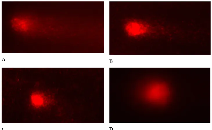
Comet assay. In this research, the DNA damage of irradiated lymphocytes was assessed by comet assay by staining the cells with ethidium bromi-de and the comet that mainly consist of single stranded DNA can be seen with a fluorescence microscope as presented in Figure 1.
by measuring the length of the comet tail using an ocular scale fitted in the eyepiece of the micros-cope or by visual scoring of degree of damage from 0 to 4 according to comet appearance [Fi-gure 1]. Alternatively, there are numerous image analysis software to quantitate additional DNA damage parameters such as percentage of DNA in head, percentage of DNA in tail, tail moment (product of tail length and percentage of DNA in tail), and tail area.
In order to avoid or inhibit these DNA da-mages we treated the blood with an antioxidant vitamin E before irradiation. In this study four concentrations of vitamin E (0.2; 0.4; 0.6; and 0.8 mM) were added to the blood 15 minutes be-fore irradiation. All of the results are presented in Figure 2.
From the visualization presented in Figu-re 2, it can be seen that theFigu-re is a shortening the tail of comet of lymphocytes treated with vitamin
E. Higher concentration of vitamin E resulted in shorter tail of comet which means that there is a reduction of DNA damages in the presence of vi-tamin E and this treatment effectively reduce the deleterous effects of gamma radiation.
The comet itself can be analysed by vi-sual scoring or computerised image analysis. In this research the comet were analyzed using CASPLab Comet Assay software which reported using a range of different endpoints. This comet assay sofware can measure 8 parameters of the digital image of comet value : Long-tailed comet (TL), DNA Tail, Tail Moment (TM), Olive tail moment (OTM), DNA head, DNA Percentage in head, and the length of comet (L.comet). Head DNA that indicating the number of DNA in head of comet is an additional parameter in compute-rized program of CASP Lab.
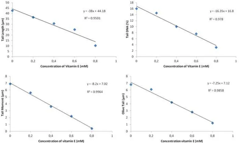
The graph in Figure 3 demonstrate the relationship between vitamin E concentration
Figure 1. Results of visualisation of DNA stained with EtBr in Comet assay of the lymphocytes of
control blood sample (left) and that exposed to 6 Gy dose of gamma radiation with tail of comet (right).
Figure 2. Visualization of comet of lymphocyte irradiated with 6 Gy and treated with vitamin E at
and DNA damages induced by 6 Gy gamma ra-diation. Here we found that each parameter has tendention to decrease by increasing the concent-ration of vitamin E added (Figure 3). The corre-lation coefficient (R2) for every parameter : (a) tail moment (TM), (b) olive tail moment (OTM), (c) tail DNA, and (d) tail length (TL) more than 0,9 indicating a very good relationship.
Figure 4. Relationship between head DNA with
the increasing of vitamin E concentration Figure 4 shows that head DNA and the concentration of vitamin E have a positive corre-lation meaning that higher concentration of vita-min E higher number of DNA in head. This fin-ding indicating that vitamin E effectively reduce DNA damage due to ionizing irradiation. Among comet tail parameters, TM gave the highest per-centage of reducing damage given with 0.8 mM of vitamin E. Based on 5 parameters in comet test it is known that vitamin E is a good radioprotec-tor in suppressing DNA damage post irradiation
with R2 more than 0.9.
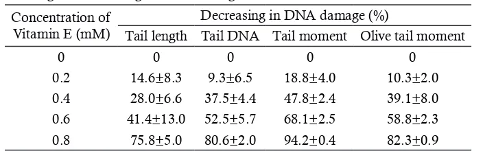
The percentage reduction in DNA damage to increase the concentration of vitamin E in the fourth comet tail parameters are shown in Table 1. Seen the higher the concentration of vitamin E is added before irradiation can reduce radia-tion-induced DNA damage. The value of TM provides the highest percentage decrease DNA damage the four parameters of comet tails. The decreasing of DNA damage percentage shown in the highest value of TM at a concentration of 0.8 mM vitamin.
The cell-type-of-choice in biomonitoring research activities is mostly the lymphocyte be-cause blood is easily collected and lymphocytes have proved to be good surrogate cells. In Comet assay the process of electrophoresis is done under the alkaline conditions where the strand breaks through their ability to relax DNA supercoiling, allow the negatively charged DNA loops to ex-tend towards the positively charged anode (Singh et al., 1988).
Similar research was performed by Singh et al. (2013) who has done an in vivo study to de-termine the effect of tocopherol succinate (TS) treatment against DNA damage caused by ion-izing radiation in peripheral blood mononuclear cells, splenocytes and thymocytes of mouse. The mice were treated with vehicle or TS (400 mg/ kg) and exposed to high dose (9.2 Gy that is the LD90/30 dose that causes hematopoietic injury) of
60Co γ-radiation 24 h after drug injection.
Periph-eral blood, spleen and thymus were collected 30
Figure 3. The effect of vitamin E addition on the four parameters of Comet assay (DNA fragment)
age by scavenging ROS before they act on cellular components. A variety of reducing agents, such as vitamin E analogs, polyphenols, thiols and su-peroxide dismutase mimetics have been described as potential radiation countermeasures in the re-cent time.
Here we tested the potentail of vitamin E as a Radioprotector to lethal dose of gamma ray. It was approved that administration of vita-min E modulated the expression of antioxidant enzymes and inhibited expression of oncogenes in irradiated cells (Singh et al., 2013). Vitamin E is a group of eight structurally related fat-soluble vitamins, four tocopherols (α, β, γ, and δ), and in more detail, acting as antioxidants by preventing the propagation of free radical reactions by do-nating hydrogen from their phenolic group to sta-bilise the radicals, and thereby break the chain of events leading to oxidative damage. The tocoph-erols protect the structure and function of human cell membranes (Vasilyeval & Bespalov, 2015; Niki, 2014; Satyamitra et al., 2011). And here we treat the blood with vitamin E 15 minutes be-fore irradiation. This is due to the fact from some other published results (Singh et al., 2013; Mau-rya et al., 2006; Ghosh et al., 2009; Citrin et al., 2010) that vitamin E did not protect mice when administered as a mitigator after irradiation and it can be used only as a radio-protector of DNA which is a target for mutagens and carcinogens, and induce changes in DNA structure giving rise to mutations and/ or cell death.
Nair et al. (2003) had been done a very comprehensive study on the efficacy of tocopher-ol monoglucoside (TMG) as radioprotector and they found that, this chemical, which is a water soluble derivative of vitamin E, offers protection against the deleterious effects of ionizing radia-tion, either in vivo or in vitro studies, to biologi-cal systems. It has a potent antioxidant and an effective free radical scavenger, so that it protects DNA from radiation-induced strand breaks for-mation. It also protected thymine glycol forma-tion induced by gamma-radiaforma-tion. It prevents gamma-radiation-induced loss of viability of EL-minutes and 4 h after irradiation, and used for
alkaline comet assay. The administration of TS significantly inhibited DNA damage in periph-eral blood cells and thymocytes compared with vehicle-treated mice, evidenced by the shorter tail length and smaller percentage of DNA in the comet tail. Another study conducted by Yassa et al. (2011) aimed to investigate potential protec-tive vitamin E against pesticide diazinon (DZN) in murine. The results showed that mice treated with vitamin E reduced DZN- induced DNA damage where the length TL shortened by up to 50%. These results suggest that vitamin E has a protective effect on DZN-induced DNA and showing that vitamin E prevent genotoxicity induced by DZN. It indicates that vitamin E is also effectively suppressing the harmfull effects induced by other chemical.
Since many types of radiation are now being frequently used in clinical treatment of pa-tients with cancer and in exprimental research, it is essential that more detailed information on the chemical capabilities in minimizing the effect of irradiation be obtained. In this research, like other studies, we are searching for the efficacy of natural chemical in suppressing the negative ef-fects of ionizing radiation. And many studies had shown that vitamin E can scavenge molecular ox-ygen, peroxide and hydroxyl radicals and atomic oxygen radicals induced by ionizing radiation.
The understanding in radiation effects has placed emphasis on the search for antioxidant agents that are suitable as radiation countermea-sures (Sing et al., 2012; Weiss & Landauer, 2009; Dumont et al., 2010). Although endogenous anti-oxidant systems (glutathione, thioredoxin, super-oxide dismutase, and catalase) normally inhibit the deleterious effects of ROS, these systems may be overwhelmed in irradiated cells. Exogenously supplemented antioxidants, or agents that stimu-late endogenous antioxidant systems within cells, have shown promise in terms of suppressing the harmful effects of irradiation. If present in the cells at the time of radiation exposure, such an-tioxidants may protect cells from radiation
dam-Table 1. Percentage of decreasing in DNA damage due to the administration of vitamin E± SD.
Concentration of Vitamin E (mM)
Decreasing in DNA damage (%)
Tail length Tail DNA Tail moment Olive tail moment
0 0 0 0 0
tumor cells and peroxidation of lipids in micro-somal and mitochondrial membranes. It reduce upto 75% embryonic mortality resulting from exposure of pregnant mice to ionizing radiation (2 Gy) by ip administration (0.6 g/kg, body wt) prior to irradiation. This chemical TMG also of-fered protection to mice against whole body gam-ma-radiation-induced lethality and weight loss. Study also showed that the LD50(30) of mice increased from 6 to 6.72 Gy upon post irradiation administration of a single dose of TMG (0.6 g/ kg, body weight) by intraperotoneal route.
Vitamin E can alleviate radiation induced decrement in delayed-type hypersensitivity and as adjuvant to other radioprotectant. A very old and simple research conducted by Mahdy (1991) from Middle Eastern Regional Radioisotope Centre for the Arab Countries, Cairo, in Egypt found that intraperitoneally injection of vitamin E before whole body gamma irradiation at the dose of 7 Gy to female albino rats remarkably recovered in the serum protein content at all post-irradiation days, while it slightly recovered in the level of serum urea.
Basically there are some mechanisms of this chemical in its action. One investigator re-vealed that vitamin E act by preventing lipid pe-roxidation which does not generally play a major role in cell killing by ionizing radiation. Other suggested the chemical by scavenging of secon-dary radicals but it needs a very high concentra-tion to effectively prevent DNA damage which is responsible for classical reproductive cell death, or by suppressing the protracted oxidative stress which is difficult to be distingusihed from its role as scavenging radicals, decreasing oxygen con-centration which is due to the fact that hypoxic cells are radioresistant so that treatments that de-cerase microenvironmental oxygen can be radio-protective, enhancing the DNA repair which is a major factor in determining the radiosensitivity (Bump, 1998).
Borek (2008) proposed that vitamin E is an antioxidant and its radiprotective action is de-pend on the oxygen partial pressure in tissue and it was shown that this chemical effectively protect tissue from deletirius effects of ionizing radiation in high oxyen pressure such as lung. Recent stu-dies have suggested several proposed alternative mechanisms: most notably, an indirect effect of tocopherols in eliciting specific species of radio-protective growth factors or cytokines such as granulocyte colony-stimulating factor (G-CSF). The vitamin E treatment in irradiated group of rat presented more acinar cells than the irradiated group, but no statistically significant difference
was observed (p>0.05). They conclude that vita-min E seems to have failed as a radioprotective agent on acinar cells in rat parotid glands (Gomes et al., 2013).
Our results demonstrate that vitamin E has the potential to protect DNA damage lyn-phocyte cell from radiation injury . As is known radiotherapy given to patients often results in munologic cell damage resulting in decreased im-munity. Lymphocyte cells have immune cells an important role in immunity. This study are useful to enrich radioprotectant information that can protect radiotherapy patients from the effects of radiation
CONCLUSIONS
Ionizing radiation at a dose of 6 Gy, which is equivalent to highly lethal doses for humans, effectively cause the DNA damage. This study shows that the addition of vitamin E is signifi-cantly suppressed the DNA damages at all con-centration tested, concon-centration of 0.8 mM of vi-tamin E could reduce DNA damage up to 94.2%. It was given just before the irradiating gamma rays
ACKNOWLEDGEMENTS
This work in part was financially supported by annual project of the Center for Technology of Radiation Safety and Metrology, National Nuclear Energy Agency (project year of 2014).
REFERENCES
Azzam, E. I., Jay-Gerin, J. P. & Pain, D. (2012). Ion-izing radiation-induced metabolic oxidative stress and prolonged cell injury. Cancer Letter, 327(0), 48–60.
Borek, C. (2008). Dietary antioxidants and phytochem-icals in radioprotection and therapy, in : Herbal Radiomodulators: Applications in Medicine, Homeland Defence and Space (Rajesh Arora ed.). pp. 141-143.
Bump, E. A. (1998). Introduction, In: Radioprotectors: chemical, biological, and clinical perspectives (Edward A. Bump and Kamal Malaker). CRC Press, Boca Raton, pp. 3-10.
Chung, D. M., Kim, J. H. & Kim, J. K. (2015). Evalu-ation of MTT and Trypan Blue assays for ra-diation-induced cell viability test. International Journal of Radiation Research, 13(1), 331-335. Citrin, D., Cotrim, A. P., Hyodo, F., Baum, B. J.,
Radia-tion countermeasure agents: an update. Expert opinion on therapeutic patents, 20(1), 73-101. Ghosh, S. P., Kulkarni, S., Hieber, K., Toles, R., Kao,
T. C., Hauer-Jensen, M. (2009). Gamma-tocot-rienol, a tocol antioxidant as a potent radiopro-tector. International journal of radiation biology,
85(7), 598-606.
Gomes, C. C., de Moraes Ramos-Perez F. M., da Cruz Perez, D. E., Novaes P. D., Bóscolo F. N., & de Almeida, S. M. (2013). Radioprotective effect of vitamin E in parotid glands: a mor-phometric analysis in rats. Brazilian Dental Jour-nal, 24(3), 183-187.
Hall, E. J. & Giaccia, A. J. (2006). Radiobiology for the Radiologist. Sixth Edition. Philadelphia: Lippincott Williams & Wilkins; pp. 5-15. Liu, S. Z. (2010). Biological effects of low level
expo-sures to ionizing radiation: theory and practice.
Human & experimental toxicology, 29(4), 275-281. Mahdy, A. M. (1991). Vitamin E as A chemical radio-protector for controlling the radiation induced changes in the levels of protein and urea in the serum of irradiated rats. Isotope and Radiation Research, 23(2), 117-123.
Maurya, D. K., Devasagayam, T. P. A., & Nair, C. K. K. (2006). Some novel approaches for radio-protection and the beneficial effect of natural products. Indian Journal of Experimental Biology, 44(2), 93-114.
Nair, C. K., Devi, P. U., Shimanskaya, R., Kunugita, N., Murase, H., Gu, Y. H. & Kagita, T. V. (2003). Water soluble vitamin E (TMG) as a Radioprotector. Indian Journal of Experimental Biology, 41(12), 1365-1371.
Niki, E. (2014). Role of vitamin E as a lipid-soluble peroxyl radical scavenger: in vitro and in vivo evidence. Free Radical Biology and Medicine, 66, 3-12.
Olive P. L. & Banáth, J. P. (2006). The comet assay: a method to measure DNA damage in individual cells, Nature protocols, 1(1), 23-29.
Palozza, P., Simone, R., Picci, N., Buzzoni, L., Cilib-erti, N., Natangelo, A. (2008). Design, synthe-sis, and antioxidant potency of novel alpha-to-copherol analogues in isolated membranes and intact cells. Free Radical Biology and Medicine,
44(7), 1452-1464.
Panda, S. K., Kumar, S., Tupperwar, N. C., Vaidya, T., George, A., Rath, S., Bal, V. & Ravindran, B. (2012). Chitohexaose activates macrophages by alternate pathway through TLR4 and blocks endotoxemia. PLoS Pathog, 8(5), e1002717. Rahman, M. M., Islam, M. B., Biswas, M. & Alam, A.
H. M. K. (2015). In vitro antioxidant and free
radical scavenging activity of different parts of
Tabebuia pallida growing in Bangladesh. BMC research notes, 8(1), 621.
Satyamitra, M., Kulkarni, S., Ghosh, S. P., Mullaney, C. P., Condliffe, D., & Srinivasan, V. (2011). Hematopoietic recovey and amelioration of radiation-induced lethality by the vitamin E isoform, delta-tocotrienol. Radiation research,
175(6), 736-745
Singh, N. P., McCoy, M. T., Tice, R. R. & Schneider, E. L. (1988). A simple technique for quantitation of low levels of DNA damage in individual cells. Experimental cell research, 175(1), 184-191. Singh, P. K., Wise, S. Y., Ducey, E. J., Fatanmi, O. O.,
Elliott, T. B., & Singh, V. K. (2012). Alpha-to-copherol succinate protects mice against radi-ation-induced gastrointestinal injury. Radiation research, 177(2), 133-145.
Singh, V. K., Ducey, E. J., Brown, D. S., & Whitnall, M. H. (2012). A review of radiation counter-measure work ongoing at the Armed Forces Radiobiology Research Institute. International journal of radiation biology, 88(4), 296-310. Singh, V. K., Singh, P. K., Wise, S. Y., Posarac, A. &
Fatanmi, O. O. (2013). Radioprotective proper-ties of tocopherol succinate against ionizing radiation in mice. Journal of Radiation Research
54(2), 210–220.
Speit, G. & Hartmann, A. (2005). The comet assay: a sensitive genotoxicity test for the detection of DNA damage. Molecular toxicology protocols. 291, 85–95.
Tetriana, D., Mailana, W., Kurnia, I., & Syaifudin, M. (2015). Preliminary Study on the Single Nucleotide Polymorphism (SNP) of XRCC1 Gene Identificationto Improve the Outcomes of Radiotherapy for Cervical Cancer. Biosainti-fika: Journal of Biology & Biology Education, 7(2), 79-86.
Vasilyeval, I. N. & Bespalov, V. G. (2015). Release of Extracellular DNA after Administration of Radioprotective Combination of α-Tocopherol and Ascorbic Acid. Radiatsionnaia biologiia, ra-dioecologiia, 55(5), 495-500.
Weiss, J. F. & Landauer, M. R. (2009). History and development of radiation-protective agents.
International journal of radiation biology, 85(7), 539-573.