Optimization of Emergence Profile of Implant Prosthesis:
A Literature Review
Minoru Sanda1, Daisuke Sato2, Kazuyoshi Baba1
1Department of Prosthodontics, School of Dentistry, Showa University – Japan 2Department of Implant Dentistry, School of Dentistry, Showa University – Japan
‘Corresponding Author:Kazuyoshi Baba, School of Dentistry, Showa University – Japan. Email: [email protected]
Received date: September 7, 2017.Accepted date:November 30, 2017.Published date:January 25, 2018.
Copyright: ©2018 Sanda M, Sato D, Baba K. This is an open access article distributed under the terms of the Creative Commons Attribution License, which permits unrestricted use, distribution, and reproduction in any medium provided the original author and sources are credited.
ABSTRACT
In order to achieve esthetically optimal outcome with implant prosthesis, appropriate topography of emergence profile is crucial. The objective of this review is to explorer current evidence regarding this topic and relevant issue. Extent of interproximal papilla is determined not by the shape of emergence profile but the length between interproximal alveolar bone prominence and interproximal contact of crowns. There have been concerned that multiple times of disconnection and reconnection of abutment enhance peri-implant marginal bone loss, but it’s certified not to be a clinically significant level. Current digital workflow makes this step faster and easier, by copying emergence profile of contralateral tooth or extracted teeth.
Keywords : aesthetic outcome, dental implant, emergence profile, prosthodontic, provisional restoration
Background
An implant prosthesis is required not only to survive, that is, remain stable inside patient’s mouth, but also to be aesthetically pleasing, whereby the restoration and the peri-implant tissue mimic the natural healthy dentogingival complex.1 According to the systematic
review and meta-analysis by Jung et al, survival of implants supporting single crowns at 5 years is up to 97.2% (95% CI: 96.3–97.9%) and 95.2% (95% CI: 91.8– 97.2%), respectively, at 10 years, whereas the cumulative 5-year aesthetic complication rate was 7.1% (95% CI: 3.6–13.6%).2In order to avoid compromised aesthetics,
the presence or absence of the papilla, level of the mucosal margin, two-dimensional and three-dimensional changes of the peri-implant tissues, as well as fabrication of a reconstruction that matches the
color, shape, and texture of the contralateral natural tooth are important factors requiring consideration.3 After
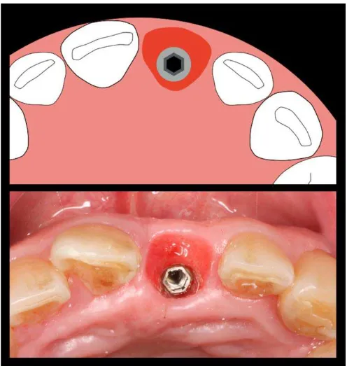
implant placement and uncovering surgery for abutment connection, soft tissue around the transmucosal part shows circular topography when observed from the occlusal aspect, as shown in Fig. 1, which is not in line with the innate shape of the gingiva.4In order to modify
this soft tissue topography so that it resembles the emergence profile of the soft tissue around a natural tooth, it should be altered to an expanded shape as shown in Fig. 2.5 To accomplish this configuration,
Figure 1. Just after uncovering surgery, peri-implant mucosa shows a circular topography.
Furze et al. conducted a study that evaluated whether tissue conditioning with provisional restoration has a significant impact on objective aesthetic outcome.6Twenty
patients were randomly allocated two groups; the test group received a provisional restoration and the emergence profile was altered, while the control group did not receive a provisional restoration before the final crown was delivered. After one year, successful integration of the implants was confirmed; the modified pink esthetic score (modPES), which assesses the peri-implant soft tissue on the basis of five variables (mesial and distal papilla, curvature of the facial mucosa, level of the facial mucosa, root convexity/soft tissue color, and texture at the facial aspect of the implant site), and the white esthetic score (WES), which evaluates the visible part of the implant restoration itself with five parameters (general tooth form, outline/volume of the crown, color, surface texture, translucency, and characterization by a score of 0, 1, 2), were evaluated.7,8The combined value of each parameter
for the test group (16.7± 2.06) was significantly higher than that for the control group (10.5 ± 3.31, p <0.05), suggesting that soft tissue conditioning by provisional restorations would be highly recommended from the point of view of aesthetics.
However, there are several concerns regarding the methods used to achieve excellent pink and white esthetics. First, since the favorable convexity/concavity has not been discussed on the basis of evidence very well, most of the clinicians or technicians design the outline of the emergence profile empirically. In addition, it has been demonstrated that multiple disconnections of the provisional restoration would undermine the peri-implant soft tissue attachment; this might lead to marginal bone loss around the implant, which has the potential to deteriorate esthetic outcomes. Regarding proximal papilla formation, apart from the provisional emergence profile considerable evidence suggests that the distance from prominence of the bone to the proximal contact plays a crucial role; thus, the clinician should have knowledge about the prerequisite for papilla formation.
For all these reasons, this narrative review discusses the basic techniques for adjustments of provisional restorations in relation with biological considerations, such as papilla formation, as well as the effect of abutment disconnection on the surrounding tissue and concavity of transmucosal part, in order to achieve aesthetically
successful outcomes. Alternative techniques by involving specific components or digital technologies are introduced as well.
Technical
Perspective:
Adjusting
Emergence Profile
The emergence profile is contoured according to the following principle steps:9
Facial emergence: Starting from the implant shoulder, with a slightly flat/concave profile, towards the height of convexity at the point where the mucosal margin will be established
Interproximal emergence: Starting from the implant shoulder, with a straight emergence, towards a slight convexity just apical to the contact area, providing support for the interproximal tissue.
Palatal emergence: Starting from the implant shoulder, with a straight to slightly convex emergence, towards the mucosal margin, focusing on matching the palatal contours of the adjacent teeth so that there is a smooth transition between the two.
In order to achieve the emergence profile described by the steps above, Wittneben et al. introduced a technique for conditioning the soft tissue around the implant prosthesis and the emergence profile, which is the so called “dynamic compression technique”.10This technique
at involves pushing and compressing the mesial and distal proximal peri-implant papillae by means of an over contoured provisional restoration. Selective pressure is applied by adding the material on selected sites, thus causing ischemic changes in the peri-implant mucosa. After two weeks, for modifying the shape of the soft tissue, some amount of material is removed from around the interproximal and cervical areas. This creates space for the soft tissue and allows the papillae to shift into the prepared space.
Convexity at The Transmucosal Part
Several studies have been conducted to identify the correlation between the convexity/concavity of emergence profile and the peri-implant tissue reaction. Huh et al. compared three types of transmucosal profiles for different implant surfaces, straight-machined implants (SM), machined implants (CM), or concave-roughened implants (CR), in beagle dogs. In radiographic and histometric evaluation, the least bone resorption was observed for CM implants, and SM implants were associated with the greatest bone resorption (p <0.05). Further, histometric analysis showed that the highest connective tissue attachment was observed around CM implants.12
From the aesthetic point of view, a straight slope of the emergence profile can cause apical migration of the free gingival margin.13 Therefore, in order to obtain
symmetric mucosal margin around implant, concavity of the root form would be suitable for the labial aspect.
However, in a study by Sancho-Puchades et al., which compared two abutment designs (concave or convex) in terms of cement remnants after cementation of prosthesis on individualized abutments and cement removal,14the concave abutments presented significantly
more cement remnants than CV abutments in the entire abutment area. This study implies that an emergence profile with excessive concavity makes it difficult to eliminate excess cement; this remaining cement may lead to adverse effects, such as peri-implant disease. Considering the above findings, it may be concluded that the transmucosal part, especially the labial aspect, should keep convexity insofar as it not too convex to cause functional problems.
Relationship With Proximal Bone
Height, Papilla Filling, and Contact
Point
It is well known that distance from the proximal contact point to the crest of the bone has significant effect on the interproximal papilla adjacent to the implant restoration. Choquet et al. conducted a clinical and radiographic retrospective evaluation of the papilla level around single dental implants, and their adjacent tooth
was performed in the anterior maxilla in 26 patients restored with 27 implants.15When the measurement from
the contact point to the crest of the bone was 5 mm or less, the papilla was present almost 100% of the time. When the distance was ≥6 mm, the papilla was present 50% of the time or less. Degidi et al. and Lops et al. evaluated the incidence of inter-proximal papilla between a tooth and an adjacent immediate implant placed into a fresh extraction socket in 1-year prospective study.16
Among forty-six patients with a total of 46 teeth scheduled for tooth extraction and immediate implant placement, when inter-implant–tooth distance was 3–4 mm, and the distance from the base of the contact point to the inter-dental bone was 3–5 mm, the inter-proximal papilla was significantly present (p <0.05). Therefore, clinicians should predict the prospects of papilla filling according to the patient’s clinical situation and discuss the expected final result with patients in advance.
Effect of Abutment Disconnection
and Reconnection
Along with the adjustment of emergence profile, the clinician needs to remove and connect the provisional restoration several times. Some experiments suggest that disconnection or reconnection of the provisional restoration or abutment can jeopardize the integrity of the peri-implant tissue.
Abrahamsson et al. in their experiment on a dog studied this effect on the marginal peri-implant tissues following repeated abutment removal and subsequent reconnection.17 They installed Branemark implants,
which has an external platform-matching connection, to the beagle dog’s bilateral mandibular premolar area. On one side, the abutment was disconnected and reconnected 5 times during the 6 months of observation period, whereas the other abutment was remained as it was. According to the histomorphometric analysis, the reconnected group showed more apical connective tissue attachment and marginal bone resorption compared to the intact group.
accompanied with 1-4 times of abutment dis/reconnection.18 Rodríguez et al. compared
platform-switched and platform-matched implant with regard to the effect on horizontal (H) and vertical (V) bone resorption with abutment dis/reconnection performed 1-4 times.18 For simplicity, we have named the groups as
follows: PM-1, platform-matched (single abutment disconnection); PM-4, platform-matched (abutment disconnection performed 4 times); PS-1, platform-switched (single abutment disconnection); and PS-4, platform-switched (abutment disconnection performed 4 times). The average horizontal and vertical bone resorption were as follows: 1 (H: 0.31, V: 0.72), PM-4 (H: 0.98 mm, V: 1.09 mm), PS-1 (H: 0 mm, V: 0.03 mm), and PS-4 (H: 0.37 mm, V: 0.41 mm). Comparing PM-1 with PM-4, there were no significant differences. Paris of PM-1/PS-1, PM-4/PS-4, and PS-1/PS-4, the extent of bone resorption was significantly different (p <0.05). However, the difference in bone resorption between PS-1 and PS-4 seems clinically insignificant.
Esposito et al. compared a repeated disconnection group and a no disconnection group in a multicenter randomized controlled trial.19 Patients requiring one
single crown or one fixed partial prosthesis supported by a maximum of three implants were treated in four centers, and each patient was followed up for 1 year after initial loading. They concluded that one year after loading, although repeated dis/reconnection of the abutment significantly increased bone loss of 0.16 mm, this difference is clinically negligible; thus, clinicians can use the procedure they prefer. Considering the results of these studies, the literature generally suggests that the shape of the connection has a more significant effect on peri-implant tissue than the number of times dis/reconnection of the abutments is performed.
Alternative Techniques for Emergence
Profile
Since the stepwise conditioning procedure requires multiple sessions and prolonged chair-time, several methods have been investigated for achieving optimal
emergence profile without involving a time-consuming procedure.
Becker et al. introduced a technique utilizing a prefabricated emergence profile.20Neoss Implant System
(Neoss Ltd, Harrogate, North Yorkshire, UK) employs a standard root measurement of six maxillary anterior teeth at the CEJ and duplicated in PEEK material (polyether ether ketone), for a customized healing abutment. This material is flexible and fits according to the patient’s specific anatomy. After implant placement surgery or uncovering surgery, the abutment is seated and the height and shape are adjusted. Thus, after the healing period, the optimal emergence profile is already shaped according to the customized abutment. This reduces chair-time and the number of sessions required for stepwise conditioning of the provisional restoration.
Joda et al. introduced a technique to fabricate individualized CAD/CAM healing abutment prior to uncovering surgery.21 According to their method, the
shape of the emergence profile of the contralateral tooth is copied from the DICOM data. After implant placement, digital impression with an intra-oral optical scan (IOS) is performed to identify the final three-dimensional position of the implant. Digitally flipped (mirrored) DICOM data of contra-lateral tooth and the STL-file of the IOS are superimposed to fabricate an individualized healing abutment using CAD/CAM from PMMA-based material, which is delivered to the patient at the uncovering surgery.
Vafiadis et al. introduced a digital fabricating method for immediate implant placement and immediate loading protocol.22 Their method involves copying the shape of
the tooth to be extracted and its emergence profile using the preoperative CBCT image. The data obtained is used to fabricate a crown-root matrix (resin shell) by CAD/CAM. The matrix is connected with the temporary abutment intra-orally and used as an immediate provisional restoration.
Conclusion
A clinician is responsible for determining the treatment needs and establishing aesthetically and functionally optimal implant prosthesis by conditioning of the emergence profile at the provisional state. According to the currently available evidence, labial emergence should have some concavity to accommodate sufficient peri-implant mucosa, while the palatal emergence may be convex in order to align harmoniously with neighboring teeth. Proximal profile should be controlled with regard to the height of the proximal bone because it has a critical role in papilla formation. Although it has been suggested that repeated disconnection of the abutments should be avoided in view of peri-implant soft tissue preservation,17literature suggests its effects in terms of
causing peri-implantitis are limited. Within the limitations of this narrative review, it is evident that comprehension of aesthetics, peri-implant tissue biology, and prosthetic procedure is crucial.
References
1. Papaspyridakos P, Chen C-J, Singh M, Weber H-P, Gallucci GO. Success criteria in implant dentistry: A systematic review. J Dent Res. 2015:242–8. DOI: 10.1177/0022034511431252
2. Jung RE, Zembic A, Pjetursson BE, Zwahlen M, Thoma DS. Systematic review of the survival rate and the incidence of biological, technical, and aesthetic complications of single crowns on implants reported in longitudinal studies with a mean follow-up of 5 years. Clin Oral Implants Res. 2012;23:2–21. DOI: 10.1111/j.1600-0501.2012.02547.x
3. Cosyn J, Thoma DS, Hämmerle CHF, De Bruyn H. Esthetic assessments in implant dentistry: objective and subjective criteria for clinicians and patients. Periodontol 2000. 2017;73:193–202.
4. Gallucci GO, Belser DMDUC, Dent PM. Modeling and characterization of the cej for optimization of esthetic implant design. Int J Periodontics Restorative Dent. 2004;24:19–29.
5. Priest G. Developing optimal tissue profiles with implant-level provisional restorations. Dent Today. 2005;
24(11):96,98,100.
6. Furze D, Byrne A, Alam S, Wittneben JG. Esthetic outcome of implant supported crowns with and without peri-implant conditioning using provisional fixed prosthesis: A randomized controlled clinical trial. Clin Implant Dent Relat Res. 2016;24:1153–62. DOI: 10.1111/cid.12416
7. Belser UC, Grütter L, Vailati F, Bornstein MM, Weber H-P, Buser D. Outcome evaluation of early placed maxillary anterior single-tooth implants using objective esthetic criteria: A cross-sectional, retrospective study in 45 patients with a 2-to 4-year follow-up using pink and white esthetic scores. J Periodontol. 2009;80:140–51. DOI: 10.1902/jop.2009.080435
8. Fürhauser R, Florescu D, Benesch T, Haas R, Mailath G, Watzek G. Evaluation of soft tissue around single-tooth implant crowns: the pink esthetic score. Clin Oral Implants Res 2005;16:639–44. DOI: 10.1111/j.1600-0501.2005.01193.x
9. Chappuis V, Martin W. Implant therapy in the esthetic zone – current treatment modalities and materials for single-tooth replacements. In:Barter S, Chen S, Wismeijer D, editors. ITI Treatment Guide. Volume 10. Basel: Quintessence Publishing Company; 2017.
10. Wittneben J-G, Buser D, Belser UC, Brägger U. Peri-implant soft tissue conditioning with provisional restorations in the esthetic zone: the dynamic compression technique. Int J Periodontics Restorative Dent. 2013;33:447–55. DOI: 10.11607/prd.1268.
11. Morton D, Chen ST, Martin WC, Levine RA, Buser D. 5th ITI consensus statements and recommended clinical procedures regarding optimizing esthetic outcomes in implant dentistry. Int J Oral Maxillofac Implants.2014;29 Suppl:216-20. DOI: 10.11607/jomi.2013.g3.
12. Huh J-B, Rheu G, Kim Y, Jeong C, Lee J, Shin S-W. Influence of implant trans mucosal design on early peri-implant tissue responses in beagle dogs. Clin Oral Implants Res. 2014;25:962–8. DOI: 10.1111/clr.12179 13. Kinsel RP, Pope BI, Capoferri D. A review of the positive
influence of crown contours on soft-tissue esthetics. Comped Contin Educ Dent. 2015; 36(5):352–7.
14. Sancho-Puchades M, Crameri D,ŐzcanM, Sailer I, Jung RE, Hämmerle CHF, et al. The influence of the emergence profile on the amount of undetected cement excess after delivery of cement-retained implant reconstructions. Clin Oral Implants Res. 2017;28(12):1515-22. DOI: 10.1111/clr.13020
dental implants. aretrospective study in the maxillary anterior region. J Periodontol. 2001;72(10):1364–71. DOI: 10.1902/jop.2001.72.10.1364
16. Lops D, Chiapasco M, Rossi A, Bressan E, Romeo E. Incidence of inter-proximal papilla between a tooth and an adjacent immediate implant placed into a fresh extraction socket: 1-year prospective study. Clin Oral Implants Res. 2008;19(11):1135–40. DOI: 10.1111/j.1600-0501.2008.01580.x
17. Abrahamsson I, Berglundh T, Lindhe J. The mucosal barrier following abutment dis/reconnection. anexperimental study in dogs. J Clin Periodontol. 1997;24:568–72.
18. Rodríguez X, Vela X, Méndez V, Segalà M, Calvo-Guirado JL, Tarnow DP. The effect of abutment dis/reconnections on peri-implant bone resorption: a radiologic study of platform-switched and non-platform-switched implants placed in animals. Clin Oral Implants Res. 2013
Mar;24(3):305–11. DOI:
10.1111/j.1600-0501.2011.02317.x
19. Esposito M, Bressan E, Grusovin MG, D'Avenia F, Neumann K, Sbricoli L, et al. Do repeated changes of
abutments have any influence on the stability of peri-implant tissues? one-year post-loading results from a multi centre randomised controlled trial. Eur J Oral Implantol. 2017;10:57–72.
20. Becker W, Doerr J, Becker BE. A novel method for creating an optimal emergence profile adjacent to dental implants. J Esthet Restor Dent. 2012;24(6):395–400. DOI: 10.1111/j.1708-8240.2012.00525.x
21. Joda T, Ferrari M, Brägger U. A digital approach for one-step formation of the supra-implant emergence profile with an individualized CAD/CAM healing abutment. J Prosthodont Res. 2016;60(3):220–3. DOI: 10.1016/j.jpor.2016.01.005
22. Vafiadis D, Goldstein G, Garber D, Lambrakos A. Immediate implant placement of a single central incisor using a cad / cam crown-root form technique: provisional to final restoration. J Esthet Restor Dent. 2017;29(1):13-21. DOI: 10.1111/jerd.12265