In Vitro Immunomodulatory Activity of Aqueous Extract of
Moringa oleifera Lam. Leaf to the CD4 +, CD8
+and
B220
+Cells in Mus musculus
Indriya Rachmawati and
Muhaimin Rifa’i
*
Department of Biology, Faculty of Mathemathic and Natural Sciences, University of Brawijaya, Malang, Indonesia
Abstract
Immune system is a system of biological structures and processes within an organism that protects against disease. It can be promoted by substance referred as immunomodulator. Usually people use synthetic drug or synthetic immunomodulator to get quick response against the disease. This habit lead to arising side effects such as nausea, bone marrow degradation, thrombocytopenic purpura and agranulocytosis. As alternative, natural immunomodulator derived from active compound in plant. The objectives of this study are to determine the effect of aqueous extract of Moringa oleifera Lam. leaf to the population of CD4+ and CD8+ T cells, and also B220+ cellon Mus musculus through in vitro study and to analyze the difference of immune response in treatment and non-treatment group (control). In this experiment we used spleen from Balb/C mice. Cells were grown in RPMI medium with three doses (10 µg/ml, 1 µg/ml, 0,1 µg/ml) of M.oleifera extract. The cells were grown for four days culture in the CO2 incubator at 37°C with 5 % CO2. The cells number and expression were analyzed by flowcytometry. Data was analyzed using one-way ANOVA with
α=0,05 by SPSS 16.0 for windows with complete randomized design. The result shows that the extract has immunostimulant activity and the low dose (0,1 µg/ml) can increase the cell number of CD4+ and CD8+, while high dose (10 µg/ml) significantly increase B220+ cells compared to the control. This result strenghten that M.oleifera has immunomodulator activity to immunity system and worth to be developed into medicinal drug.
Keywords: immunomodulator, in vitro, Moringa oleifera, T cell
INTRODUCTION
Human health can not be separated from the surrounding environtment. The number of infectious pathogens such as viruses, bacteria and fungi can easily attack if the body is in an unfit state due to weak immune system (deficiency) and will lead to arising many diseases. People often deal the emerging diseases with antibiotics or other synthetic drug treatment to get quick healing effect. This habit will lead to another problem such as antibiotic and syntetic drug resistance. Besides that, certain drugs can produce side effects such as nausea, bone marrow damage, thrombocytopenic purpura, and agranulocytosis that can lead to other diseases. Herbs are safe to use with no side effects and in some cases is the most effective choice [1]. Because of that case, many species of medicinal plants are widely used in herbal medicine [2].
Correspondence author:
Muhaimin Rifa’i
Email : [email protected]
Address : Laboratory of Animal Physiology, University of Brawijaya, Jl. Veteran, Malang, 65145
Prevention of diseases and free radicals which attack the body can be done by modulating the immune system. Immunomodulator is a substance which able to modulate the function and activity of the immune system. Moringa oleifera Lam. is one of the species which known for having the immunomodulatory activity to the immune system [3].
Moringa oleifera Lam. is Family Moringaceae
that most known and widely distributed in the world. This plant has a high value because almost all parts of the plant (leaf, roots, stems, flowers, fruit peel and seeds) can be used as highly nutritious food and also has been reported to have antimicrobial compound [4]. This plant also serves as an immune system builder and is used in some countries to overcome malnutrition and malaria [5]. This herb was choosen due to its rich phytochemical compound, which including saponins, carotenoids, phenolic compounds and flavonoids. Saponin and flavonoid serve as natural immunomodulator that expected to enhance lymphocytes cells development which is very important in immunity system [6].
showed immunomodulatory effects because it was able to increase the number of the cell population of HSC (CD34+), B lymphocytes (B220+), precursors of erythrocytes (TER119+ VLA-4+), mature erythrocytes (TER119+), expression of naive T cells (CD62L+) and proinflammatory cytokines IFN-γ. Therefore, this research aims to assess the effect of aqueous extract of M. oleifera Lam. leaf to the population of T cell CD4+, CD8+ and B220+ on Mus musculus through in vitro study and to analyze the difference of immune response in treatment and non-treatment group (control).
MATERIALS AND METHODS
This research was conducted on September 2013 to March 2014 at Laboratory of Animal Physiology, Department of Biology and Biomedical Laboratory, Faculty of Medicine, University of Brawijaya.
Material and Equipments
The materials which is used are aqueous extract of M. oleifera leaf, Mus musculus Balb/C, RPMI 1640 medium, sterile water, α-CD3, FBS (fetal bovine serum), 2-mercaptoethanol, antibiotics (penicillin - streptomycin), PBS, 70 % alcohol, and monoclonal antibodies (FITC conjugated rat anti-mouse CD4, PE conjugated rat anti - mouse CD8, PE/Cy5 conjugated rat anti - mouse B220.
The equipments used for this research are erlenmeyer glass, medium bottles (Schott), CO2
incubator, freeze drying machine, autoclave, microscope, microtube, milliphore membrane, micropipette, syringe, propylene tube, centrifuge, BD FACS Calibur ™ flow cytometer, petri dish, wire, surgical scissor, cuvettes, LAF (laminar air flow), haemocytometer and 48 wells culture dish.
Moringa oleifera extraction
Moringa oleifera leaf was obtained from Tirtomarto Village, District Ampelgading, Malang in August 2013. This method was performed in the Laboratory of Biochemistry, Department of Chemistry, Brawijaya University. Moringa oleifera
leaf was successively extracted with water by maceration method. Extraction process was done by taking the mature leaf, washed and air-dried, then crushed into powder to obtain the simplisia form. The result of this extraction process was in gel form. To obtain the dry extract, it was freeze dried for ± 24 hours.
Cell Isolation
Mus musculus used in this study were male mice, Balb/C strain. It was killed by neck dislocation. After dislocation, the mice were dissected using surgical scissor on a surgical board in the dorsal part, and then the spleen was taken and washed with PBS.
Spleen was placed in a petri dish containing PBS ± 2ml. The organ then crushed with a syringe base, clockwise until the organ was destroyed. The homogenat was put in a 15 ml propylene tube filtered with wire to take out the debris. PBS was added to the tube untill 12 ml. After that, cell was centrifuged at 2500 rpm for 5 min and 4⁰C. The supernatant was discarded and the pellet was taken. Pellet was resuspended by micropippete in 1 ml of RPMI media. Then it was taken 20 μl into a microtube, homogenized with 80 μl evans blue dye, and placed in a haemocytometer to count the number of living cells by microscope. Living cells were characterized by cells that were not stained blue. The number of cells needed for culture was 3x10⁶ cells for each well.
Cell Culture and Treatment
The cells were grown in RPMI 1640 media supplemented with 10% fetal bovine serum (FBS), antibiotic (penicillin & streptomycin), 1% α -CD3, 1 μl 2-mercaptoethanol, respectively. The media was mixed with three doses of M.oleifera
extract (0,1 μg/ml, 1 μg/ml, 10 μg/ml) and 0 μg/ml (control), and seeded into 48 wells culture dish. Each well contains approximately 1 ml of medium and cells suspension. All process were done in Laminar Air Flow (LAF) with sterile room and aseptic methods to avoid contamination. The equipments for cell culture were under sterile condition by autoclaving in 121⁰ C. Total of 70% alcohol was used for sterilized the LAF surface. Furthermore, the dish was kept at CO2 incubator
with temperature 37⁰C, 5% CO2 for four days.
Cell Harvesting
discarded and the pellet was taken. Pellet ready to be labelled with specific antibody.
Antibody staining
The extracellular staining was conducted by antibody and divided into two groups of staining, group A (CD4+, CD25+, B220+) and group B (CD4+, CD8+, CD62L+). It was done with added 50 mL specific antibody to the pellet of second centrifugation, pipetting, incubated in an ice box, added 300 mL PBS and transferred to flow cytometry cuvet.
Flow Cytometry Analysis
Flowcytometric analysis was to determine the cell number of CD4+, CD8+ and B220+. After transfer into the flow cytometry cuvet, samples were ready for analysis according to the parameters that had been set in the flow cytometry FACS Calibur machine. Obtained result analyzed by Cell Quest ProTM software and data was analyzed using one-way ANOVA with α=0,05 by SPSS 16.0 for windows with complete randomized design.
RESULT AND DISCUSSION
Absolute Cells Number of CD4+, CD8+ and B220+
in the Spleen of Mus musculus
The aqueous extract of M.oleifera leaf that was given in lymphocytes of Balb/C mice showed immunomodulatory activity as immunostimulant. This activity can be seen through the increase in the cells number of CD4+ (T helper cells), CD8+ (T cytotoxic cells) and B220+ cells in different doses. CD4+ and CD8+ are surface molecules which present in the activated-T cells. T cell activation can be caused by antigen infection.
In normal cells, the CD4+ and CD8+ molecule should not be excessive expressed. Excessive CD4+ and CD8+ molecules expression in normal cells indicated reactivity is present and that should be under apoptotic to mantain the homeostatic condition in the body.
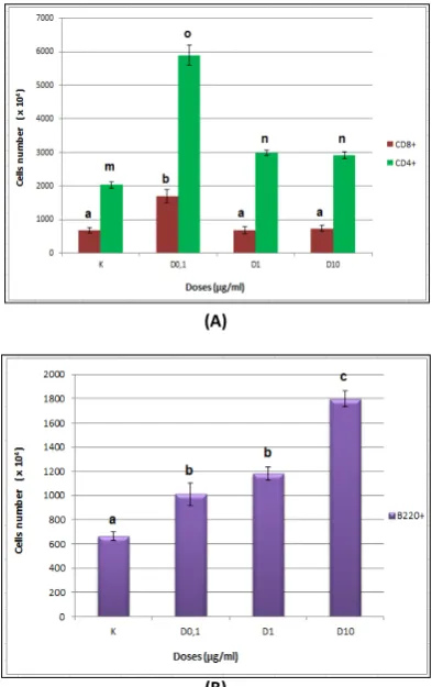
The effective dose to stimulate highest cell proliferation in CD4+ and CD8+ cells is in the lowest dose (0,1 µg/mL), but the effective dose in B220+ is in the highest dose (10 µg/ml) instead. The number of CD4+ cells in control is 20x106 cells, in 0,1 µg/ml, 1 µg/ml and 10 µg/ml is 59x106, 30x106 and 29x106 cells, respectively. The number of CD8+ cells in control is 6,82x10⁶ cells, in 0,1 µg/ml, 1 µg/ml and 10 µg/ml is 17x106, 6,8x10⁶ and 7,3x10⁶ cells, respectively. While the number of B220+ cells in control is 6,6x10⁶ cells,
in 0,1 µg/ml,1 µg/ml and 10 µg/ml is 10x106, 12x106 and 18x106 cells.
(A)
(B)
Figure 1. Effect of aqueous extract of M. oleifera leafto the absolute cells number of A (CD4+ & CD8+) and B (B220+) after four days culture with dose 0 µg/ml (control), 0,1 µg/ml, 1 µg/ml, 10 µg/ml. Cells were isolated from the spleen of M.
musculus. Number of cells were analyzed by
flowcytometry.
Based on ANOVA, the absolute cell number of CD4+ T cell in all dose treatment shows significant difference compared to the control (p<0,05). However, dose 1 µg/ml and 10 µg/ml have no significant difference (p>0,05). The lowest dose (0,1µg/ml) stimulate the highest cell proliferation and in higher doses, the number of cell is decrease. It indicated that absolute cells number of CD4+ is affected by the difference of treatment (doses).
CD25+ through the cytokine secretion. It also stimulates B cell to secrete antibody [8].
One of the cytokines produced by CD4+ is interleukin-2 (IL-2), which is very essential in increasing proliferation of T cells and B cells [9]. IL2 will regulate cell communication to activate the other cells. The cytokine synthesis is initiated by gene transcription and occurs due to the presence of a stimulus, in this case the stimulus derived from active compounds in the extract of
M. oleifera. IL-2 is one of the earliest immune response of T helper cells after activated by a stimulus. These cells will also bind to the APC, which generally are macrophages after doing phagocytosis. It rise the immune response in the body [10].
The immunomodulatory activity of M.oleifera
also present in CD8+ cell. The lowest dose (0.1 µg/ml) can stimulate the highest cells proliferation. The high number of CD8+ cells also affected by CD4+ because it secrete IL2 and IFN-γ to induce the proliferation of CD8+ [11]. Figure 1 also tells us that the significant difference only identified as cytoplasmic antigen and will recognize and killed infected cells, so it is called cytotoxic T cells [12]. When these cells destruct the targets – which considered as foreign antigens, will cause systemic inflammation [9].
Cytotoxic T cells directly attack cells that contain foreign antigens or abnormal molecules on the surface. Cytotoxic T cells are particularly useful for attacking viruses because viruses often hiding from the immune system as it grow within infected cells. These cells recognize small fragments of the virus which is visible from the cell membrane and then kill the infected cells [13]. CD8+ has many subset with diverse function, e.g. subset CD8+CD122+ is a regulatory T cells which are naturally occurring [14]. This cell is indispensably important in the maintenance of immune system homeostasis and expressed in mice, but not identified in human. Human showed CD8+CXCR3+ instead, with same function with CD8+CD122+ [15].
Aqueous extract of M.oleifera leaf also shows immunostimulant activity in B cells (B220+). All of the three doses can increase the number of B220+ cells compared to the control. The number of B220+ cells increased significantly (p<0,05) due to higher dose given and the number of cells is the highest at the highest dose treatment (10 ug/ml). B220+ (CD45R) are a cell surface marker on B cells and expressed mostly in all cell B lineages. CD45 is a tyrosine phosphatase that participates in modulating the immune response in both B cells and T cells [16].
One of the factors that influence the activation of B cells is IL-2 produced by CD4+. When cells expressed high number of CD4+, which is in dose 0,1 mg/ml, it showed the lowest number of B220+ expression. Otherwise, when decreased expression of B220+ (downregulated) [17]. In the other word, B220+ is a marker for naive B cells. Naive B cells are the B cells that have not been activated by the presence of antigen. IL-2 secreted by CD4+ help the B cell activation, resulting in the higher secretion of IL-2, then the cell surface expression of B220+ will decrease. This is because the activated B cell will have decrease expression of B220+.
Immunostimulatory activity found in the increasing number of CD4+, CD8+ and B220+ due to the active substances such as saponins and flavonoids in the aqueous extract of M. oleifera
leaf [18]. Cell proliferation induced by lymphocyte respond can be caused by exogenous stimuli in the form of the active compounds of plants which act as immunostimulator [11,19]. The active compounds in M.oleifera act as MAPK (mitogen activated protein kinase), which will stimulate T and B cell proliferation through the expression of IL-2 [7].
secretory molecules in macrophages and neutrophils and this compound also plays role in activation of these cells [23].
Cells Expression Profile of CD4+, CD8+ and B220+
by Flow Cytometry Analysis
The profiles showed the expression of relative cell number of CD4+, CD8+ and B220+ (Fig. 2 and 3). The expression of CD4+ on Fig. 2 shows that the lower dose (0,1 µg/ml) can stimulate CD4+ expression (54,09%), but the effective dose which can stimulate the highest cell number is dose 1 µg/ml (58, 71 %).
The lowest expression is in the dose 10 µg/ml (51,50%), while in control is 50,20%. Therefore, in CD8+, the dose with highest cells expression is in the lower dose (15, 57%), compared with dose 1 µg/ml (13,43%) and 10 µg/ml (12, 86 %), while in control is 16,86%.
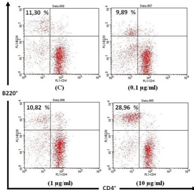
Expression of B220+ shows that the high dose of M. oleifera extract is effective to increase the B220+ expression (Fig. 3). The highest cell expression number is in the highest dose (10 µg/ml) (28,96%), while in dose 0,1 µg/ml is 10,82%, in 0,1 µg/ml is 9, 89 % and control is 11,30%. From those data, we can see the immunostimulant activity in the CD4+, CD8+ and B220+ cells.
Figure 2. Cells expression profiles of CD4+ and CD8+. This result obtained through flow cytometry and analyzed by Cell Quest ProTM software.
Figure 3. Expression profile of B220+ cells. This result obtained through flow cytometry and analyzed by Cell Quest ProTM software.
CONCLUSION
The aquoeus extract of M. oleifera leaf has activity as immunomodulator through it’s active compound, such as saponin and flavonoid, which act as an immunostimulant on CD4+ (T helper cell) and CD4+ (T cytotoxic cell), as well as B220+. The low dose (0, 1 µg/ml) is effective to increase the number of CD4+ and CD8+ cells, while high dose (10 µg/ml) is effective to increase the number of B220+ cells.
ACKNOWLEDGEMENT
The author thank Dewi Satwika, S.Si, M.Si, Ahmad Sony, S.Si, M.Si, Bambang P., S.Si, Dinia R.D., S.Si and animal physiology laboratory team for the support in conducting this research.
REFERENCES
[1] Sharififar, F., S. Pournourmohammadi, M. Arabnejad. 2009. Immunomodulatory activity of aqueous extract of Achillea wilhemsii C. Koch in mice. Indian Jounal of Experimental Biology. 47. 668-671.
[2] Koruthu, D.P., N.K. Manivarnan, A. Gopinath, A. Abraham. 2011. Antibacterial evaluation, reducing power assay and phytochemical screening of M. oleifera leaf extract: effect of solvent polarity.
International Journal of Pharmaceutical Sciences and Research. 2 (11). 2991-2995. [3] Gupta, A., M.K. Gautam, R.K. Singh, M.V.
2010. Immunomodulatory effect of M. oleifera extract on Cyclophosphamide induced toxicity in mice. Indian Jounal of Experimental Biology. 48. 1157-1160. [4] Moyo, B., P.J. Masika, V. Muchenje. 2012.
Antimicrobial activities of Moringa oleifera
Lam leaf extracts. African Journal of Biotechnology. 11(11). 2797-2802.
[5] Thilza, I.B., S. Sanni, Z.A. Isah, F.S. Sanni, M. Talle, M.B. Joseph. 2010. In Vitro antimicrobial activity of water extract of M. oleifera leaf stalk on bacteria normally implicated in eye diseases. Academia arena. 2(6).
[6] Wagner, H. 1999. Immunomodulator agents from plants. Institut fur Pharmazeutische Biologie. Munchen.
[7] Hefni, M. 2013. Imunomodulator activity of aqueous extract of M. oleifera Lam on immunity response of mice (Mus musculus)
which infected with Salmonella typhi. Master Thesis. University of Brawijaya. Malang.
[8] Bell, J., D. Gray. 2003. Antigen-capturing Can Masquerade as Memory B Cells. Journal Exp. Med. 197 (10). 1233-1244.
[9] Rifa'i, M. 2011. Immunology and Bioregulator. UB Press. Malang.
[10] Campbell, N.A., J.B. Reece, L.G. Mitchell. 2003. Biology Fifth Edition. Benjamin Cummings. New York.
[11] Rifa'I, M., Z. Shi, S.Y. Zhang, Y.H. Lee, H. Shiku, K. Isobe, H. Suzuki. 2008. CD8+CD12+ regulatory T cells recognize activated T cells via conventional MHC class I–αβTCR interaction and become IL-10-producing active regulatory cells. International immunology. 20 (7). 937-947.
[12] Rifa’i, M., Y. Kawamoto, I. Nakashima, H. Suzuki, 2004. Neuroprotective Effects of Ginsenoisides. Journal Acta. Neurobiol. Exp (Wars). 66. 369-375.
[13] National Institute of Health. 2008. Immune system: T cells. U.S Department of Health and Human Services. http://nih.com. [14] Shi, Z., M. Rifa’i, Y.H. Lee, H. Shiku, K. Isobe,
and H. Suzuki. 2008. Importance of CD80/CD86–CD28 interactions in the recognition of target cells by CD8+CD122+ regulatory T cells. Immunology. 124. 121-128.
[15] Shi Z., Y. Okuno, M. Rifa'i, A.T. Endharti, K. Akane, K. Isobe, H. Suzuki 2009. Human CD8+CXCR3+ T cells have the same function
as murine CD8+CD122+ Treg. European Journal of Immunology. 39 (8). 2106-2119. [16] Cascalho, M., J. Wong, J. Brown, H. Jack, C.
Steinberg, M. Wabl. 2000. A B220-, CD19 -Population of B Cells in The Peipheral Blood of Quasimonoclonal Mice. International Immunology. 12(1). 29-35.
[17] Rolink, A., E.T. Boekel, F. Melchers, D.T. Fearon, I. Krop, J. Anderson. 1996. A subpopulation of B220 + Cells in murine bone marrow does not express CD19 and contains natural killer cell Progenitors. J. Exp. Med. 183. 187-194.
[18] Biswas, S.K., A. Chowdury, D. Joysre, R. Ajoy, H. Zahid. 2012. Harmacological potential of
Moringa oleifera Lam. a review.
International Journal of Pharmaceutical Sciences and Research. 3(2). 305-310. [19] Endharti, A.T., M. Rifa'I, Z. Shi, Y. Fukuoka,
Nakahara, Y. Kawamoto, K. Takeda, K. Isobe, H. Suzuki. 2005. Cutting edge: CD8+CD122+ regulatory T cells produce IL-10 to suppress IFN-gamma production and proliferation of CD8+ T cells. Journal of immunology, 175 (11). 7093-7097.
[20] Reichert, T.E., S. Nagashima, Y. Kashii, J. Stanson, G. Gao, P.Q. Dou, T.L. Whiteside. 2000. Interleukin-2 expression in human carcinoma cell lines and its role in cell cycle progression. Journal of Oncogene. 19. 514-525.
[21] Middleton, E., C. Kandaswami, T.C. Theoharide. 2000. The effects of plant Flavonoids on mammalian cells: implications for inflammation, heart disease,and cancer. The American Society for Pharmacology and Experimental Therapeutics Pharmacol. Rev52. 673–751. [22] Rao, V.S.N., L.A.F. Paiva, R.M. Santos, L.A.
Da Silva, E.T. Gurgel, E.T. De Sousa, E.R. Silveira. 2003. Anti-inflammatory effect of Ternatin, a Flavonoid from Egletes viscosa
Less., in the rat model of Colitis induced by acetic acid. Boletín Latinoamericano y del Caribe de Plantas Medicinales y Aromáticas. 2(4). 48-51.