DOI: 10.5994/jei.10.2.93
KOMUNIKASI SINGKAT
The effect of DMSO on ITS2 amplification in the
molecular identification of
Anopheles farauti
Laveran
(Diptera: Culicidae), from a colony established
in the laboratory
Pengaruh DMSO terhadap amplifikasi ITS2 dalam identifikasi
molekular
Anopheles farauti
Laveran (Diptera: Culicidae), dari
koloni yang dikembangbiakan di laboratorium
Ign. Joko Suyono1,2*, Jesmandt Situmorang2, R.C. Hidayat Soesilohadi2,
Rarastoeti Pratiwi2
1Departemen Biologi, Fakultas Matematika dan Ilmu Pengetahuan Alam,
Universitas Cenderawasih, Jalan Kamp Walker Waena, Jayapura 99358
2Fakultas Biologi, Universitas Gadjah Mada
Jalan Teknika Selatan, Yogyakarta 55281
(diterima Oktober 2012, disetujui Mei 2013)
ABSTRACT
Sibling species identification is very important in the understanding of malaria epidemiology. Morphological criteria are usually used in the identification of anopheline species, but this fails when sibling or cryptic species occur. Analysis by PCR-RFLP of rDNA ITS2 is currently the most reliable and sensitive method for distinguishing between members of the Anopheles punctulatus group. The objective of this study was to investigate the effect of DMSO concentration on ITS2 amplification of An. farauti from the colony maintained at BATAN Jakarta using PCR-RFLP based on the rDNA ITS2. The results showed that the addition of 6 % and 7 % DMSO produced ITS2 amplification products in the size 750 bp. DMSO could be used in PCR to relieve secondary structures when amplifying high GC templates. Molecular identification of An. farauti is found to be Anopheles farauti sensu stricto.
Key words: molecular identification, sibling species, PCR-RFLP, rDNA ITS2, secondary structure
ABSTRAK
Identifikasi spesies sibling sangat penting untuk mempelajari epidemiologi malaria. Ciri morfologi secara umum digunakan dalam identifikasi spesies Anopheles, namun tidak sesuai ketika digunakan untuk spesies sibling dan spesies kriptik. Analisis dengan menggunakan PCR-RFLP dari ITS2 rDNA pada saat ini merupakan metode yang paling cocok dan sensitif untuk membedakan
spesies Anopheles punctulatus group. Tujuan penelitian ini adalah untuk mengetahui pengaruh
konsentrasi DMSO terhadap amplifikasi ITS2 dalam mengidentifikasi An. farauti dari koloni di BATAN Jakarta dengan PCR-RFLP berdasarkan ITS2 rDNA. Hasil penelitian menunjukkan bahwa penambahan DMSO dengan konsentrasi 6% dan 7% menghasilkan produk amplifikasi ITS2 dengan ukuran 750 pb. DMSO dapat digunakan dalam PCR untuk mencegah terbentuknya struktur sekunder
*Penulis korespondensi: Ign. Joko Suyono. Departemen Biologi, Fakultas Matematika dan Ilmu Pengetahuan Alam, Universitas Cenderawasih, Jalan Kamp Walker Waena, Jayapura 99358
Tel: +6281328886652, Faks: 0967-571115, Email: [email protected]
INTRODUCTION
The main malaria vectors in the southwest Pacific islands of Moluccas, Papua, Papua New Guinea, the Bismarck Archipelago, the Solomon and Vanuatu are the members of the Anopheles punctulatus group. The species belong to this group
originally are An. farauti, An. koliensis and An. punctulatus. These species could be differentiated
morphologically based on the pale scales of the proboscis. The proboscis of An. farauti entirely
dark scaled except for a narrow light ring at
extreme apex. An. koliensis with a ventral patch
of white scales on the apical third of the proboscis
and variable size, and An. punctulatus with the
apical half of the proboscis almost entirely white scaled (Rozebbom & Knigh 1946 cit Beebe et al. 2000a). Crossmating, cytogenetic, DNA probe and allozyme analyses now identified 12 species
within Anopheles punctulatus group: Anopheles farauti 1–7 (An. farauti complex), An. punctulatus,
An. koliensis, An. clowi, An. rennellensis and An.
species near punctulatus (Beebe et al. 2000a; Beebe et al. 2000b; Schmidt et al. 2003).
Identification of An. punctulatus group
commonly based on the proboscis morphology
and sector spot on the costa is proving to be quite
unreliable in differentiating them, making field studies on their biology and behavior of each of the members of the group difficult. Analysis by polymerase chain reaction-restriction fragment length polymorphism (PCR-RFLP) of the ribosomal DNA (rDNA) internal transcribed spacer 2 (ITS2) is currently the most reliable and sensitive method for distinguishing between members of the group. It is based on specific banding pattern for each of the sibling species (Beebe & Saul 1995; Benet et al. 2004).
The ITS2 region of An. punctulatus group has rapid evolution compared with the coding region.
This conserved sequence especially 3’ to the 5.8 rDNA and 5’ to the 28S rDNA can pair and form stable stem loops. Due to this strong secondary structure, 10% dimethyl sulfoxide (DMSO) was
used in the amplification reaction to eliminate the DNA secondary structure (Beebe & Saul 1995). But there is limited discussion on DMSO function and no information is given about how to run this
experiment.
Mosquito colonies maintained at The National Agency of Atomic Energy (BATAN) Jakarta were obtained from The Naval Medical Research Unit 2 (NAMRU 2) Jakarta. Especially for this An. farauti
colony which was maintained at BATAN, the name was based on its morphological identification, therefore the exact name of this sibling species remains unclear. In the former study conducted by Benet et al. (2004) reported that specimens that were identified morphologically as An. farauti
were consisted of An. farauti sensu stricto, An. farauti 4, An. farauti 2, An. koliensis and An. punctulatus by PCR-RFLP analysis, therefore is
very important to identify the An. farauti colony
from BATAN.
The aim of this study is to investigate the effect of DMSO concentration on ITS2 amplification in the identification of An. farauti complex species
from the colony maintained at BATAN Jakarta using PCR-RFLP.
METHODOLOGY
Sample preparation
An. farauti was obtained from a colony
maintained at BATAN Jakarta. Mosquitoes were preserved in vials and kept dry in tins containing silica gel for molecular identification using PCR-RFLP. The investigation was performed from July to September 2010 at the Laboratory of Genetic Engineering of Research Center for Biotechnology, University of Gadjah Mada Yogyakarta.
DNA extraction
The DNA was extracted by following the
procedure described in Li et al. (2005). Each
of mosquito specimens were individually homogenized in 50 µl of TE buffer consisting of 10 mM Tris-HCl, 1mM EDTA, pH 8. These ketika mengamplifikasi template dengan kandungan GC yang tinggi. Identifikasi molekular An. farauti dengan
menggunakan PCR-RFLP merupakan Anopheles farauti sensu stricto.
tubes were incubated at 100 °C for 10 minutes
and centrifuged at 9,500 rpm for 10 minutes. Supernatant was transferred to new tube and stored
at -20 °C for further use.
PCR for effect of DMSO on ITS2 amplification
DNA template of 3 µl was added directly to PCR mixture in final reaction volume of 25 µl that contained 50 mM KCl, 10 mM Tris-HCL, pH 8.3, 2.5 mM MgCl2, 0.2 mM dNTP, various
concentrations (4%, 6%, 7%, 8% and 10%) of DMSO, 1.25 unit of Taq polymerase and 0.6 µM
of each primer. The internal transcribed spacer 2 (ITS2) was amplified using forward primer
ITS2A, 5’-TGTGAACTGCAGGACACAT-3’
and reverse primer ITS2B, 5’-TATAGCTTAAA TTCAGGGGGT-3’ (Beebe & Saul 1995). A Perkin-Elmer type GeneAmp System 2400 thermocycler was used for all reactions with following parameters: an initial denaturation
at 94 °C for five minutes, and then 35 cycles at 94 °C for one minute, 53 °C for one minute, and 72 °C for two minutes, followed by a final extention
of 7 minutes at 72 °C. The ITS2 products were subjected to electrophoresis on a 1.5% agarose gel containing 0.5 µg/ml of ethidium bromide and visualized on an ultraviolet transilluminator. The ITS2 amplification results with optimum DMSO (6%) then was used for subsequent enzyme reaction analysis.
Product digestion and visualization
The ITS2 products were subjected to electrophoresis on a 1.5% agarose gel containing 0.5 µg/ml of ethidium bromide and visualized on an ultraviolet transilluminator. The PCR products were then digested with five units of Msp I (final
volume 20 µl) and the resulting products resolved
by electrophoresis on a 2% agarose gel containing 0.5 µg/ml of ethidium bromide and visualized on an ultraviolet transilluminator. The patterns of bands were compared to known patterns for An. punctulatus group (Beebe & Saul 1995).
RESULTS AND DISCUSSION
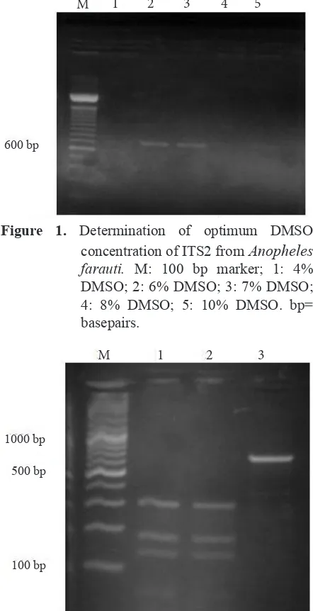
Various concentration of DMSO (4%, 6%, 7%, 8% and 10%) were investigated to define conditions resulting consistent amplification of
ITS2 product. Figure 1 shows that amplification of ITS2 had DMSO optima in 6% and 7%.
DNA from individual mosquitoes was used as a template for PCR amplification of the ITS2 after 35 cycles delivered products of approximately 750 bp (Figure 2 lane 3). Species-specific banding patterns were generated from an Msp I digestion
using one-fifth of the PCR mixture and could be visualized on a 2% agarose gel containing ethidium bromide. All the above samples produced bands at ≈ 300, 190, and 150-bp. (Figure 2 lane 1-2).
Figure 1. Determination of optimum DMSO
concentration of ITS2 from Anopheles farauti. M: 100 bp marker; 1: 4% DMSO; 2: 6% DMSO; 3: 7% DMSO; 4: 8% DMSO; 5: 10% DMSO. bp=
basepairs.
Figure 2. Polymerase chain reaction-restriction frag
-ment length polymorphism pattern for in
-ternal transcribed spacer 2 of Anopheles farauti from BATAN separated on a 2% agarose gel. M: 100 bp marker; 1-2: An. farauti sensu stricto; 3: Undigested ITS2 amplification product. 6% DMSO was used in the PCR reaction. bp= basepairs.
600 bp
500 bp
100 bp 1000 bp
M 1 2 3 4 5
Molecular identification of An. farauti from the
colonymaintained at BATAN Jakarta using
PCR-RFLP is An. farauti sensu stricto (formerly An. farauti No.1), confirming the PCR-RFLP result by
Beebe & Saul (1995).
Amplification of ITS2 occurred at 6 % and 7% of DMSO. The PCR products of ITS2 approximately 750 bp size. The PCR product of ITS2 also was detected at 10% DMSO with faint band. This result showed that the apparent inhibitory effect of increasing DMSO concentrations in PCR is presumably due to the inactivation of Tag DNA
polymerase (Gelfand 1989), so that more enzymes were required. In this study, 6% DMSO can be used to amplify PCR products ITS2 required 1.25
U Tag polymerase, but another study by Beebe &
Saul (1995) showed that used 10% DMSO for ITS2 amplification was required more Tag polymerase
enzyme concentration (2.5 U).
DMSO is an organosulfur compound with a high polarity and high dielectric constant, that is used in PCR to facilitate strand separation of double helix DNA by altering its melting characteristics, especially acts by disrupting inter and intrastrand re-annealing and then to disrupt secondary structure formation in the DNA template (Simon et al. 2009; Jansen et al. 2010). This is particularly useful in templates with high GC content, because the increased hydrogen bond strength increases the difficulty of denaturing the template and causes intermolecular secondary structures to form more readily, which can compete with primer annealing (Chakrabarti & Schutt 2001). Thus the addition of DMSO can greatly improve yields and specificities of PCR priming reactions.
The effect of DMSO that we observed in this study may be related in part to the destabilizing influence of DMSO on dsDNA. The presence of a high GC ratio would stabilize dsDNA in both PCR products and intramolecular secondary structures, and could inhibit the PCR. Moreover Bower et al. (2009) reported that the GC ratio ITS2 from An. punctulatus group varied between 64-71.3%. The
5’ end of ITS2 usually has multiple tandem copies of G-C rich repeat (63.5-73%), 109-188 bp that folds into a conserved stem structure.
The ITS2 from several anophelines, e.g. sibling species of the members of An. maculipennis
complex, An. funestus group and An. minimus
group could be amplified without addition of DMSO in PCR (Djadid et al. 2007; Garros et al. 2004; Van Bortel et al. 2000), because the ITS2 from these species has slightly low GC content, e.g. the ITS2 of An. maculipennis complex has a
GC content of 49.33-54.76% (Djadid et al. 2007). Therefore, the finding in this experiment may be applied by researcher who will do research on the
An. punctulatus group.
PCR-RFLP analysis based on the ITS2 region of the rDNA is successfully applied to mosquito colony from BATAN, although that technique has been modified by Suyono et al. (2011). Molecular identification result is An. farauti sensu stricto
(formerly An. farauti 1) which belongs to An. farauti complex included in An. punctulatus group. Sibling species within An. farauti complex are
very similar and can only be rediably differentiated by allozyme and DNA-based technology (Folley et al. 1993 cit Mueller et al. 2002; Beebe & Saul 1995). At present there is no reliable way of morphollogically identifying any of the members of An. punctulatus group and identification using
proboscis morphology should be approached with
great caution (Beebe et al. 2000a).
DMSO concentration is an important factor in the PCR reaction to amplify ITS2 of An. punctulatus group. DMSO could be used in PCR
to relieve secondary structures when amplifying high GC templates. Molecular identification of
An. farauti from the colonymaintained at BATAN
Jakarta using PCR-RFLP of ITS2 revealed to be
Anopheles farauti sensu stricto (formerly An. farauti 1).
ACKNOWLEGMENTS
We appreciate the assistance of Drs Nigel W. Beebe and Michael J. Bangs for their advice and suggestion to this research. We also like to thank Mr. Tony Ruwaedi for technical work in the laboratory and Mr. Ali Rahayu for providing us with mosquito’s laboratory colony in BATAN. This study was a part of the research project funded by DGHE Republic of Indonesia.
REFERENCES
Beebe NW, Cooper RD, Morrison DA, Ellis JT. 2000a. A phylogenetic study of Anopheles punctulatus group of malaria vectors comparing rDNA sequence alignments derived from the
mitochondrial and nuclear small ribosomal subunits. Molecular Phylogenetics and Evolution 17:430-436. doi: http://dx.doi.org/10.1006/ mpev.2000.0853.
Beebe NW, Cooper RD, Morrison DA, Ellis JT. 2000b. Subset partitioning of the ribosomal DNA small subunit and its effect on the phylogeny of the Anopheles punctulatus group. Insect Molecular Biology 9:515-520. doi: http://dx.doi.
org/10.1046/j.1365-2583.2000.00211.x.
Beebe NW, Saul A. 1995. Discrimination of all members of the Anopheles punctulatus complex
by polymerase chain reaction-restriction fragment length polymorphism analysis. American Journal of Tropical Medicine and Hygiene 53:478-481.
Benet A, Mai V, Bockarie V, Lagog M, Zimmerman P, Alpers MP, Reeder JC, Bockarie MJ. 2004.
Polymerase chain reaction diagnosis and the changing pattern of vector ecology and malaria transmission dynamic in Papua New Guinea. American Journal of Tropical Medicine and Hygiene 71:277-284.
Bower JE, Cooper RD, Beebe NW. 2009.Internal
repetition and intravidual variation in the rDNA ITS1 of the Anopheles punctulatus Group
(Diptera: Culicidae) multiple unit and rate turnover. Journal of Molecular Evolution. 68:66-79. doi: http://dx.doi.org/10.1007/s00239-008-9188-z
Chakrabarti R, Schutt CE. 2001. The enhancement of PCR amplification by low molecular-weight sulfones. Gene 274:293-298. doi: http://dx.doi. org/10.1016/S0378-1119(01)00621-7.
Djadid ND, Gholizadeh S, Tafsiri E, Romi R, Gordeev M, Zakeri S. 2007. Molecular identification of Paleartic members of Anopheles maculipennis in northern Iran. Malaria Journal 6:6. doi: http:// dx.doi.org/10.1186/1475-2875-6-6.
Garros C, Koekemoer LL, Coetzee M, Coosemans M, Manguin S. 2004. A single multiplex assay to identify major malaria vectors within the African Anopheles funestus group and the oriental An. minimus groups. American Journal of Tropical Medicine and Hygiene 70:583-590.
Gelfand DH. 1989. Thermus aquaticus DNA polymerase. In: Erlich HA, Gibbs R, Kazazian HH. (Eds.), Current Communications in Molecular Biology: Polymerase Chain Reactions. pp. 11-17. New York: Cold Spring Harbor Laboratory, Cold Spring Harbor.
Jansen MA, Fukushima M, Davis RW. 2010. DMSO and Betanine greatly improve amplification of GC-rich construct in de novo synthesis. PLoS ONE 5:e11024. doi: http://dx.doi.org/10.1371/ journal.pone.0011024.
Li C, Lee JS, Groebner JL, Kim HC, Klein TA, O’Guinn ML, Wilkerson RC. 2005. A newly
recognized in Anopheles Hyrcanus Group and molecular identification of related species from the Republic South Korea (Diptera: Culicidae). Zootaxa 939:1-8.
Mueller I, Taime J, Ibam E, Kundi J, Lagog M, Bockarie M and Reeder JC. 2002. Complex patterns of malaria epidemiology in the highland region of Papua New Guinea. Papua New Guinea Medical Journal 45:200-205.
Schmidt ER, Foley DH, Bugoro H, Bryan JH. 2003. A morphological study of the Anopheles punctulatus group (Diptera: Culicidae) in the
Solomon Islands, with a description of Anopheles (Cellia) irenicus Schmidt, sp.n. Bulletin of Entomological Research 93:515-526. doi: http:// dx.doi.org/10.1079/BER2003267.
Simon LS, Grierson LM, Naseer Z, Bookman AA, Shainhouse JZ. 2009. Efficacy and safety of topical diclofenac containing dimethyl sulfoxide (DMSO) compared with those of topical placebo, DMSO vehicle and oral diclofenac for knee
osteoarthritis. Pain 143:238-245. doi: http:// dx.doi.org/10.1016/j.pain.2009.03.008.
Suyono IJ, Rarastoeti, Hidayat RC, Situmorang J. 2011. Pengembangan PCR-RFLP berdasarkan ITS2 rDNA untuk identifikasi spesies sibling
anggota Anopheles punctulatus group dari Papua. Vektora 3:44-53.
Van Bortel W, Trung HD, Roelants P, Harbach RE, Backeljau T, Coosemans M. 2000. Molecular identification of Anopheles minimus s.l. beyond distinguishing the members of the species
complex. Insect Molecular Biology