Green synthesis of silver nanoparticles using latex extract of Thevetia peruviana: a novel
approach towards poisonous plant utilization
This article has been downloaded from IOPscience. Please scroll down to see the full text article.
2013 J. Phys.: Conf. Ser. 423 012032
(http://iopscience.iop.org/1742-6596/423/1/012032)
Download details:
IP Address: 114.79.58.234
The article was downloaded on 16/04/2013 at 13:20
Please note that terms and conditions apply.
Green synthesis of silver nanoparticles using latex extract of
Thevetia peruviana: a novel approach towards poisonous plant
utilization
N Nyoman Rupiasih1,3,4, Avinash Aher2, Suresh Gosavi2 and P B Vidyasagar2
1
Department of Physics, Faculty of Mathematics and Natural Sciences, Udayana University, Kampus Bukit Jimbaran, Badung, Bali 80362, Indonesia
2
Department of Physics, University of Pune, Ganeshkhind, Pune-411007, India
E-mail: [email protected]
Abstract. The development of green synthesis of nanoparticles has received increasing
attention due to ease of preparation, less chemical handling, and eco-friendly. In the present study, silver nanoparticles (AgNPs) were successfully synthesized from AgNO3 through a simple green synthesis route using the latex of Thevetia peruviana as reducing as well as capping agent. Synthesized silver nanoparticles were characterized using UV-vis, FTIR, SEM-EDS, XRD and HRTEM. UV-vis spectra showed absorption at 570 nm with shoulder at 395 nm. FTIR spectra analysis confirmed the functional groups involved in the AgNPs formation. X-ray diffraction pattern of AgNPs exhibited the diffraction at 2θ = 34.48°, 40.12° and 45.39° corresponding to (222), (400), and (420) Braggs reflection planes of silver nanoparticles. TEM analysis showed the particles size is spherical in nature with its size distribution between 10- 30 nm. The HRTEM image showed 2.59Å interplaner spacing.
Keywords: Green synthesis, Thevetia peruviana, Latex, Silver nitrate, Silver nanoparticles
1.Introduction
The field of nanotechnology is one of the most active areas of research in recent materials science. Nanoparticles are often referred to as clusters, nanospheres, nanorods and nanocups of the size ranges from 1 to 100 nm which exhibit completely new or improved properties based on size-distribution, and morphology [1-3].
Nanoparticles with controlled size and composition are of fundamental and technological interest as they provide solutions to technological and environmental challenges in the areas of solar energy conversion, catalysis, medicine, and water treatment. Global warming and climate change have induced a worldwide awareness and efforts to reduce generated hazardous wastes. Thus, “green”
3
Author to whom any correspondence should be addressed: [email protected]; 4Grant: the Ministry of Education and Culture Republic of Indonesia, No.: 0542/023.04.1.01/00/2011 date 5 June 2011 the 2nd DIPA Revision
ScieTech 2013 IOP Publishing
chemistry and chemical processes are progressively being integrated in science and industry for sustainable development [4-5]. One of them is synthesis of nanoparticles by biological process which implemented to develop safe, cost-effective and environmentally friendly technologies [4].
A great deal of effort has been put into the biosynthesis of inorganic material, especially metal nanoparticles using microorganisms and plants [1-2, 6]. Use of plant extract for the synthesis of nanoparticles could be advantageous over other environmental friendly biological processes by eliminating the elaborate process of maintaining cell cultures [4, 6-7]. Gardea-Torresdey and co-workers first reported the formation of gold and silver nanoparticles by living plants [8-9]. Their synthetic protocol by plant extract or biomass exemplifies the promising application of the green synthesis of metal nanoparticles. Very recently green silver nanoparticles have been synthesized using various natural products like green tea (Camellia sinensis), neem (Azadirachta indica) leaf broth, natural rubber, starch, aloe vera plant extract, lemongrass leaves extract, etc [7, 10]. The shape and size of the nanoparticles synthesized using plants can be controlled and modulated by changing the pH [2]. The reduction of silver ions (Ag+) in aqueous solution generally yields colloidal silver with particle diameters of several nanometers.
Medicinal herbs are the local heritage with global importance. It has curative properties due to the presence of various complex chemical substances of different composition, which are found as secondary plant metabolite in one or more parts of the plants [4-5, 7, 11]. These plant metabolites according to their composition are grouped as alkaloids, glycosides, corticosteroids, essential oils, etc.
Thevetia peruviana is an ever-green ornamental dicotyledonous plant that belongs to Apocyanaceae family. It is commonly found in the tropics and sub-tropics where as it is native to Central and South America [12]. The whole plant contains a milky juice (white latex) containing a compound called thevetin that is used as a heart stimulant but in its natural form is extremely poisonous. It’s poisonous owing to the presence of cardiac glycosides or cardiac toxins, which acts directly in the heart [12-14]. Ingestion of these plant parts could lead to death [12-13]. It is cultivated as ornamental tree standards in gardens and parks in temperate climates [15]. In the West Indies half a leaf is known as an emetic and purgative. In Java Indian immigrants are reported to smoke using the dried leaves. Tincture of bark is also emetic and purgative and has been used as a febrifuge, but tests with bark and fruit extracts have proved inactive against avian malaria. In Indonesia it is reported used as a fish-poison [16].
Studies have indicated that biomolecules like protein, phenols, and flavonoids not only play a role in reducing the ions to the nanosize, but also play an important role in the capping of the nanoparticles [17]. The reduction of Ag+ ions by combinations of biomolecules found in these extracts such as vitamins, enzymes/proteins, organic acids such as citrates, amino acids, and polysaccharides [17–18] is environmentally benign, yet chemically complex.
The present paper demonstrate method for the synthesis of silver nanoparticles by the reduction of aqueous silver ions using latex from Thevetia peruviana, a poisonous as well as medicinal herbs plant [15]. It resulted in the extracellular formation of AgNPs at room temperature which was further harvested by centrifugation and simple natural drying evaporation.
2.Materials and methods
2.1.Preparation of latex extracts
Fresh fruits of Thevetia peruviana were collected, washed thoroughly 2-3 times with tap water, continued with distilled water, and used for extraction. Milky white latex was obtained by cutting the green stems of the fruits. 100 ml of distilled water was added with 100 µl of fresh latex extract, mixed gently and filtered through Whatman No.1 filter paper. The solution was stored at 4°C for further used.
ScieTech 2013 IOP Publishing
Journal of Physics: Conference Series423(2013) 012032 doi:10.1088/1742-6596/423/1/012032
2.2.Synthesis of silver nanoparticles
A 50 µl of aqueous solution of 1 M silver nitrate (AgNO3) was added into 10 ml of extract solution for
reduction of silver nitrate into Ag+ ions and kept at room temperature. After around 6 hours, the colour of the mixture turned into light purple and continuously changed to purple-brown indicating the formation of silver nanoparticles.
2.3.Characterization techniques
UV-Vis absorption spectra were measured using JASCO V-670 UV-Vis spectrophotometer in the wavelength range 200-800 nm. The progress of the reaction between metal ions and the latex extract solution were monitored in extract solution at different reaction time between 0-52 hours. Elemental analysis was done using Energy Dispersive X-ray (EDS) JEOL JSM 6360A. Crystalline nature of silver nanoparticles was examined by X-ray diffraction analysis using X-ray diffractometer (BRUKER AXS D8) with a Cu-Kα (λ = 1.54 Å) in the range 20-800. Morphology and size of silver nanoparticles were investigated using transmission electron microscopy using TEM-TECNAI G2 20U-TWIN instrument. FTIR measurements were done using JASCO (FT/IR-6100) spectrophotometer in the range 400-4000 nm.
3.Results and discussion
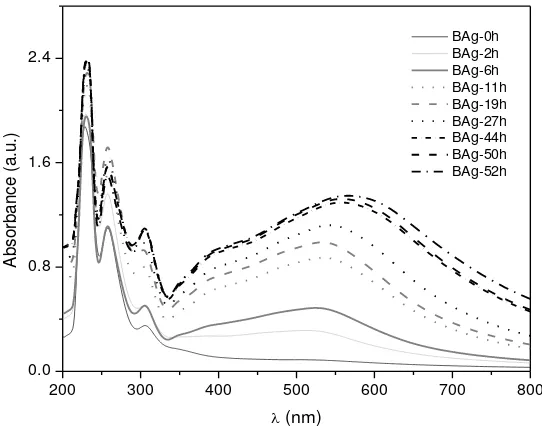
Figure 1 shows the UV-Vis absorption spectra of silver nanoparticles (AgNPs) synthesized using latex extract of Thevetia peruviana at different time interval. It shows the characteristic of surface plasmon resonance (SPR) absorption band at around 570 nm for AgNPs (purple-brown solution). It is observed that the intensity of SPR bands increases as the reaction time progresses from 0-44 h and after 44 h of reaction time the SPR intensity nearly constant indicating that the reaction process is almost completed. The observed absorption spectra are broad in nature with a tail at longer wavelength and shoulder at around 395 nm indicating the formation of polydispers AgNPs. Figure 1 also shows the red shifted band from 510 nm to 570 nm occurred with increasing reaction time from 0-52 h.
200 300 400 500 600 700 800
Figure 1. UV-vis absorption spectra of silver nanoparticles synthesized at room temperature
using latex extract of Thevetia peruviana at different time durations: 0-52 h.
ScieTech 2013 IOP Publishing
The strong absorption peak at around 230 nm indicates the presence of several organic compounds which are known to interact with silver ions. An absorption band at around 260 nm is attributed to the aromatic amino acids of proteins. It is well known that the absorption band at 260 nm arises due to electronic excitations in tryptophan and tyrosine residues in the proteins. This observation indicates the release of proteins into solution by latex of Thevetia peruviana and suggests a possible mechanism for the reduction of the metal ions present in the solution.
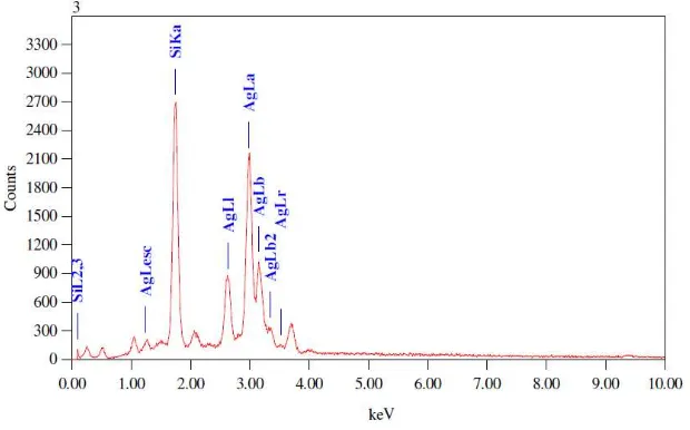
Fig.2 shows representative EDX profile for synthesized silver nanoparticles. The energy dispersive X-ray analysis (EDS) reveals strong signal in the silver region and confirms the formation of silver nanoparticles. There is also a strong signal for Si, which due to Si (111) wafers that has used as substrate to prepare thin film. Other elemental signals are recorded possibly due to elements from enzymes or proteins present within the latex of Thevetia peruviana.
Figure 2. EDS spectrum for silver nanoparticles synthesized at room temperature using latex
extract of Thevetia peruviana.
A thorough study on the literature of Thevetia peruviana reveals that the major components of the plant are protein, phenols, oil, mome inositol, Ethyl palmitate, Ethyl linolenate, and lesser amount of glycosides with the major component is thevetin. The presence of theveridoside, digitoxigenin, cerberin, peruvoside and theveside has been established in the plant [14]. It has been reported by Yao Zhouet al. [18] that the carbonyl groups from the amino acid residues and peptides of proteins have a strong affinity to bind metals. So that protein can act as encapsulating agent and thus protect the nanoparticles from agglomeration. For this phenomenal, FTIR measurements were carried out to identify the possible biomolecules responsible for the reduction of the Ag+ ions and the capping of the bioreduced silver nanoparticles synthesized by the latex extract of Thevetia peruviana. The solution after complete reduction of Ag+ was centrifuged at 15000 rpm for 10 minutes to isolate the silver nanoparticles free from proteins or other compounds present in the solution. The representative spectra of nanoparticles obtained (figure 3) manifest absorption bands located at 3426, 1640, 1390, 1338, 1104, and 1030 cm-1. The absorption peak at around 1030 cm-1 can be assigned as absorption peaks of -C-O-C- or -C-O-. The peak at around 1640 cm-1 is assigned to the amide I bonds of proteins. The bonds or functional groups such as -C-O-C-, -C-O- and -C=C- derived from heterocyclic compounds and the amide I bond derived from the proteins which are present in the extract are the capping ligands of the nanoparticles [1, 7, 10, 17]. The band at 1390 and 1104 cm-1, as well as a strong band at 1338 cm-1 indicating presence of amine groups, as would be expected due to plant-origin of the samples. The peak at around 3426 cm-1 was characteristic of -N-H stretching of amide I band [17].
ScieTech 2013 IOP Publishing
Journal of Physics: Conference Series423(2013) 012032 doi:10.1088/1742-6596/423/1/012032
500 1000 1500 2000 2500 3000 3500 4000
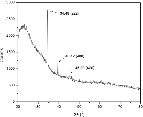
Figure 4 shows that the XRD patterns of natural dried silver nanoparticles synthesized using latex of Thevetia peruviana. A number of Bragg reflections with 2θ values of 34.480, 40.120, and 45.390 sets of lattice planes are observed which may be indexed to the (222), (400), and (420) faces of silver respectively. XRD pattern thus clearly illustrates that the silver nanoparticles formed in this present synthesis are crystalline in nature and having Face Cantered Cubic crystal structure.
20 30 40 50 60 70 80
Figure 5 shows the TEM micrograph of the Ag nanoparticles synthesized by latex of Thevetia peruviana. Figure 5a is the representative TEM image of synthesized silver nanoparticles. The morphology of AgNPs is found to be predominantly spherical. The nanoparticles are polydispers having two dominant size distributions ranges from 10-20 nm and 20-30 nm as shown in figure 5b. The figure 5c shows HRTEM image of silver nanoparticle. The fringe spacing is found to be 2.59Å. These results were in agreement with those reported by Kiruba Daniel et al. [1], Harekrishna Bar et al. [7] and Naheed Ahmad et al. [17].
Figure 5. a) Representative TEM image of silver nanoparticles synthesized at room temperature using
latex extract of Thevetia peruviana. b) Histogram of particle size distribution and C) High Resolution Transmission electron microscopy (HRTEM) image of silver nanoparticles.
4.
ConclusionThe rapid synthesis of silver nanoparticles using latex extract of Thevetia peruviana has been demonstrated. Present green synthesis showed that the environment friendly and renewable latex of Thevetia peruviana could be used as an effective capping as well as reducing agent for the synthesis of silver nanoparticles. This eco-friendly route for Synthesis of silver nanoparticles is a challenging alternative to chemical synthesis.
Acknowledgments
The author thanks the Higher Education Ministry (DIKTI), Jakarta, Indonesia for “Abroad SAME Program 2012 Funding” and to Dept. of Physics, University of Pune, India as the university based of research conducted. The author also thanks to Miss Jyotsana Dixit, Biophysics Lab. Dept. of Physics, University of Pune, India for valuable helps.
ScieTech 2013 IOP Publishing
Journal of Physics: Conference Series423(2013) 012032 doi:10.1088/1742-6596/423/1/012032
References:
[1] Kiruba Daniel S C G, Kumar R, Sathish V, Sivakumar M, Sunitha S and Anitha Sironmani T 2011 Green Synthesis (Ocimum tenuiflorum) of Silver Nanoparticles and Toxicity Studies in Zebra Fish (Danio rerio) Model International Journal of NanoScience and Nanotechnology
2(2) 103-117
[2] Sangiliyandi Gurunathana, Kalimuthu Kalishwaralala, Ramanathan Vaidyanathana,
Venkataraman Deepaka, Sureshbabu Ram Kumar Pandiana, Jeyaraj Muniyandia, Nellaiah Hariharana, Soo Hyun Eom 2009 Biosynthesis, purification and characterization of silver nanoparticles using Escherichia coli Colloids and Surfaces B: Biointerfaces 74 328-335 [3] Guangquan Li, Dan He, Yongqing Qian, Buyuan Guan, Song Gao, Yan Cui, Koji Yokoyama
and Li Wang 2012 Fungus-Mediated Green Synthesis of Silver Nanoparticles Using Aspergillus terreus Int. J. Mol. Sci. 13 466-476
[4] Asmita J Gavhane1, Padmanabhan P, Suresh P Kamble and Suresh N Jangle 2012 Synthesis of silver nanoparticles using extract of Neem leaf and triphala and evaluation of their
Antimicrobial activities Int. J. Pharm. Bio. Sci. 3(3) 88-100
[5] Geethalakshmi R and Sarada D V L 2010 Synthesis of plant-mediated silver nanoparticles using Trianthema decandra extract and evaluation of their anti microbial activities International Journal of Engineering Science and Technology 2(5) 970-975
[6] Ponarulselvam S, Panneerselvam C, Murugan K, Aarthi N, Kalimuthu K and Thangamani S 2012 Synthesis of silver nanoparticles using leaves of Catharanthus roseus Linn. G. Don and their antiplasmodial activities Asian Pacific Journal of Tropical Biomedicine 574-580 [7] Harekrishna Bar, Dipak Kr Bhui, Gobinda P Sahoo, Priyanka Sarkar, Sankar P De, Ajay Misra
2009 Green synthesis of silver nanoparticles using latex of Jatropha curcas Colloids and Surfaces A: Physicochem. Eng. Aspects 339 134-139
[8] Gardea-Torresdey J L, Parsons J G, Dokken K, Peralta-Videa J, Troiani H E, Santiago P and Jose-Yacaman M 2002 Formation and growth of Au nanoparticles inside live Alfalfa plants. Nano Lett 2 397-401
[9] Gardea-Torresdey J L, Gomez E, Peralta-Videa E, Parsons J G, Troiani H E and Jose-Yacaman M 2003 Alfalfa sprouts: a natural source for the synthesis of silver nanoparticles. Langmuir
19 1357-1361
[10] Manish Hudlikar, Shreeram Joglekar and Mayur Dhaygude Kisan Kodam 2012 Latex-mediated synthesis of ZnS nanoparticles: green synthesis approach J Nanopart Res 14 865
[11] Venkataraman Deepak, Kalimuthu Kalishwaralal, Sureshbabu Ram Kumar Pandian and Sangiliyandi Gurunathan 2011 An Insight into the Bacterial Biogenesis of Silver Nanoparticles, Industrial Production and Scale-up Rai M and Duran N (eds.), Metal Nanoparticles in Microbiology (Berlin Heidelberg: Springer-Verlag) chapter 2 pp 173-214 [12] Usman L A, Oluwaniyi O O, Ibiyemi S A, Muhammad N O and Ameen O M 2009 The
potential of oleander (thevetia peruviana) in African agricultural and industrial development: a case study of nigeria Journal of Applied Biosciences 24 1477-1487
[13] Mathuravalli K and Eswara Lakshmi R 2012 Analysis of phytochemical components and anti- microbial activity of the toxic plant -Thevetia Peruviana Indian J. Innovations Dev. 1 (2) 97- 101
[14] Sangodare R S A, Agbaji A S, Dakare M A, Usman Y O, Magomya A, Paul E D, Ibraheem A S, Etim N A, Ibrahim A, Aribido O and Shikeola O 2012 Investigation of the Chemical Constituent of Extracts of Thevetia peruviana Seed Using GC-MS and FT-IR International
Journal of Food Nutrition and Safety2(1) 27-36
[15] Garima Zibbu and Amla Batra 2011 Thevetia peruviana (Pers.) Schum. : A plant with enormous therapeutic potential Journal of Pharmacy Research 4(12) 4461-4464
[16] Burkill HM 1985 Entry for Thevetia Neriifolia Juss. [Family Apocynaceae] The Useful Plants of West Tropical Africa, Jstor Plant Science Vol 1 Http://Plants.Jstor.Org/Upwta/1_411 (Accessed On 09-10-2012)
ScieTech 2013 IOP Publishing
[17] Naheed Ahmad, Seema Sharma, Singh V N, Shamsi S F, Anjum Fatma and Mehta B R 2011 Biosynthesis of Silver Nanoparticles from Desmodium triflorum: A Novel Approach TowardsWeed Utilization Biotechnology Research International 2011 8 pages
[18] Yao Zhou, Wenshuang Lin, Jiale Huang, Wenta Wang, Yixian Gao, Liqin Lin, Qingbiao Li, Ling Lin and Mingming Du 2010 Biosynthesis of Gold Nanoparticles by Foliar Broths: Roles of Biocompounds and Other Attributes of the Extracts Nanoscale Res Lett 5 1351- 1359
ScieTech 2013 IOP Publishing
Journal of Physics: Conference Series423(2013) 012032 doi:10.1088/1742-6596/423/1/012032
![Figure 5 [7] and Naheed Ahmad et al. [17]. peruvianamorphology of AgNPs is found to be predominantly spherical](https://thumb-ap.123doks.com/thumbv2/123dok/684247.279211/7.595.116.473.241.531/figure-naheed-ahmad-et-peruvianamorphology-agnps-predominantly-spherical.webp)
