Green synthesis of silver nanoparticles using
latex extract of
Thevetia peruviana
: a novel
approach towards poisonous plant utilization
Ni Nyoman Rupiasih1, Avinash Aher2, Suresh Gosavi2 and P B Vidyasagar2
1 Department of Physics, Faculty of Mathematics and Natural Sciences, Udayana University,
Kampus Bukit Jimbaran, Badung, Bali 80362, Indonesia
2
Department of Physics, University of Pune, Ganeshkhind, Pune-411007, India Email: [email protected]
Abstract The development of green synthesis of nanoparticles has received in-creasing attention due to ease of preparation, less chemical handling and of eco-friendly nature. It is of considerable importance to expand their biological applica-tions. Currently, a variety of inorganic nanoparticles with well-defined chemical composition, size and morphology have been synthesized by using different mi-croorganisms, fungus, plant extracts and their applications in many cutting-edge technological areas have been explored. In the present study, silver nanoparticles (AgNPs) were successfully synthesized from AgNO3 through a simple green
syn-thesis route using the latex of Thevetia peruviana as reducing as well as capping agent. Synthesized silver nanoparticles were characterized using UV-Vis spectro-photometer, FTIR, SEM-EDS, XRD and HRTEM. UV-Vis spectra showed ab-sorption at 570 nm with a shoulder at 395 nm. FTIR spectra analysis confirmed the functional groups involved in the silver nanoparticles formation. X-ray diffrac-tion pattern of silver nanoparticles exhibited the diffracdiffrac-tion angle at 34.48°, 40.12° and 45.39° which is corresponding to (222), (400) and (420) Braggs reflection planes respectively with interplaner spacing of 2.59Å obtained by HRTEM. TEM analysis showed the particles are spherical in nature with its size distribution be-tween 10-30 nm.
1 Introduction
single, complex, aggregated or agglomerated forms with spherical, tubular and irregular shapes.
Nanoparticles exhibit completely new or improved properties compared to their bulk counterparts. These novel properties are derived due to the variation in specific characteristics such as size, distribution and morphol-ogy of the particles [1-3]. The two main reasons that materials at nano scale can have different properties are increased relative surface area and quantum size effects [4]. Nanoparticles have much greater surface area to volume ratio than their bulk forms, which can lead to greater chemical re-activity and affect their strength. Also at the nano scale, quantum effects can become much more important in determining the materials properties and characteristics, leading to novel optical, electrical and magnetic behav-iours [4]. Technologically nanomaterials are resources designed at the mo-lecular (nano meter) level to take advantage of their small size and novel properties which are generally not seen in their conventional, bulk coun-terparts.
The emergent properties of the nanoparticles have its potential for great impacts in electronics, medicine and other fields. In medicine, nanoparti-cles delivery systems had been used for tumor targeting [5]. The specific surface area is relevant for catalytic activity and other related properties such as antimicrobial activity [2]. As the specific surface area of nanopar-ticles is increased, their biological effectiveness can also increase on the account of a rise in surface energy. P Gupta, et al, 2008 reported that in cotton fabrics, silver nanoparticles show biocidal action against the bacte-ria E. coli, which is showing great potential to be used as an antiseptic dressing or bandage, which is in high demand for biomedical applications [6].
Nanoparticles with controlled size and composition are of fundamental and technological interest as they provide solutions to technological and environmental challenges in the areas of solar energy conversion, catalysis, medicine and water treatment. Global warming and climate change have induced a worldwide awareness and efforts to reduce generated hazardous wastes. Thus, “green” chemistry and chemical processes are progressively being integrated in science and industry for sustainable development [7-8]. One of them is synthesis of nanoparticles by biological process which is implemented to develop safe, cost-effective and environmentally friendly technologies [7].
use of toxic chemicals are toxic enough to pollute the environment if large scale nanoparticles are produced. It greatly limits their biomedical applica-tions, in particular in clinical fields. Also, these methods are quite expen-sive due to low material conversions, high energy requirements, difficult and wasteful purifications [11]. Therefore, development of reliable, non-toxic and eco-friendly methods for synthesis of nanoparticles is of utmost importance to expand their biomedical applications as well as saving the environment. Thus, among biosynthetic methods employing biological things such as microorganisms or plant extracts have emerged as a simple, clean, eco-friendly, nontoxic and viable alternative to physical and chemi-cal methods.
A great deal of effort has been put into the biosynthesis of inorganic material, especially metal nanoparticles using microorganisms and plants [1-2, 12]. Biosynthesis methods are methods that use biological systems to synthesize nanoparticles. So far, different biological resources including herbal extracts [11-13], microalgae [14], fungus [3, 15] and bacteria [2, 16-17] have been used for synthesis of metal nanoparticles. The use of plant extracts for the synthesis could be more advantageous, because it does not require elaborate processes such as intracellular synthesis and multiple pu-rification steps or the maintenance of microbial cell cultures [7, 11-12].
Up to now, metallic nanoparticles are mostly prepared from noble met-als such as Ag, Pt, Au and Pd, among those; silver (Ag) is the metal of choice in the field of biological system, living organisms and medicine [18]. Among inorganic antibacterial agents, silver has been employed most extensively since ancient times to fight infections and control spoilage. The antibacterial and antiviral actions of silver, silver ion and silver com-pounds have been thoroughly investigated [19].
Gardea-Torresdey and co-workers first reported the formation of gold and silver nanoparticles by living plants [20-21]. Their synthetic protocol by plant extract or biomass exemplifies the promising application of the green synthesis of metal nanoparticles. Recent research reported that silver nanoparticles have been synthesized using various natural products like green tea (Camellia sinensis), neem (Azadirachta indica) leaf broth, natu-ral rubber, starch, aloe vera plant extract, lemongrass leaves extract, etc [11, 13]. The shape and size of the nanoparticles synthesized using plants can be controlled and modulated by changing and controlling the pH of the solution [2]. The reduction of silver ions (Ag+) in aqueous solution gener-ally yields colloidal silver with particle diameters of several nanometers.
such as vitamins, enzymes/proteins, organic acids such as citrates, amino acids and polysaccharides [22-24] is environmentally benign, yet chemi-cally complex.
Medicinal herbs are the local heritage with global importance. It has cu-rative properties due to the presence of various complex chemical sub-stances of different composition, which are found as secondary plant me-tabolite in one or more parts of the plants [7-8, 11, 25]. These plant metabolites according to their composition are grouped as alkaloids, gly-cosides, corticosteroids, essential oils, etc.
Thevetia peruviana is an ever-green ornamental dicotyledonous plant that belongs to Apocyanaceae family. It is native of tropical America espe-cially Mexico and West Indies and a close relative to Nerium oleander. It has naturalized in tropical regions [26-27].
The whole plant contains a milky juice (white latex) containing a com-pound called thevetin that is used as a heart stimulant but in its natural form is extremely poisonous. It’s poisonous owing to the presence of ca r-diac glycosides or carr-diac toxins, which acted directly in the heart [26-28]. Ingestion of these plant parts could lead to death [26-27]. The seed con-tains oil with high content of fatty acid. It is believed that Thevetia oil is rich in antioxidants with some specific components of vitamin E groups [27]. It is cultivated as ornamental tree standards in gardens and parks in temperate climates [29]. In the West Indies half a leaf is known as an emetic and purgative. In Java, Indian immigrants are reported to smoke us-ing the dried leaves. Tincture of bark is also emetic and purgative and has been used as a febrifuge, but tests with bark and fruit extracts have proved inactive against avian malaria. In Indonesia it is reported being used as a fish-poison [30].
2 Materials and Methods
2.1 Preparation of Latex Extracts
Fresh fruits of Thevetia peruviana were collected, washed thoroughly 2-3 times with tap water, continuously rinse with distilled water, and used for the extraction. Milky white latex was obtained by cutting the green stems of the fruits. 100 ml of distilled water was added with 100 µl of fresh latex extract, mixed gently and filtered through Whatman No.1 filter paper. The solution was stored at 4°C for further used.
2.2 Synthesis of Silver Nanoparticles
A 50 µl of aqueous solution of 1 M silver nitrate (AgNO3) was added into
10 ml of latex extract solution and was kept at room temperature. The re-action of silver ions with the extract solution leads to the formation of sil-ver nanoparticles (AgNPs). After around 6 hours, the colour of the mixture turned into light purple and continuously changed to purple-brown indicat-ing the formation of silver nanoparticles.
2.3 Characterization Techniques
3 Results and Discussion
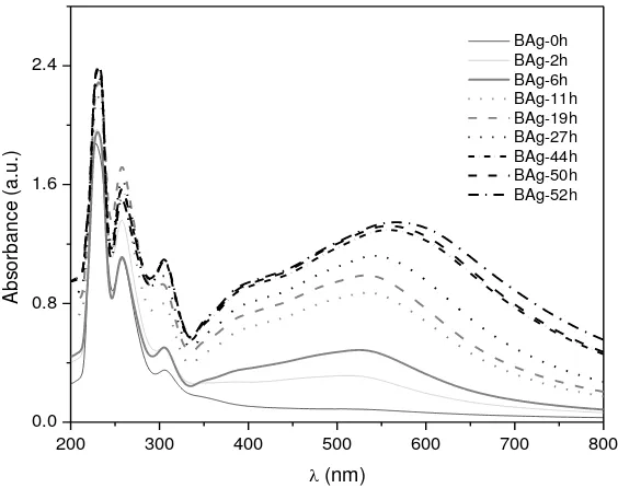
Figure 1 shows the UV-Vis absorption spectra of silver nanoparticles (AgNPs) synthesized using latex extract of Thevetia peruviana at different time interval. It shows the characteristic of surface plasmon resonance (SPR) absorption band at around 570 nm for silver nanoparticles (purple-brown solution). It is observed that the intensity of SPR bands increases as the reaction time progresses from 0-44 h and after 44 h of reaction time the SPR intensity is nearly constant indicating that the reaction process is al-most completed. The observed absorption spectra are broad in nature with a tail at longer wavelength and a shoulder at around 395 nm indicating the formation of polydispers AgNPs. Figure 1 also shows the red shifted band from 510 nm to 570 nm which occurred with increasing reaction time from 0-52 h.
Fig. 1. UV-vis absorption spectra of silver nanoparticles (AgNPs)
synthe-sized at room temperature using latex extract of Thevetia peruviana at dif-ferent time: 0-52 h.
The strong absorption peak at around 230 nm observed indicated the presence of several organic compounds which are known to interact with
silver ions. Also an absorption band observed at around 260 nm is attribut-ed to the aromatic amino acids of proteins. It is well known that the ab-sorption band at 260 nm arises due to electronic excitations of tryptophan and tyrosine residues in the proteins. This observation indicates the release of proteins into solution by latex of Thevetia peruviana and suggests a possible mechanism for the reduction of the silver ions present in the solu-tion.
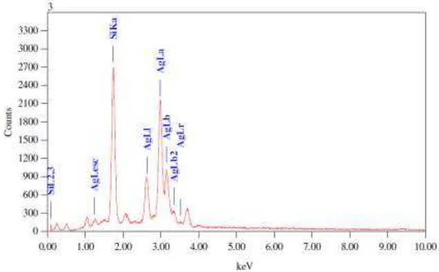
Figure 2 shows representative EDS profile for synthesized silver nano-particles. The energy dispersive X-ray analysis reveals strong signal in the silver region and confirms the formation of silver nanoparticles. There is also a strong signal for Si, which due to Si (111) wafers which have been used as substrate to prepare thin film. Other elemental signals are recorded possibly due to elements from enzymes or proteins present within the latex of Thevetia peruviana.
Fig. 2. EDS spectrum of silver nanoparticles (AgNPs) synthesized at room
temperature using latex extract of Thevetia peruviana.
of proteins have a strong affinity to bind metals. So that protein can act as encapsulating agent and thus protect the nanoparticles from agglomeration.
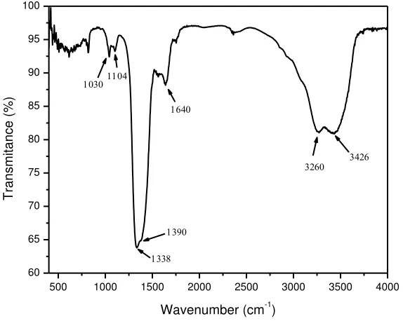
Thevetia oil contains high proportion of fatty acid and is known to be rich in antioxidants with some specific components of vitamin E groups [27]. This compound may also contribute to the reduction of Ag ions to silver nanosize. Ascorbic acid (vitamin E) is a reducing agent and can reduce, and thereby neutralize, reactive oxygen species leading to the formation of ascorbate radical and an electron. This free electron reduces the Ag+ ion to Ago [22]. FTIR measurements were carried out to identify the possible bi-omolecules responsible for the reduction of the Ag+ ions and the capping of the bioreduced silver nanoparticles synthesized by the latex extract of
Fig. 3. FTIR spectra of silver nanoparticles (AgNPs) synthesized at room temperature using latex extract of Thevetia peruviana.
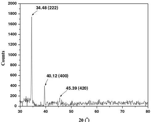
Figure 4 shows the XRD patterns of natural dried silver nanoparticles synthesized using latex of Thevetia peruviana. A number of Bragg reflec-tions with 2θ values of 34.48o, 40.12o and 45.39o sets of lattice planes are observed which may be indexed to the (222), (400) and (420) faces of sil-ver respectively. XRD pattern thus clearly illustrates that the silsil-ver nano-particles formed in this present synthesis are crystalline in nature and hav-ing Face Cantered Cubic crystal structure.
30 40 50 60 70 80 0
200 400 600 800 1000 1200 1400 1600 1800 2000
45.39 (420) 40.12 (400) 34.48 (222)
C
o
u
n
ts
2 (o)
Fig. 4. XRD patterns of silver nanoparticles (AgNPs) synthesized at room
temperature using latex extract of Thevetia peruviana.
Fig. 5. a) Representative TEM image of silver nanoparticles (AgNPs) syn-thesized at room temperature using latex extract of Thevetia peruviana. b) Histogram of particle size distribution and C) High resolution transmission electron microscopy (HRTEM) image of silver nanoparticles.
6 Concluding remarks
The rapid synthesis of silver nanoparticles (AgNPs) using latex extract of
(420) Braggs reflection planes respectively, with interplaner spacing of 2.59Å obtained by HRTEM image. TEM analysis showed the particles are spherical with size between 10-30 nm. The present green synthesis showed that the environment friendly and renewable latex of Thevetia peruviana
could be used as an effective capping as well as reducing agent for the syn-thesis of silver nanoparticles. This eco-friendly route for the synsyn-thesis is a challenging alternative to chemical synthesis.
References
[1] Kiruba Daniel SCG, Kumar R, Sathish V, Sivakumar M, Sunitha S, Anitha Sironmani T (2011) Green Synthesis (Ocimum tenuiflorum) of Silver Nanoparticles and Toxicity Studies in Zebra Fish (Danio rerio) Model. International Journal of NanoScience and Nanotechnology2(2):103-117
[2] Sangiliyandi Gurunathana, Kalimuthu Kalishwaralala, Ramanathan Vaidyanathana, Venkataraman Deepaka, Sureshbabu Ram Kumar Pandiana, Jeyaraj Muniyandia, Nellaiah Hariharana, Soo Hyun Eom (2009) Biosynthesis, purification and characterization of silver nanoparticles using Escherichia coli. Colloids and Surfaces B: Biointerfaces 74:328-335
[3] Guangquan Li, Dan He, Yongqing Qian, Buyuan Guan, Song Gao, Yan Cui, Koji Yokoyama, Li Wang (2012) Fungus-Mediated Green Synthesis of Silver Nanoparticles Using Aspergillus terreus. Int J Mol Sci 13:466-476
[4] Cristina Buzea, Ivan I Pacheco, Kevin Robbie (2007) Nanomaterials and Nanoparticles: Sources and Toxicity. Biointerphases 2(4):MR17-MR172 http://arxiv.org/ftp/arxiv/papers/0801/ 0801.3280.pdf (Accessed on: 02-06-2013)
[5] VJ Mohanraj, Y Chen (2006) Nanoparticles - A Review. Tropical Journal of Pharmaceutical Research 5(1):561-573
[6] P Gupta , M Bajpai, SK Bajpai (2008) Investigation of Antibacterial Properties of Silver Nanoparticle-loaded Poly (acrylamide-co-itaconic acid)-Grafted Cotton Fabric. The Journal of Cotton Science12:280-286
[7] Asmita J Gavhane1, Padmanabhan P, Suresh P Kamble, Suresh N Jangle (2012) Synthesis of silver nanoparticles using extract of Neem leaf and triphala and evaluation of their Antimicrobial activities. Int J Pharm Bio Sci 3(3):88-100
silver nanoparticles using Trianthema decandra extract and evaluation of their anti microbial activities International. Journal of Engineering Science and Technology 2(5):970-975
[9] Maribel G Guzmán, Jean Dille, Stephan Godet (2009) Synthesis of silver nanoparticles by chemical reduction method and their antibacterial activity. International Journal of Chemical and Biological Engineering 2(3):104-111
[10] Mukunthan KS, Elumalai EK, Trupti N Patel, V Ramachandra Murty (2011) Catharanthus roseus: a natural source for the synthesis of silver Nanoparticles. Asian Pacific Journal of Tropical Biomedicine:270-274
[11] Harekrishna Bar, Dipak Kr Bhui, Gobinda P Sahoo, Priyanka Sarkar, Sankar P De, Ajay Misra (2009) Green synthesis of silver nanoparticles using latex of Jatropha curcas. Colloids and Surfaces A: Physicochem Eng Aspects 339:134-139
[12] Ponarulselvam S, Panneerselvam C, Murugan K, Aarthi N, Kalimuthu K, Thangamani S (2012) Synthesis of silver nanoparticles using leaves of Catharanthus roseus Linn. G. Don and their antiplasmodial activities. Asian Pacific Journal of Tropical Biomedicine:574-580
[13] Manish Hudlikar, Shreeram Joglekar, Mayur Dhaygude Kisan Kodam (2012) Latex-mediated synthesis of ZnS nanoparticles: green synthesis approach. J Nanopart Res14:865
[14] G Singaravelu, JS Arockiamary, V Ganesh Kumar, K Govindaraju (2007) A novel extracellular synthesis of monodisperse gold nanoparticles using marine alga, Sargassum wightii Greville. Colloids and Surfaces B: Biointerfaces 57:97-101
[15] Amrita Mishra, Suraj Kumar Tripathy, Rizwan Wahab, Song-Hoon Jeong, Inho Hwang, You-Bing Yang, Young-Soon Kim, Hyung-Shik Shin, Soon-Il Yun (2011) Microbial synthesis of gold nanoparticles using the fungus Penicillium brevicompactum and their cytotoxic effects against mouse mayo blast cancer C2C12
cells. Appl Microbiol Biotechnol 92:617-630
[16] MI Husseiny, M Abd El-Aziz, Y Badr, MA Mahmoud (2007) Biosynthesis of gold nanoparticles using Pseudomonas aeruginosa. Spectrochimica Acta Part A 67:1003-1006
[17] Kannan Natarajan, Subbalaxmi Selvaraj, V Ramachandra Murty (2010) Microbial Production of Silver Nanoparticles. Digest Journal of Nanomaterials and Biostructures 5(1):135-140
Biochemistry and Bioinformatics IPCBEE 5:334-337. IACSIT Press, Singapore
[19] Parameswari E, Udayasoorian C, S Paul Sebastian, RM Jayabalakrishnan (2010) The bactericidal potential of silver nanoparticles. International Research Journal of Biotechnology 1(3):044-049
[20] Gardea-Torresdey JL, Parsons JG, Dokken K, Peralta-Videa J, Troiani HE, Santiago P, Jose-Yacaman M (2002) Formation and growth of Au nanoparticles inside live Alfalfa plants. Nano Lett 2:397-401
[21] Gardea-Torresdey JL, Gomez E, Peralta-Videa E, Parsons JG, Troiani HE, Jose-Yacaman M (2003) Alfalfa sprouts: a natural source for the synthesis of silver nanoparticles. Langmuir 19:1357-1361
[22] Naheed Ahmad, Seema Sharma, Singh VN, Shamsi SF, Anjum Fatma, Mehta BR (2011) Biosynthesis of Silver Nanoparticles from Desmodium triflorum: A Novel Approach Towards Weed Utilization. Biotechnology Research International:8 pages [23] Yao Zhou, Wenshuang Lin, Jiale Huang, Wenta Wang, Yixian Gao,
Liqin Lin, Qingbiao Li, Ling Lin, Mingming Du (2010) Biosynthesis of Gold Nanoparticles by Foliar Broths: Roles of Biocompounds and Other Attributes of the Extracts. Nanoscale Res Lett 5:1351-1359.
[24] Krzysztof Szczepanowicz, Joanna Stefańska, Robert P Socha, Piotr Warszyński (2010) Preparation of Silver Nanoparticles Via Chemical Reduction And Their Antimicrobial Activity. Physicochemical Problems Mineral Processing45:85-98
[25] Venkataraman Deepak, Kalimuthu Kalishwaralal, Sureshbabu Ram Kumar Pandian, Sangiliyandi Gurunathan (2011) An Insight into the Bacterial Biogenesis of Silver Nanoparticles, Industrial Production and Scale-up Rai M and Duran N (eds.), Metal Nanoparticles in Microbiology (Berlin Heidelberg: Springer-Verlag). Chapter 2:173-214
[26] Usman LA, Oluwaniyi OO, Ibiyemi SA, Muhammad NO, Ameen OM (2009) The potential of oleander (thevetia peruviana) in African agricultural and industrial development: a case study of Nigeria. Journal of Applied Biosciences 24:1477-1487
[27] Mathuravalli K, Eswara Lakshmi R (2012) Analysis of phytochemical components and anti-microbial activity of the toxic plant -Thevetia Peruviana. Indian J Innovations Dev 1(2):97-101
Paul ED, Ibraheem AS, Etim NA, Ibrahim A, Aribido O, Shikeola O (2012) Investigation of the Chemical Constituent of Extracts of Thevetia peruviana Seed Using GC-MS and FT-IR. International Journal of Food Nutrition and Safety 2(1):27-36 [29] Garima Zibbu, Amla Batra (2011) Thevetia peruviana (Pers.)
Schum.: A plant with enormous therapeutic potential. Journal of Pharmacy Research4(12):4461-4464