Lateral Extracavitory Approach (LECA) For Thoracic Vertebral
Lession
Rully Hanafi Dahlan (1,2) Farid Yudoyono (1,2) Sevline Estethia Ompusunggu (1,2) Muhammad Zafrullah Arifin (1)
1. Spine Research Working Group, Neurospine, Pain and Peripheral Nerve Division, Department of Neurosurgery, Faculty of Medicine Universitas Padjadjaran–Dr. Hasan Sadikin Hospital, Bandung, Indonesia;
2. Department of Neurosurgery, Faculty of Medicine Universitas Padjadjaran–Dr. Hasan Sadikin Hospital, Bandung, Indonesia
Abstract
Introduction. The Lateral Extracavitory Surgical Approach (LECA) to the thoracic is
challenging since anterior interventions to this region are quite complicated with the presence of major vascular elements or important visceral and soft-tissue structures.
Method. Operative technique was performed initially on three consecutive patients.
Costotransvesectomy was performed on the right side and pediculectomy were added in two patient to achieve wide visual angle during corpectomy and the stabilization procedure was completed with posterior instrumentation with the exception of one patient who had a hardening of bone due to receiving
Results. The initial clinical presenting symptom was paraparesis in three patients Total
excision was achieved in 3 patients (1 schwanommas, 2 tuberculosis). No significant postoperative complications occurred and discharge was achieved in all patients with an average hospital stay of 5 days.
Conclussion. A one-step approach through a posterior midline incision is feasible, safe and
efficient for complete excision of thoracic lession. This approach facilitates laminectomy, costotransversectomy, pediculectomy and instrumentation under flouroscopy guidance but excessive blood loss, operative time and postoperative pain. Despite that early mobilization with a reduced hospital stay were achieved.
Introduction
Lateral Extracavitory Approach (LECA) is a technique that is useful in spinal surgery to remove lesions located in the ventral and ventrolateral portion of the spinal cord or spine such as burst fracture, neoplasm, infection, or herniated disc. LECA is mostly effective for spine surgery vertebrae level T3 to L1 but generally there are several surgical approaches for lesions located in ventral and ventrolateral namely, costotransversectomy, Leca, or transpedicular. (3-5)
Case 1
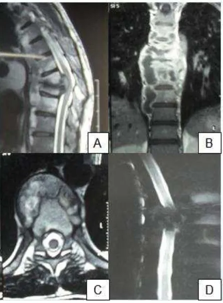
Figure 1. MRI A). Isointens on T1WI MRI picture on thorakal V Th 2-5 B). T2WI MRI hyperintense
signal pressing spinal cord to the posterior C, D) MRI axial T1WI mass fills the spinal canal in the right
ventrolateral (2).
Case 2
Male 31 years complained of weakness both lower extremities with motor strength 0/5 since 6 months ago. He denied smoking history and family history of similar disease. History of pulmonary Tb (+).Magnetic Resonance Imaging (MRI) examination show vertebrae thorakal destruction > 50% accompanied with an epidural mass and paravertebrae (Fig 2).
Figure 2. A) On T2WI MRI reveal hipointens on extradura which compress the spinal cord and bone
destruction > 50%. B, C) Coronal and axial view shows paravertebral abscess. D) Myelography
Case 3
Male 36 years complained of weakness both lower extremities with motor strength 0/5 since 9 months ago. He denied smoking history and family history of similar disease. History of pulmonary Tb (+). Preoperative MRI show thoracic vertebrae destruction > 50% accompanied with an epidural mass and paravertebrae.
Operation Procedure
Preoperative preparation is similar with preparation for other major spinal surgery. Vein and artery access and folley catheter are used. Operator (RD, FY, SV) used Wilson frame to reduce intra-abdominal venous pressure to reduce bleeding during surgery. Once induced, intubated, infused and ETT is confirmed safe, four or five assistants are needed to transfer the patient into a prone position to reduce the manipulation of the spinal cord that may cause injury. Intraoperative C-arm is used to confirm the anatomical level (3, 6-8).
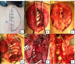
Upper arm and knee placed above a soft pad. Operating table can be tilted from side to side so that the operating field can be seen clearly by neurosurgeons and assistants. With semilunar incision, a skin flap was then made (Figure 3A). Blunt dissection is conducted right on thoracodorsal fascia. Retractors are used to maintain a good exposure of the operating field. Muscle dissection is performed laterally until the edge of the transverse process (Figure 3B). A T-shape incision was made on thoracodorsal fascia. Erector spinae muscle can be observed after retraction of the fascia. A dissection is performed from medial to bottom lateral of the erector spinae muscles, separated from quadratus lumborum muscle on the waist area, ribs and intercostal muscles in the back of the chest (Figure 3C). Trapezius, rhomboid, and latissimus dorsi muscles can be incised to maximize visual field (Figure 3D).
of the surrounding soft tissues (including the neurovascular bundle (Figure 3E). Cutter rib is used for cutting lateral ribs. Further dissection is needed to release the parietal pleura from the rib and vertebral corpus. The procedure is done carefully to avoid injury to the intercostal nerves and pleura. This dissection, including ribs removal, allows access to the lateral aspect of the vertebral bone. In thorakal area, intercostal nerves may be sacrificed to maximize exposure, but not in lumbar region. Pathological pedicle of vertebral are partially removed with a rongeur then dissection of the nerve roots to the medial aspect of the pedicle is performed. Removing the pedicle expose the lateral aspect of the dura. (3, 4, 7-10). For extradura lesions will appear pathological masses, for intradura lesions after opening the dura mater using bysturi No. 11 tumor mass can be observed (Figure 3F). Tumor mass is removed peacemeal to decompress the spinal canal.
Figure 3. Leca Operation A). Semilunar incision on Th3-Th5 B). Plane incision of the thoracodorsal
fascia C). Muscle dissection performed lateral to the edge of the transverses D).
Costotransversectomy on three levels and partial removal of pedicle on the tumor E). After
durotomy, tumor is visible in ventrolateral F). Unilateral Posterior stabilization after total tumor
removal.
Discussion
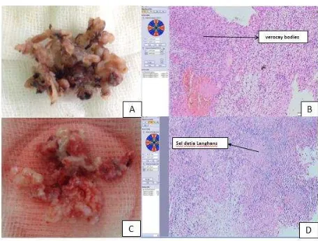
Figure 4. A). Picture of the tumor after removal B). Histopathology result show schwannoma antoni
mix A & B (H & E 40 x) Figure 9 C). Abscess paravertebrae D). Histopathology result reveal Datia
Langhans cells which indicate spondylitis tuberculosis (H & E 40 x)
Posterior approach consisting of transpedicular, costotransversectomy, extracavitary lateral approach, and modifications are used to access the ventral and ventrolateral thoracal vertebrae through the posterior approach, however there are some weaknesses of extracavitary lateral approach on thoracal and thoracolumbar which is associated with the manipulation of the visceral pleura in the chest wall (8,12,13). Selection of the operating technique requires experience and skill. Blood loss, patient positioning and exposure area is also a problem. Blood loss from the epidural venous plexus can be reduced by releasing the compression of
the stomach using a Wilson frame.
Macroscopic and histopathological analysis showed a verocay bodies; mixed type schwanomma in case 1 (Figure 4A, B) and presence of Datia Langhans cells indicating tuberculosis spondylitis infection in cases 2 and 3 (Figure 4C, D).
Conclusion
Daftar Pustaka
1. McLain RF IC. Spinal Tuberculousis deserves a PLace on Radar Screen. Cleveland Clinic Journal Of
Medicine 2004;71(7):537-49.
2. Verdelhan OD HC, Carsin-Nicol B, Riffaud L, Amlashi SFA, Brassier G,et al. MR ImagingFeatures of
spinal Schwannomas and Meningiomas. JNeuroradiol. 2005;32:42-29.
3. Chou D EM, Yang I,Lu D,Manley G. Rib head disarticulation for multilevel transpedicular thoracic
corpectomies and expandable cage reconstruction. Neurology India 2009;57(4).
4. Thorat JD RT, Thirugnanam A, Ivan H. B. . Single-stage posterior midline approach for dumbbell
tumors of the thoracic spine, with intraoperative CT guidance. Surgical Neurology International.
2011;2(31).
5. Dakwar E SD, Malone KT , Uribe JS. Minimally invasive lateral extracavitary resection of foraminal
neurofibromas. Journal of Clinical Neuroscience 2011;18:1510-2.
6. Edward C. Benzel MD, Division of Neurosurgery LSUMC, Shreveport, Louisiana. The lateral
extracavitary approach to the spine using the three-quarter prone position. J Neurosurgery.
1989;71:837-41.
7. Jandial R CM. Modified lateral extracavitary approach for vertebral column resection and expandable
cage reconstruction of thoracic spinal metastases. Surgical Neurology International. 2012;3:136.
8. Larson SJ HR, Hemmy DC, Sances A. Lateral extracavitary approach to traumatic lesions of the
thoracic and lumbar spine. J Neurosurg. 1976;45.
9. Güzey KF BS, Emel E,Özkan N, Güzey D,Alatafi B. Planning of Surgical Management of Giant Spinal
Schwannomas: Report of Four Cases. Turkish Neurosurgery 2006;16(3):139-44.
10. Kim SD SJ, Ha SK, Kim JH, Cho TH, Park JY, Kim H. Surgical Anatomy of Lateral Extracavitary
Approach to the Thoracolumar Spine J Korean Neurosurg Society. 2001;30:1187-92.
11. Akgül AG ÇU, Yurt ZK. An asymptomatic schwannoma originating from an intercostal nerve: A case
12. Singh K PD. Lumbar Extracavitary Corpectomy With a Single Stage Circumferential Arthrodesis:
Surgical Technique and Clinical Series. Am J Orthop. 2012;41(7):316-20.
13. Edward C. Benzel. Technique, Complication Avoidance, and Management. Spine surgery 2nd Edition.