RET EXPRESSION AND ITS CORRELATION
WITH CLINICOPATHOLOGIC DATA
IN PAPILLARY THYROID CARCINOMA
Ante Punda1, Vladimir Bedeković2, Ana Barić1, Mirko Kontić3, Zaviša Čolović3, Lucija Vanjaka Rogošić4, Hrvoje Punda5, Nenad Kunac6, Leo Grandić7 and Valdi Pešutić Pisac61Department of Nuclear Medicine, Split University Hospital Centre, Split, Croatia; 2Department of ENT, Sestre milosrdnice University Hospital Centre, Zagreb, Croatia;
3Department of ENT, Split University Hospital Centre, Split, Croatia; 4Dermatovenereology Private Surgery, Split, Croatia;
5Department of Diagnostic and Interventional Radiology, Split University Hospital Centre, Split, Croatia; 6Department of Pathology, Forensic Medicine and Cytology, Split University Hospital Centre, Split, Croatia;
7Department of Surgery, Split University Hospital Centre, Split, Croatia
SUMMARY – Th e purpose of this study was to analyze the possible prognostic value of RET mutation in papillary thyroid carcinoma and its incidence in the past few decades in our population, due to the increasing incidence of papillary thyroid carcinoma. Th e present study included 180 pa-tients operated for papillary thyroid carcinoma. Th e clinical and histopathologic characteristics were analyzed. Paraffi n sections of the selected histologic slides were cut again and immunohistochemi-cally stained by the Clone 3F8 P (HIER) from Novocastra (Vision Bio Systems Europe, Newcastle upon Tyne, UK) monoclonal antibody to RET oncoprotein. Univariate analysis indicated sex (p=0.01), histologic subtype (p=0.075) and capsular invasion (p=0.010) to be statistically signifi cant predictors of lymph node metastases, whereas age (p=0.796), tumor size (p=0.556) and intraglandular dissemina-tion (p=0.131) showed no such correladissemina-tion. Th e presence of RET mutation (p=0.704) was not a statis-tically signifi cant predictor of the tumor metastasizing potential. RET mutation (p=0.500) showed no statistically signifi cant correlation with papillary thyroid carcinoma classifed into prognostic groups according to clinicopathologic features either. RET mutation was detected in 30% of 180 papillary thyroid carcinomas. Th is is the fi rst large study demonstrating that RET mutation incidence in papil-lary thyroid carcinoma in Croatian population is consistent with the classic distribution of sporadic cases, despite the increased prevalence of papillary thyroid carcinoma in the past few decades.
Key words: Mutation; Th yroid cancer, papillary; Chernobyl nuclear accident; Lymphatic metastasis; Radiation
Correspondence to: Ana Barić, MD, Department of Nuclear Medi-cine, Split University Hospital Centre, Spinčićeva 1, HR-21000 Split, Croatia
E-mail: ana.baaric@gmail.com
Received March 6, 2017, accepted October 11, 2018
Introduction
Rearrangement of the tyrosine kinase receptor gene (RET gene named RET/PTC) is the most com-mon structural genetic alteration which, however,
shows great geographical variability ranging from 0 to 80% in diff erent studies1. In addition, BRAF point
mutation, TRK gene rearrangement and RAS muta-tion have been found in papillary carcinoma2-4.
RET proto-oncogene has been identifi ed in he-reditary MEN2 syndrome, and is involved in sporadic medullary and papillary thyroid carcinoma (PTC) and Hirschsprung’s disease5. RET proto-oncogene is
lo-cated on the long arm of chromosome 10. Genetically activated RET rearrangements are implied in the
pathogenesis of PTC6. Variation in the prevalence of
RET activation is believed to be attributable to diff er-ent methods and sensitivity of the techniques used, and to environmental factors, ionizing radiation expo-sure in particular1.
Th ere is no RET immunoreactivity in normal thy-roid tissue7. Th e mean frequency of RET mutation in
sporadic papillary carcinoma is 20%-30% in adults, rising up to 45%-50% in pediatric and young patients, and being highest (50%-80%) in patients with a his-tory of accidental or therapeutic radiation exposure8-10.
Accordingly, it appears quite reasonable to believe that the pathomorphologically identical papillary mas must have followed diff erent routes of carcino-genesis characterized by diff erent clinical manifesta-tion. In the last decades, there was a signifi cant increase in the number of PTC, so one of the possibilities was the infl uence of radiation like that due to Chernobyl nuclear power plant disaster11. Knowing that RET
mutation has been proven to be a good marker for its infl uence, we decided to analyze this relationship and to evaluate if there is increament in the RET mutation incidence following the rising PTC incidence9.
Patients and Methods
Patients
Th e present study included 180 PTC patients op-erated between January 1, 1999 and December 31, 2001 at ENT Department, Split University Hospital Centre in Split. Clinical data were collected from pa-tient medical records from Department of Nuclear Medicine fi les and histology fi ndings from Depart-ment of Pathology and Cytology database. Clinical and pathologic data were analyzed and classifi ed into two groups according to study criteria (age, sex, tumor size, histologic subtype, intraglandular dissemination, extrathyroid dissemination, and metastases): group 1 with favorable prognosis (low risk) included patients aged <45, female, tumor size up to 2 cm, papillary and follicular subtype limited to the thyroid, no intraglan-dular dissemination, and absence of metastases; and group 2 with poor prognosis (high risk) included all those that did not meet all group 1 criteria, i.e. age >45, male, tumor size exceeding 2 cm, all subtypes except for papillary and follicular ones, capsular invasion, in-traglandular dissemination, and metastases to lymph
nodes. At the time of the Chernobyl accident, 33% and 66% of patients were aged <30 and >30 years, re-spectively.
Methods
Tissue for histologic analysis was obtained from operative material, fi xed in 4% buff ered formalin for 24 h, paraffi n embedded, cut into 3- to 5-m sections and stained by the standard hemalaun eosin method (HE). Paraffi n sections of the selected histologic prep-arations were cut again and immunohistochemically stained. Antigen retrieval was performed in 10 mM citrate buff er (pH 6.0) inside a microwave pressure cooker. Slides were immediately placed into a bath of tap water. Sections were washed in TBS buff er for 1x5 minutes, then placed in diluted normal serum for 10 minutes. Th en the slides were incubated with primary antibody. Th e Clone 3F8 P (HIER) from Novocastra (Vision Bio Systems Europe, Newcastle upon Tyne, UK) monoclonal antibody to RET oncoprotein was used, at 1:40 dilution. Th en the slides were washed in TBS buff er for 2x5 minutes, incubated in biotinylated secondary antibody, washed in TBS buff er for 2x5 minutes again, incubated in ABC reagent, washed in TBS buff er for 2x5 once more, incubated in DAB and washed thoroughly in running tap water. Slides were counterstained in hematoxylin, dehydrated and mounted.
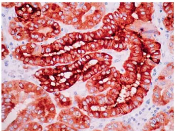
Th e presence of RET/PTC was determined by the presence of diff use cytoplasmic staining on the normal measuring scale (Fig. 1). Th e absence of staining
indi-Fig. 1. Diff use cytoplasmic staining indicates the presence of RET/PTC.
cated absence of the study mutation. Small intestine ganglial cells were used as positive control12.
Statistics
Descriptive statistics was employed for distribution of data obtained in the patient sample, whereas Pear-son’s 2-test, Fisher exact test and multivariate analysis
of binary logistic regression were used on correlation of the results obtained. Th e level of statistical signifi -cance was set at p<0.05.
Results
In the study population, patients aged >45 as an unfavorable prognostic factor prevailed, with a 2:1 older to younger group ratio. Sex distribution of the study population showed as many as 79.4% of female patients, yielding a 1:3.8 male to female ratio.
According to the new TNM classifi cation, T1 tu-mor has a borderline size of 2 cm, which was also ad-opted in the present study. Using this size criterion, carcinomas of up to 2 cm in size predominated (n=130; 72.2%). Based on the defi ned prognostic features of histologic subtypes and trying to facilitate analysis, papillary carcinomas were classifi ed into two catego-ries of favorable and unfavorable subtypes.
Papillary and follicular subtypes were classifi ed into favorable category and accounted for as many as 146 (81.1%) cases, whereas unfavorable category in-cluded high cellular, diff usely sclerosing and oncocytic subtypes, found in 34 (18.9%) cases. Intraglandular dissemination (multifocality) was observed in 44.4% of study carcinomas. Current state-of-the-art indi-cates capsular invasion as a defi nitely poor prognostic factor. Th is lesion was recorded in 28 (15.6%) and lymph node metastases in 38 (21.1%) carcinomas. Al-though distant metastases from PTC are extremely rare, sporadic cases have been reported from similar large studies. In our study, however, distant metastases were not recorded at all. Analysis of immunohisto-chemical staining for RET mutation revealed its pres-ence in 57 (31.7%) patients (Table 1).
Analyzing particular characteristics of papillary carcinoma in our patients, we assessed the impact of each clinical and histologic feature on the presence of metastases. Patient age had no eff ect on the prevalence of metastases, as indicated by the results of 2-test
(p=0.796) and Fisher exact test (p=0.849) (Table 2).
Metastases to lymph nodes were detected in 25 of 143 female patients and 13 of 37 male patients. Results of 2-test (p=0.019) and Fisher exact test (p=0.025)
re-vealed the prevalence of metastases to be statistically signifi cantly higher in male patients (Table 2).
Considering the borderline tumor size of 2 cm, the ratio of non-metastasizing to metastasizing tumors was 104:26 in the <2 cm group and 38:12 in the >2 cm group. Results of 2-test (p=0.556) and Fisher exact
test (p=0.547) showed that tumor size had no
statisti-Table 1. Frequency of RET mutation in papillary carcinoma
RET n %
Absent 123 68.3
Present 57 31.7
Total 180 100
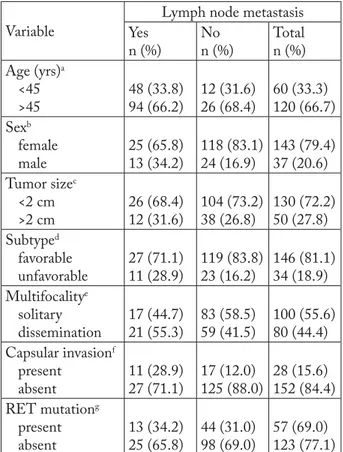
Table 2. Lymph node metastasis in relation to clinical and histopathologic parameters
Variable
Lymph node metastasis Yes n (%) No n (%) Total n (%) Age (yrs)a <45 >45 48 (33.8) 94 (66.2) 12 (31.6) 26 (68.4) 60 (33.3) 120 (66.7) Sexb female male 25 (65.8) 13 (34.2) 118 (83.1) 24 (16.9) 143 (79.4) 37 (20.6) Tumor sizec <2 cm >2 cm 26 (68.4) 12 (31.6) 104 (73.2) 38 (26.8) 130 (72.2) 50 (27.8) Subtyped favorable unfavorable 27 (71.1) 11 (28.9) 119 (83.8) 23 (16.2) 146 (81.1) 34 (18.9) Multifocalitye solitary dissemination 17 (44.7) 21 (55.3) 83 (58.5) 59 (41.5) 100 (55.6) 80 (44.4) Capsular invasionf present absent 11 (28.9) 27 (71.1) 17 (12.0) 125 (88.0) 28 (15.6) 152 (84.4) RET mutationg present absent 13 (34.2) 25 (65.8) 44 (31.0) 98 (69.0) 57 (69.0) 123 (77.1)
a2-test p=0.796; Fisher exact test p=0.849; b2-test p=0.019; Fisher
exact test p=0.025; c2-test p=0.556; Fisher exact test p=0.547; d2-test p=0.075; Fisher exact test p=0.101; e2-test p=0.131; Fisher
exact test p=0.145; f2-test p=0.010; Fisher exact test p=0.021; g2-test p=0.704; Fisher exact test p=0.699
cally signifi cant impact on the occurrence of metasta-ses (Table 2).
Division of PTCs into favorable and unfavorable subtypes revealed the presence of metastasizing poten-tial in 27 of 146 and 11 of 34 patients, respectively. Th e 2-test (p=0.075) and Fisher exact test (p=0.101)
yielded a statistically signifi cantly greater rate of me-tastases in the group of unfavorable subtypes (Table 2). Th ere was no statistically signifi cant diff erence in the prevalence of metastases according to the presence of intraglandular dissemination (multifocality) (2
-test: p=0.131; Fisher exact -test: p=0.145) (Table 2). Metastases were detected in 27 of 152 cases of PTC without capsular invasion and in as many as 11 of 28 cases with capsular invasion, yielding a statisti-cally signifi cant between-group diff erence (2-test:
p=0.010; Fisher exact test: p=0.021). Accordingly, the prevalence of metastases was infl uenced by the pres-ence of capsular invasion (Table 2).
Th ere was no statistically signifi cant diff erence in the prevalence of metastases according to the presence or absence of RET mutation as the mechanism of car-cinogenesis (2-test: p=0.704; Fisher exact test:
p=0.699) (Table 2).
According to the set criteria for good and poor prognosis, 21 PTC cases met all the criteria for good prognosis, whereas 159 high-risk cases were associated with poor prognosis.
Th ere was no statistically signifi cant diff erence in the frequency of RET mutation as the mechanism of carcinogenesis between the two prognostic groups of PTC patients (2-test: p=0.500; Fisher exact test:
p=0.618) (Table 3).
Univariate analysis was followed by multivariate analysis of the PTC characteristics according to the occurrence of metastases.
Binary logistic regression identifi ed two variables that statistically signifi cantly infl uenced the occur-rence of metastases, i.e. male sex associated with a 2.409 risk of metastases recorded in female patients (p=0.058) and capsular invasion associated with a 2.519 risk of metastases found in patients free from invasion (p=0.083) (Table 4).
Discussion
Immunohistochemical methods of staining that enable detection of specifi c markers of particular cell types of normal and tumor tissue have found applica-tion in the diagnosis of thyroid carcinomas. It is known that no RET immunoreactivity is found in normal thyroid tissue7. In the pathogenesis of PTC, RET
ac-tivation is frequently induced by radiation, mostly at younger age but also in adults11-13 . RET mutation
ac-counts for 20%-30% of sporadic papillary carcinomas in adults, 45%-50% in children and adolescents, and 50%-80% in patients with a history of accidental or therapeutic radiation exposure8-10. In our study
popu-lation, RET mutation was detected in 57 (31.7%) pa-tients (Table 1), which is consistent with most studies on sporadic papillary carcinoma. Th ese results con-fi rmed our hypothesis that radiation exposure like the one caused by Chernobyl disaster did not contribute to the increased incidence of papillary carcinoma in the population.
Investigating the incidence of PTC during the last four decades, Jung et al. found that the proportion of
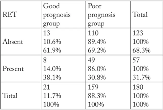
Table 3. Immunoreactivity to RET mutation in low- and high-risk patient groups
RET Good prognosis group Poor prognosis group Total Absent 13 10.6% 61.9% 110 89.4% 69.2% 123 100% 68.3% Present 8 14.0% 38.1% 49 86.0% 30.8% 57 100% 31.7% Total 21 11.7% 100% 159 88.3% 100% 180 100% 100%
2-test p=0.500; Fisher exact test p=0.618
Table 4. Multivariate analysis of lymph node metastasis predictors in thyroid papillary carcinoma
Variable Signifi cance Odds ratio
Age (yrs) 0.384 0.660 Sex (male/female) 0.058 2.409 Tumor size 0.366 0.648 Tumor subtype 0.569 1.343 Intraglandular dissemination 0.301 1.556 Capsular invasion 0.083 2.519 RET 0.828 1.102
RET/PTC rearrangements decreased, so the increased incidence of PTC was not due to environmental or therapeutic radiation15.
Analysis of correlation between the classic param-eters of PTC and their metastasizing potential re-vealed sex, tumor size, carcinoma variant and capsular invasion to infl uence the occurrence of metastases, whereas age and multifocality had no statistically sig-nifi cant impact on the tumor metastasizing potential (Table 2). Th ese results are consistent with the current state-of-the-art on the role of these pathologic and clinical characteristics in papillary carcinoma2, thus
our study group represented an appropriate model for additional analyses.
Analyzing the correlation between RET mutation and classic prognostic parameters in papillary carci-noma, some authors demonstrated RET mutation to be present at a higher frequency in small, slow pro-gressing and less aggressive tumors, and conclude that it may serve as a favorable marker in these tumors16,17.
Th is is supported by Sugg et al., who identifi ed this mutation mostly in occult microcarcinomas18. An
op-posite concept has been proposed by Miki et al., based on the fi nding of higher RET expression in association with extrathyroid tumor invasion19.
Sassolas et al. report similar results in a population of young adults, where the presence of RET/PTC (1 and 3) was associated with histologic and clinical short-therm aggressiveness, but no such relationship was found in children and adolescents20.
In the present study, RET association with papil-lary carcinomas divided into prognostic categories (Table 3) according to the classic clinical and patho-logic parameters was investigated. Categorization into prognostic groups by use of very strict criteria for fa-vorable prognosis (low-risk group): age <45, female sex, tumor size up to 2 cm, papillary or follicular vari-ant confi ned to the thyroid, no intraglandular dissemi-nation, and no metastases; and poor prognosis (high-risk group): those not meeting all the former group criteria, showed as many as 88.3% of study patients to belong to the high-risk group, thus highlighting the possible factors for poor prognosis. However, there was no statistically signifi cant diff erence in the prevalence of RET mutation between these prognostic groups of PTC patients (2-test: p=0.500; Fisher exact test:
p=0.618) (Table 3). Th is fi nding is highly comparable with the report by Shin et al. on the association of
RET expression with the clinicopathologic parameters of age, sex, tumor size, intraglandular dissemination, lymph node metastases and extrathyroid invasion14.
Th e researchers investigating the post-Chernobyl prevalence of papillary carcinoma in Ukraine and in young population exposed to therapeutic irradiation in childhood found a high percentage of RET mutation associated with an increased rate of lymph node me-tastases7,9,21. In contrast, Collins et al. found the
asso-ciation of young age and lymph node metastases to be more pronounced in the group of patients without a history of radiation exposure. In their study, age diff er-ence between patients with and without lymph node involvement was only recorded in the group of patients without a history of radiation exposure, thus the in-creased prevalence of metastases could be directly re-lated to younger age7. Th e association between RET
mutation and metastasizing potential has been investi-gated in many studies. Jhiang et al. suggest that RET might also be associated with distant metastases in sporadic cases22, while Mayr et al. postulate it to
cor-relate with early dissemination to lymph nodes23,
which is less probable in case of distant metastases. More recent results reveal connection of a higher rate of RAS mutation with the less aggressive nature of encapsulated follicular variant of PTC, which dif-fers from the more aggressive infi ltrative follicular variant, whereas opposite holds for the BRAF muta-tion pattern24.
In our study, there was no association of RET mu-tation with lymph node metastases, thus there was no statistically signifi cant diff erence in the prevalence of metastases according to the presence or absence of RET mutation as the mechanism of carcinogenesis (2-test: p=0.704; Fisher exact test: p=0.699) (Table 2).
Th e more so, RET mutation was the fi rst parameter excluded by the stepwise method, implying its lowest possible impact (Table 4).
Considering obvious variation in the results re-ported from diff erent studies including ours, it appears quite reasonable to presume that some other factors may also infl uence the prognostic signifi cance of RET mutation. Also, contribution to the increased inci-dence of papillary carcinoma in our population is probabbly due to diff erent factors such as better diag-nostic procedures and more sophisticated medical technology.
References
1. Santoro M, Carlomagno F, Hay ID, Hermann MA, Grieco M,
et al. Ret oncogene activation in thyroid neoplasms is restricted
to the papillary cancer subtype. J Clin Invest. 1992;89:1517-22. doi:10.1172/JCI115743
2. Soares P, Trovisco V, Rocha AS, Lima J, Castro P, et al. BRAF mutations and RET/PTC rearrangements are alternative events in the etiopathogenesis of PTC. Oncogene. 2003;22 (29):4578-80. doi:10.1038/sj.onc.1206706
3. Musholt TJ, Musholt PB, Khaladj N, Schulz D, Scheumann GF, et al. Prognostic signifi cance of RET and NTRK1 rear-rangements in sporadic papillary thyroid carcinoma. Surgery. 2000;128(6):984-93. doi:10.1067/msy.2000.110845
4. Şahpaz A, Önal B, Yeşilyurt A, Han Ü, Delibaşı T. BRAF(V600E) mutation, RET/PTC1 and PAX8-PPAR gamma rearrangements in follicular epithelium derived thyroid lesions – institutional experience and literature review. Balkan Med J. 2015;32(2):156-66.
doi:10.5152/balkanmedj.2015.15101
5. Lian EY, Maritan SM, Cockburn JG, Kasaian K, Crupi MJ, et
al. Diff erential roles of RET isoforms in medullary and
papil-lary thyroid carcinomas. Endocr Relat Cancer. 2017;24(1): 53-69. doi: 10.1530/ERC-16-0393
6. Fischer AH, Bond JA, Taysavang P, Battles OE, Wynford-Th omas D. Papillary thyroid carcinoma oncogene (RET/PTC) alters the nuclear envelope and chromatin structure. Am J Pathol. 1998;153:1443-50.
doi:10.1016/S0002-9440(10)65731-8
7. Collins BJ, Chiapetta G, Schneider AB, Santoro M, Pentimal-li F, et al. RET expression in papillary thyroid cancer from pa-tients irradiated in childhood for benign conditions. J Clin Endocrinol Metab. 2002;87(8):3941-6.
doi: 10.1210/jcem.87.8.8748
8. Bounacer A, Wicker R, Caillou B, Cailleux AF, Sarasin A, et al. High prevalence of activating ret proto-oncogene rearrange-ments in thyroid tumors from patients who had received exter-nal radiation. Oncogene. 1997;15:1263-73.
doi: 10.1038/sj.onc.1200206
9. Rabes HM, Demidchik EP, Sidorow JD, Lengfelder E, Beim-fohr C, et al. Pattern of radiation-induced RET and NTRK1 rearrangements in 191 post-Chernobyl papillary thyroid carci-nomas: biological, phenotypic, and clinical implications. Clin Cancer Res. 2000;6:1093-103.
10. Sadetzki S, Calderon-Margalit R, Modan B, Srivastava S, Tut-tle RM. Ret/PTC activation in benign and malignant thyroid tumors arising in a population exposed to low-dose external-beam irradiation in childhood. J Clin Endocrinol Metab. 2004;89(5):2281-9. doi: 10.1210/jc.2003-030481
11. LiVolsi VA, Abrosimov AA, Bogdanova T, Fadda G, Hunt JL,
et al. Th e Chernobyl thyroid cancer experience: pathology. Clin Oncol (R Coll Radiol). 2011;23(4):261-7. doi: 10.1016/j. clon.2011.01.160
12. Cardis E, Hatch M. Th e Chernobyl accident – an epidemio-logical perspective. Clin Oncol (R Coll Radiol). 2011;23(4): 251-60. doi: 10.1016/j.clon.2011.01.510
13. Kenigsberg J, Buglova E. Health consequences. In: Smith J, Beresford N, editors. Chernobyl: Catastrophe and Conse-quences. New York: Springer-Verlag; 2005: p. 217-37. 14. Shin E, Chung WY, Yang WI, Cheong SP, Hong SW. RET/
PTC and CK19 expression in papillary thyroid carcinoma and its clinicopathologic correlation. J Korean Med Sci. 2005;20: 98-104. doi:10.3346/jkms.2005.20.1.98
15. Jung CK, Little MP, Lubin JH, Brenner AV, Wells SA Jr, et al. Th e increase in thyroid cancer incidence during the last four decades is accompanied by a high frequency of BRAF muta-tions and a sharp increase in RAS mutamuta-tions. J Clin Endocrinol Metab. 2014;99(2):E276-85. doi: 10.1210/jc.2013-2503 16. Soares P, Fonseca E, Wynford-Th omas D, Sobrinho-Simoes
M. Sporadic ret-rearranged papillary carcinoma of the thyroid: a subset of slow growing, less aggressive thyroid neoplasms? J Pathol. 1998;185:71-8. doi: 10.1002/(SICI)1096-9896 (199805)185:1<71::AID-PATH42>3.0.CO;2-S
17. Rodrigues AC, Penna G, Rodrigues E, Castro P, Sobrinho-Simões M, et al. Th e genetics of papillary microcarcinomas of the thyroid: diagnostic and prognostic implications. Curr Ge-nomics. 2017;18(3):244-54.
doi: 10.2174/1389202918666170105094459
18. Sugg SL, Zheng L, Rosen IB, Freeman JL, Ezzat S, et al. Ret/ PTC-1, -2, and -3 oncogene rearrangements in human thyroid carcinomas: implications for metastatic potential? J Clin En-drocinol Metab. 1996;81:3360-5.
doi: 10.1210/jcem.81.9.8784097
19. Miki H, Kitaichi M, Masuda E, Komaki K, Yamamoto Y, et al. Ret/PTC expression may be associated with local invasion of thyroid papillary carcinoma. J Surg Oncol. 1999;71:76-81. 20. Sassolas G, Hafdi-Nejjari Z, Ferraro A, Decaussin-Petrucci M,
Rousset B, et al. Oncogenic alterations in papillary thyroid can-cers of young patients. Th yroid. 2012;22(1):17-26.
doi:10.1089/thy.2011.0215
21. De Lellis RA, Williams ED. Tumours of the thyroid and para-thyroid. In: De Lellis RA, Lloyd RV, Heitz PU, Eng C, editors. Pathology and Genetics of Tumours of Endocrine Organs. Lyon: IARC; 2004:8:49-66.
22. Jhiang SM, Caruso DR, Gilmore E, Ishizaka Y, Tahira T, et al. Detection of the PTC/retTPC oncogene in human thyroid cancers. Oncogene. 1992;7:1331-7.
23. Mayr B, Goretzki P, Ruschoff J, Dralle H. Ret/PTC-1, -2, and -3 oncogene rearrangements in human thyroid carcinomas: im-plications for metastatic potential? [Letter to the Editor]. J Clin Endocrinol Metab. 1997;82:1306-7.
doi: 10.1210/jcem.82.4.3891-5
24. Rivera M, Ricarte-Filho J, Knauf J, Shaha A, Tuttle M, et al. Molecular genotyping of papillary thyroid carcinoma follicular variant according to its histological subtypes (encapsulated vs infi ltrative) reveals distinct BRAF and RAS mutation patterns. Mod Pathol. 2010;23(9):1191-200.
Sažetak
UČESTALOST RET MUTACIJE U PAPILARNOM KARCINOMU ŠTITNJAČE I KORELACIJA S KLINIČKO-PATOLOŠKIM KARAKTERISTIKAMA A. Punda, V. Bedeković, A. Barić, M. Kontić, Z. Čolović, L. Vanjaka Rogošić, H. Punda,
N. Kunac, L. Grandić i V. Pešutić Pisac
Cilj ovoga rada bio je ispitati moguće prognostičko značenje RET mutacije u papilarnom karcinomu štitnjače i učestalost mutacije u odnosu na porast učestalosti papilarnog karcinoma štitnjače u posljednjih nekoliko desetljeća. U istraživanje je bilo uključeno 180 bolesnika operiranih zbog papilarnog karcinoma štitnjače. Analizirane su kliničke i patohistološke osobitosti. Histološki rezovi iz parafi nskih blokova odabranih uzoraka imunohistokemijski su obojani monoklonskim protutijelom na RET onkoprotein Clone 3F8 P (HIER) proizvođača Novocastra (Vision Bio Systems Europe, Newcastle upon Tyne, UK). Univarijatnom analizom utvrđena je statistički značajna povezanost spola (p=0,01), histološkog podtipa (p=0,075) i kapsu-larne invazije (p=0,010) kao prediktora pojave metastaza u limfne čvorove vrata, dok takva povezanost nije zabilježena za dob (p=0,796), veličinu tumora (p=0,556) i intraglandularnu diseminaciju tumora (p=0,131). Prisutnost RET mutacije nije bila statistički značajan prediktor metastatskog potencijala tumora (p=0,704). Također, RET mutacija nije bila statistički značaj-no povezana s progznačaj-nostičkim skupinama papilarznačaj-nog karciznačaj-noma koje su sastavljene na temelju kliničko-patološkh osobitosti (p=0,500). RET mutacija bila je prisutna u 30% od 180 papilarnih karcinoma štitnjače. Ovo je prvo veće istraživanje kojim je dokazano da je RET mutacija u papilarnom karcinomu štitnjače u Hrvatskoj u skladu s učestalošću pojave spontane mutacije, iako se bilježi porast učestalosti papilarnog karcinoma štitnjače tijekom posljednjih desetljeća.