ISSN- 0975 1556
Research Article
Foam-Cell Signified Blood Vessel Endhotel Repair and Histopatology
of Abdominal Aorta through Stem Cell Allogenous Therapy to Rats
(Rattus norvegicus) with Atherosclerosis
Rahayu Setiyaningsih
1*, Hening Laswati
2, Ferdiansyah
3, Fedik Abdul Rantam
4,
Aulanni’am
Aulanni’am
51Departement of Physic and Rehabilitation, Haji Hospital, Surabaya, Indopnesia
2Departement of Physic and Rehabilitation, Soetomo Hospital, Medical Faculty, Airlangga University 3Departement of Orthopedic, Soetomo Hospital, Medical Faculty, Airlangga University
4Faculty of Veterinary Medicine, Airlangga University 5Faculty of Veterinary Medicine, Brawijaya University
Available Online: 25th January, 2017
ABSTRACT
Atherosclerosis is a chronic inflammation process of endothel cell layer of blood vessels which is initiated by the disfunction of the endothel. This research aimed at understanding the repairment mechanism of the function of endothel in cardiac blood vessels with ateroskleroris case after being given medium-intenity physical exercises, mesenchymal stem cell
and combination of the medium-intensity physical exercises and mesenchymal stem cell by lookin into the foam cell of abdominal aorta. This research employed true experimental research design with post test only control group design. The sample of this reseach were 24 male Wistar rats (Rattus norvegicus) furrow that were controlled its homogeneity using inclusive criteria; confirming ateroclerosis, 20 week age, weight ranged from 180-200 gram, inhybrid, and healthy that were indicated by good desire for food and behaved normally. The Rattus norvegicus which fulfilled the inclusive criteria were divided into three groups which first group was the control group (atheroscleoris rats). The second group was ateroclerosis rats and received regular medium-intensity physical exercises. The third group atherosclerosis which received combination of regular medium-intensity physical exercises and received mesenchymal stem cell. The result of manova test showed value p < 0.001 which indicated the existence of different foam cell found in the control group, exercise group,
stem cell group and combined exercise and stem cell group. It can be concluded that attempt to decrease the risk factor of aterosclerosis is one of the ways to protect the endothel of the blood vessels. Deep understanding on this mechanism is expected to give new insights to do preventive action and treatments toward ateroclerosis by combination theraphy of regular medium-intensity physical exercises and received mesenchymal stem cell.
Keywords: aterosclersosis, physical exercise, stem cell, foam cell.
INTRODUCTION
Coronary heart disease is a disease with high mortality level which case keeps increasing especially in the developing coutries. The coronary heart disease is also the main cause of the death in the world either for men or women (Rilantoro, 2014). In America, there was 550.000 people died of this disease every year. In Europe, it was estimated that around 20.000 to 40.000 people out of 1 million population died because of coronary heart disease. In 1999, heart disease placed in the third rank of the leading causes of death after diarrhea and stroke (Salim and Nurrohmah, 2013). The main cause that triggers coronary heart disease is atherosclerosis which is a multi-factor process (Setiawan et al., 2011).
Atherosclerosis is a chronic inflammation process to the endothel cell layer of blood vessels which is initiated by the disfunction of endothel cell (Rohman, 2007). Endothel disfucntion is a broad terminology that refers to any
decrease on the production or supply of nitric oxide (NO) and/or imbalance among the relaxation and contraction factors that come from the endothel (Santoso, et al., 2009). A research done in United States of America showed that atherosclerosis was the main factor that caused troubles to blood circulation system that was experienced by 10% of the population of western countries whose age were around 65 year old. This frequency increased to 20% to older people above 75 year old. The incidency number of atherosclerosis reached up tp 1.7 case per 10.000 population in a year. A reseach conducted in Italy showed incidency number of 4% to people around 34 to 44 year pd and 18% to people above 65 year old (Husin 2006). The early process of atheroscleroris includes the infiltration of LDL-cholesterol to the tunica sub-intima and the obstruction of LDL-cholesterol inside the tunica sub-intima. The obstruction of this LDL-cholesterol is caused by the interaction between apo-B which has positive
element and proteoglycan of arterial wall that has negative element, causing the LDL-cholesterol to get blocked. After that, there will be modification or oxidation of the LDL-cholesterol to be oxidated LDL-LDL-cholesterol. The oxidated LDL-cholesterol is swallowed by macrofag that will create the foam cell. This oxidated LDL-cholesterol also stimulates the endothel to create adhesive molecules that get leukocytes to enter the sub-endothel area (Santoso et al., 2009).
The adhesive molecules which were found in the atheroslclerotic lesi is are the intercellular adhesion molecules-1 (ICAM-1) and vascular cell adhesion molecules (VCAM-1). These two adhesion molecules are the part of immunoglobulin family which is the glycoprotein in the cell membrane. The ICAM-1 molecule is created by endothel as the result of Cytoxan exposure (for example the IL-1 and/or TNF-ɑ). This molecule then bounds the monocytes and the T-lymphocytes. Those two adhesive molecues can be emitted into the blood circulation which is identified in the peripheral plasma as
soluble-ICAM-1 (s-ICAM-1) and soluble-VCAM-1 (s-VCAM-1) (Santoso et al., 2009).
The adhesion process between the monocytes and the T-lyphocytes occurred in three steps that were the tethering, activation and attachment. Then the LDL-cholesterol is oxidated that also stimulates the production of monocyte chemothatic protein- 1 (MCP- 1) and macrophage colony stimulating factor (M-CSF(by the endothel. Finally, the monocytes turn into macrophage that creates scabenger receptor to swallow (fagocytosis) the LDL-cholesterol. The macrophage that swallowed the oxidated LDL-cholesterol causes the accumulation of fatty/foamy macrophage and T-lymphocytes in the tunica intima. The macrophage and the T-lymphocytes then produce the Cytoxan and growth factors that will create fatty streak. The core of the fatty streak contains esther cholesterol (Santoso et al., 2009). Fatty streak is a yellowish flat stratch which contains foam cells that can be visualized through miscroscopic examination (macrophage with fat) (Hartono et al., 2013).
Physical exercise training is physical activity which is done regularly, repeatedly, periodically and measurably. Regular physical exercise training will give positive effects to the the function of endothel either for healthy individuals or for people with illness (Wahl and Bloch, 2010; Palmerfors et al., 2014). In addition, the effect of aerobic training with different intensity is different from the medium-exercise (50% maximum VO2) improves the
endothel vasodilation. High-instense physical exercise (75% VO2 maximum) and low one (25% VO2 maximum)
do not give any benefit to the endothel function. Related to the high intensity that is triggered by oxidative stress that gives destructive impact to the arterial wall, medium-intensity exercise seems to be more appropriate for sedentary individuals who are seeking for benefits from regular aerobic exercise for the healthy heart and blood vessels.
The role of stem cell in an organ is to maintain the turn over of the organ in which quantity of the stem cell increases when there is any damage to the organ. Yet, this
mechanism is not yet enough to trigger regeneration (Bongso and Hin, 2005). Regular and measured physical training is expected to repair the microenvironment of the stem cell. The regular and measured physical exercise is closesly related to the repair of the biomarker inflammation which is showed by the circulation level. Yet, some recent studies failed to explain this effect (Tatjana, et al., 2007). The physical exercise is expected to improve the microenvironment of the endogeneous stem cell.
This reseach aimed at understanding the repair mechanism of the endothel functions in the cardiac blood vessels with atherosclerosis after being given medium-intensity physical exercise, mesenchuma stem cell and combination of the medium-intensity physical exercise and mesenchymal stem cell by looking at the image of foam cell in the abdominal aorta.
MATERIALS AND METHODS
The sample of this study was male Wistar (Rattus norvegicus) rats furrow with atherosclerosis which age around 20 weeks, weight 180 to 200 grams and healthy condition that was shown by good appetite and normal behavior.
Preparation of Atherosclerosis Animals
Atherosclerosis modelling was done by injecting adrenaline to the intravenous at 0.006mg/200g weight, which was given one in the first day into the tail of the Wistar rat. In the second day, yolk dietary was given by giving 10 gram yolk to 200g of weight by force feeding which was started intermittently from the second day for 6 weeks. The atherosclerosis modelling is the early process of atherosclerosis which is indicated by endothel disfucntion and the appearance of foam cells.
Physical exercise treatment
Rat were given measurable and regular medium-intensity physical exercise in the form of medium-impact aerobic training (45-59% VO2 maximum) for 5 days in a week, in
which each of the exercise lasted for 30 minutes and was done for 6 weeks.
Stem Cell Theraphy
Mesencymal stem cell therapy was given to rat with atherosclerosis through the intravenous with 400.000 mesenchymal stem cell/rat.
RESULTS
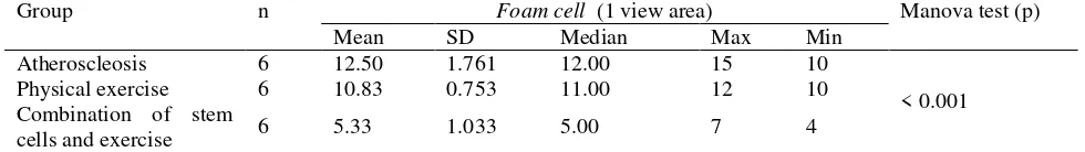
number of foam cells (Table 1). The result of the research also showed that the combination of stem cell therapy and physical exercise to individuals with atherosclerosis repaired the histopathology of the blood vessels in the abdominal aorta (Figure 2).
DISCUSSION
From the result of this study, there was a meaningful difference of value (p<0.05) after the therapy given to the control group and treatment group that received physical exercise, stem cell and the combination of the two to individuals with atherosclerosis. Plaque of atherosclerosis was located in the sub-intima which consisted of plain tendon cells, T-lymphocytes, collagen connective tissue, elastin, proteoglycan, macrophage, and dead foam cells, cholesterol residue, and calcium. The least number of foam
cells were found in the stem cells therapy group, followed by the combination of stem cell therapy and physical exercise group. It shows the changes on those indicators indicate the improvement of the blood vessels which is signified by the decreasing number of foam cells. Foam cells are formed by the macrophage which phagocytes the cholesterol. The existence of foam cells is the indicator of the early stage of endothel disfunction process which is the intial stage of atheorosclerosis.
This research employed mesenchymal stem cell. The source of mesenchymal stem cell is available in all of organs in the body especially in the perivascular areas. There are three biggest source of mesenchymal stem cells which are the adiphose tissue, blood of the umbilical cord, homeostatics, and the improvement of some tissues. The stem cell treatment, especially the balanced self
Figure 1: Histophatologycal abdominal aorta tissue of atherosclerosis Rats. Notes: Arrows show foam cells in the sub-intima area of aorta tissue.
(A) 100 times of magnification; (B) 400 times of magnification.
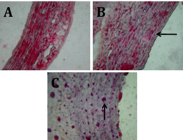
Figure 2: Histopathology of abdominal aorta hued using oil red o) (Magnification of 400x).
Notes: (A) Atherosclerosis group; (B) Physical exercise group; (C) Combination of Physical exercise and stem cell group. It can be seen in the Figure that the foam cells are shown in red color as pointed by the arrow.
A
C
improvement and differentiation which are controlled by intrinsic and extrinsic factors supported by the surrounding microenvironment is known as niche stem cell (Caplan, 2005). Niche an anatomic structure which belongs to cellular and acellular components which integrate local and systemic factors in controlling the stem cell phroliferation, differentiation, life system and homing process. Specific tissues have roles in supporting the function of the stem cells and also have responsibility to the growth, homeostatics and improvements of tissues of individuals. The repair and the defensive mechanism of stem cell are controlled by the local microenvironment inputs (Caplan, 2005; Jones and Magers, 2008).
The result of this research (Figure 2) showed that there was histopathologic repair to the abdonminal aorta after on atheroscleoris rats after given physical exercise therapy and combination of physical exercise and stem cells therapy. The physical activites triggered muscle contraxion which was indicated by inflamations that caused the mobilization of the stem cells from its source which is the bone marrow into the circulatory area (Laufs
et al., 2004; Iskandarsyah, 2007). The endogenous stem cell mobilization will provide improvements to the microenvironment which is indicated by the increasing number of SDF-1 (stroma cell derived factor 1).
In this research, the treatment of exogenous stem cell intravenous injection entered the circulatory system which improved the progenitory cells mobilization inside the circulatory sytem that was indicated by the CXCR4 (chemokine receptor 4). The treatment of exogeneous stem cell in this study is believed to trigger paracrine effects to the damaged endothel tissue. The effects of the paracrine stimulated the mobilization of endogenous stem cells which improved the microenvironment of the stem cells (Hicok and Hedrik, 2006; Bronckaers, et al., 2014). Good microenvironment condition stimulated differentiation and proliferation which created regenerative improvements on the vascular endothel which was indicated by the apoptosis process and the increasing number of VEGF which in this study was confirmed through the decreased number of foam cells (Table 1, Figure 1).
CONCLUSION
In conclusion, regular and measured medium-intensity physical exercise and the combination of the physical exercise and stem cell therapy were able to decrease the number of foam cells and stimulated repair on the histopathology of abnominal aorta of atherosclerosis rats.
REFERENCES
1. Bongso A, Hin LE, 2005. Stem Cells: Their Definition, Classification and Sources. In Stem Cells from Bench
to Bedside. World Scientific Publishing Co. Pte.Ltd, Singapore, pp.1-13.
2. Bronckaers A, Hilkens P, Martens W, Gervois P., Ratajczak J., Struys T., and Lambricths I,2014. Mesenchymal Stem/Stromal Cell as a Pharmacological and Therapeutic Approach to Accelerate Angiogenesis.Pharmacological & Therapeutics143, pp. 181-196.
3. Caplan AI, 2005. Mesenchymal Stem Cells: Cell Based Reconstructive Therapy in Orthopaedics. Tissue Eng, Vol.11, pp. 1198-1211.
4. Hartono A, and Gunardi S, 2013.Sinopsis Organ System Kardiovaskular Pendekatan dengan Sistem Terpadu dan Disertai Kumpulan Kasus Klinik. Tangerang: Karisma Publishing Group.
5. Hicok KC, Hedrik MH, 2006. Stem Cells and the Art of Mesenchymal Maitenance. In (Bronner F, Farach-Carson MC, Mikos AG, eds). Engineering of Functional Skeletal Tissue Springer-Verlag, London, pp.1-16.
6. Husni W, Hudaja O, and Kristian Y, 2006. Oklusi Arteri Perifer pada Ekstrimitas Inferior.JKM Vol.6, No.1, pp. 40-52.
7. Iskandarsyah K, 2007. The Effect Short-Term, Low-Intensity Exercise Training on the Levels of High-Sensitive C-Reactive Protein in the Patients with Acute Myocardial Infarction.J Kardiol Ind, Vol. 28, pp. :100-105.
8. Jones DL, and Wagers AJ, 2008. No Place Like Home: Anatomy and Function of the Stem Cell Niche.Nature Review, Vol. 9, pp. :11-21.
9. Laufs U, Werner N, Andreas L, Endres M, Wassman S, Jurgens K, Miche E., Bohm M, Georg N, 2004. Physical training increases endothelial progenitor cells, inhibits neointima formation, and enhances angiogenesis. Circulation, pp.:220-226.
10.Rilantono LI, 2013.Penyakit Kardiovaskular (PKV) 5 Rahasia. Jakarta: Badan Penerbit Fakultas Kedokteran Universitas Indonesia.
11.Salim AY, Nurrohmah A, 2013.Hubungan Olahraga dengan Kejadian Penyakit Jantung Koroner di RSUD Dr. Moewardi.Gaster Vol.10 No. 1, pp.: 48-56. 12.Santoso A, Erwinanto, Andriantoro H, Suryawan IGR,
Rifki S, Soerianata S, and Kasiman S, 2009.Lipid dan Penyakit Jantung Koroner.Jakarta: Centra Communications.
13.Setiawan I, Wardhani V, and Sargowo D, 2011.Akurasi Fibrinogen dan Hs-CRP sebagai Biomarker pada Sindroma Koroner Akut.Jurnal Kedokteran Brawijaya, Vol.26, No.4, pp.: 233-239.
14.Tatjana I, Vitosevic B, Milosevic L, Stevic L, and Savic T, 2007. The Effect of Physical Activity on
Table 1: The number of foam cells that were created in the abdominal aorta tissue.
Inflammatory Markers. The Risk of New Coronary Event in Coronary Heart Disease Patients. Acta Medica Medianae, Vol.4, pp. :10-14.