Principles and Practice of
Clinical Virology
F
IFTH
E
DITION
Principles and Practice of
Clinical Virology
F
IFTH
E
DITION
Edited by
Arie J. Zuckerman
Royal Free and University College Medical School, London, UK
Jangu E. Banatvala
Guy’s, King’s and St Thomas’ School of Medicine, London, UK
John R. Pattison
Department of Health, London, UK
Paul D. Griffiths
Royal Free and University College Medical School, London, UK
Barry D. Schoub
Copyrightu1987, 1990, 1994, 2000, 2004 John Wiley & Sons Ltd,
The Atrium, Southern Gate, Chichester, West Sussex PO19 8SQ, England Telephone (+44) 1243 779777
Email (for orders and customer service enquiries): [email protected] Visit our Home Page on www.wileyeurope.com or www.wiley.com
All Rights Reserved. No part of this publication may be reproduced, stored in a retrieval system or transmitted in any form or by any means, electronic, mechanical, photocopying, recording, scanning or otherwise, except under the terms of the Copyright, Designs and Patents Act 1988 or under the terms of a licence issued by the Copyright Licensing Agency Ltd, 90 Tottenham Court Road, London W1T 4LP, UK, without the permission in writing of the Publisher. Requests to the Publisher should be addressed to the Permissions Department, John Wiley & Sons Ltd, The Atrium, Southern Gate, Chichester, West Sussex PO19 8SQ, England, or emailed to [email protected], or faxed to (+44) 1243 770620. This publication is designed to provide accurate and authoritative information in regard to the subject matter covered. It is sold on the understanding that the Publisher is not engaged in rendering professional services. If professional advice or other expert assistance is required, the services of a competent professional should be sought.
Other Wiley Editorial Offices
John Wiley & Sons Inc., 111 River Street, Hoboken, NJ 07030, USA Jossey-Bass, 989 Market Street, San Francisco, CA 94103-1741, USA Wiley-VCH Verlag GmbH, Boschstr. 12, D-69469 Weinheim, Germany
John Wiley & Sons Australia Ltd, 33 Park Road, Milton, Queensland 4064, Australia
John Wiley & Sons (Asia) Pte Ltd, 2 Clementi Loop #02-01, Jin Xing Distripark, Singapore 129809 John Wiley & Sons Canada Ltd, 22 Worcester Road, Etobicoke, Ontario, Canada M9W 1L1
Wiley also publishes its books in a variety of electronic formats. Some content that appears in print may not be available in electronic books.
British Library Cataloguing in Publication Data
A catalogue record for this book is available from the British Library
ISBN 0-470-84338-1
Typeset in 9/11pt Times by Dobbie Typesetting Ltd, Tavistock, Devon. Printed and bound in Great Britain by Antony Rowe Ltd, Chippenham, Wilts.
Contents
List of Contributors . . . vii
Preface . . . xi
Preface to the Fourth Edition. . . xii
Preface to the Third Edition. . . xiii
Preface to the Second Edition. . . xiv
Preface to the First Edition . . . xv
Plates . . . xvii
1 Diagnostic Approaches. . . 1
Katie Jeffrey and Deenan Pillay 2 The Herpesviridae. . . 23
Graham M. Cleator and Paul E. Klapper 2A Herpes Simplex. . . 27
Graham M. Cleator and Paul E. Klapper 2B Varicella Zoster. . . 53
Judith Breuer 2C Cytomegalovirus . . . 85
Paul D. Griffiths 2D Epstein–Barr Virus . . . 123
Dorothy H. Crawford 2E Roseoloviruses: Human Herpesviruses 6 and 7. . . 147
Ursula A. Gompels 2F Kaposi’s sarcoma-associated Herpesvirus (Human Herpesvirus 8). . 169
Abel Viejo-Borbolla, Cornelia Henke-Gendo and Thomas F. Schulz 3 Hepatitis Viruses . . . 199
Tim J. Harrison, Geoffrey M. Dusheiko and Arie J. Zuckerman 4 Viruses Associated with Acute Diarrhoeal Disease. . . 249
Ulrich Desselberger and Jim Gray 5 Influenza. . . 271
Chris W. Potter 6 Parainfluenza Viruses. . . 299
Stelios Psarras, Nikolaos G. Papadopoulos and Sebastian L. Johnston 7 Respiratory Syncytial Virus. . . 323
Caroline Breese Hall 8 Adenovirus. . . 343
Marcela Echavarria 9 Rhinoviruses . . . 361
Nikolaos G. Papadopoulos and Sebastian L. Johnston 10 Coronaviruses and Toroviruses. . . 379
David Cavanagh 11 Measles. . . 399
Sibylle Schneider-Schaulies and Volker ter Meulen 12 Rubella. . . 427
Jennifer M. Best and Jangu E. Banatvala 13 Mumps. . . 459
Pauli Leinikki 14 Enteroviruses. . . 467
15 Poxviruses. . . 491 Inger Damon, Peter Jahrling and
James LeDuc
16 Alphaviruses . . . 509 Graham Lloyd
17 Flaviviruses. . . 531 Barry D. Schoub and Nigel K.
Blackburn
18 Bunyaviridae. . . 555 Robert Swanepoel
19 Arenaviruses . . . 589 Colin R. Howard
20 Filoviruses. . . 611 Susan P. Fisher-Hoch
21 Rabies and Other Lyssavirus
Infections . . . 631 Mary J. Warrell
22 Papillomaviruses. . . 661 Dennis McCance
23 Human Polyomaviruses. . . 675 Kristina Do¨rries
24 Human Parvoviruses. . . 703 Kevin E. Brown
25 Human Immunodeficiency Viruses . . . 721 Robin A. Weiss, Angus G. Dalgleish, Clive Loveday and Deenan Pillay
25A The Human T Cell Lymphotropic
Viruses. . . 759 Graham P. Taylor
26 Human Prion Diseases . . . 779 John Collinge
27 GBV-C and TTV. . . 813 Shigeo Hino
28 Emerging Virus Infections. . . 825 Brian W. J. Mahy
29 Hospital-acquired Infections. . . 835 29A Infections Acquired via the
Blood-borne Route. . . 837 Anthea Tilzey
29B Infections Acquired via Other
Routes . . . 843 Philip Rice
Contributors
Jangu E. Banatvala Emeritus Professor of Clinical Virology, Guy’s, King’s and St Thomas’ School of Medicine, Lambeth Palace Road, London SE1 7EH, UK
Jennifer M. Best Reader in Virology, Guy’s, King’s and St Thomas’ School of Medicine, Lambeth Palace Road, London SE1 7EH, UK
Nigel K. Blackburn Senior Consultant Virologist, National Institute for Communicable Diseases, Sandringham, South Africa
Judith Breuer Reader and Consultant in Virology, Skin Virus Laboratory, St Bartholomew’s and the Royal London School of Medicine and Dentistry, 25–29 Ashfield Street, London E1 1BB, UK
Kevin E. Brown Senior Investigator, Virus Discovery Group, Hematology Branch, National Heart, Lung and Blood Institute, Bethesda, MD, USA
David Cavanagh Principal Scientist, Institute for Animal Health, Compton Laboratory, Compton, Newbury, Berks RG20 7NN, UK
Graham M. Cleator Reader in Medical Virology, Laboratory Medicine Academic Group, Department of Virology, 3rd floor, Clinical Sciences Building, Manchester Royal Infirmary, Oxford Road, Manchester M13 9WL, UK
John Collinge Head, Department of
Neurodenegerative Disease and Director, MRC Prion Unit, Institute of Neurology, University College London, London, UK
Dorothy H. Crawford Professor of Medical
Microbiology and Head of the School of Biomedical and Clinical Laboratory Sciences, The University of Edinburgh, Hugh Robson Building, George Square, Edinburgh EH8 9XD, UK
Angus G. Dalgleish Professor of Oncology, St George’s Hospital Medical School, Cranmer Terrace, London SW17 0RE, UK
Inger Damon Chief, Poxvirus Section, Division of Viral and Rickettsial Diseases, National Center for Infectious Diseases, Centers for Disease Control and Prevention, 1600 Clifton Rd NE, Mailstop G-18, Atlanta, GA 30333, USA
Ulrich Desselberger Consultant Virologist and Director, Clinical Microbiology and Public Health Laboratory, Addenbrooke’s Hospital, Hills Road, Cambridge CB2 2QW, UK
Kristina Do¨rries Senior Scientist and Group Leader, Institute for Virology and Immunobiology, Julius-Maximilians-University Wu¨rzburg, Versbacher Strasse 7, D-97078 Wu¨rzburg, Germany
Geoffrey M. Dusheiko Professor of Medicine, Department of Medicine, Royal Free and University College Medical School, Rowland Hill Street, London NW3 2PF, UK
Marcela Echavarria Assistant Professor of Microbiology, Centro de Educacio´n Me´dica e Investigaciones Clı´nicas, CEMIC University Hospital, Galvan 4102, (C1431FWO), Buenos Aires, Argentina
Susan P. Fisher-Hoch Professor of Biological Sciences, University of Texas, Houston School of Public Health at Brownsville, 80 Fort Brown, Brownsville, TX 78520, USA
Ursula A. Gompels Senior Lecturer in Molecular Virology, Pathogen Molecular Biology Unit,
Department of Infectious and Tropical Diseases, London School of Hygiene and Tropical Medicine, University of London, Keppel Street, London WC1E 7HT, UK
Paul D. Griffiths Professor of Virology, Royal Free and University College Medical School, Rowland Hill Street, London NW3 2PF, UK
Caroline Breese Hall Professor of Pediatrics and Medicine in Infectious Diseases, University of Rochester School of Medicine, 601 Elmwood Avenue, Box 689, Rochester, NY 14642, USA
Tim J. Harrison Reader in Molecular Virology, Royal Free and University College Medical School, Royal Free Campus, Rowland Hill Street, London NW3 2PF, UK
Cornelia Henke-Gendo Clinical Virologist, Department of Virology, Hannover Medical School, Carl-Neuberg Strasse 1, 30625 Hannover, Germany
Shigeo Hino Department of Virology, Faculty of Medicine, Tottori University, 86 Nishi, Yonago 683-8503, Japan
Colin R. Howard Vice-Principal for Strategic Development and Professor of Microbiology, Royal Veterinary College, University of London, Royal College Street, London NW1 0TU, UK
Peter Jahrling USAMRIID, Fort Detrick, Frederick, MD 21702-5001, USA
Katie Jeffery Consultant Virologist, Department of Microbiology, John Radcliffe Hospital, Headington, Oxford OX3 9DU, UK
Sebastian Johnston Professor, Department of Respiratory Medicine, National Heart and Lung Institute, Imperial College, London, UK
Paul E. Klapper Consultant Clinical Scientist and Honorary Senior Lecturer, Health Protection Agency, Leeds Laboratory, Bridle Path, York Road, Leeds LS15 7TR, UK
James LeDuc Director, Division of Viral and Rickettsial Diseases, National Center for Infectious Diseases, Centers for Disease Control and Prevention, 1600 Clifton Road, Mail-stop A-30, Atlanta, GA 30333, USA
Pauli Leinikki Professor, Department of Infectious Diseases Epidemiology, National Public Health Institute (KTL), Mannerheimintie 166, FIN-00300 Helsinki, Finland
Graham Lloyd Head, Special Pathogens, Centre for Applied Microbiology and Research, Porton Down, Salisbury, Wiltshire SP4 0JG, UK
Clive Loveday Clinical Director, International Clinical Virology Centre, Great Missenden, UK
Brian W. J. Mahy Senior Scientific Research Advisor, National Center for Infectious Diseases, CDC, 1600 Clifton Road, Mailstop C12, Atlanta, GA 30333, USA
Dennis McCance Department of Microbiology and Immunology, Head, Virology Unit, University of Rochester, Box 672, 601 Elmwood Avenue, Rochester, NY 14642, USA
Philip D. Minor Head, Division of Virology, National Institute for Biological Standards and Control, Potters Bar, Hertfordshire, UK
Peter Muir Health Protection Agency South West, Myrtle Road, Bristol BS2 8EL, UK
Nikolaos G. Papadopoulos Lecturer, Allergy Unit, 2nd Department of Pediatrics, University of Athens, Greece
Deenan Pillay Reader in Virology, Royal Free and University College Medical School, Windeyer Building, 46c Cleveland Street, London W1P 6DB, UK
Chris W. Potter Emeritus Professor, University of Sheffield, Division of Genomic Medicine, School of Medicine and Biomedical Sciences, F Floor, Beech Hill Road, Sheffield S10 2RX, UK
Stelios Psarras Research Associate, Allergy Unit, 2nd Pediatric Clinic, University of Athens, Greece
Philip Rice Consultant Virologist, St George’s Hospital Medical School, Cranmer Terrace, London SW17 0RE, UK
Sibylle Schneider-Shaulies Institute for Virology and Immunobiology, University of Wu¨rzburg, Versbacher Strasse 7, D-97078, Wu¨rzburg, Germany
Barry D. Schoub Executive Director, National Institute for Communicable Diseases, Sandringham, South Africa
Thomas F. Schulz Head of the Department of Virology, Hannover Medical School, Carl-Neuberg Strasse 1, 30625 Hannover, Germany
Robert Swanepoel National Institute for
Communicable Diseases, Sandringham, South Africa
Volker ter Meulen Institute for Virology and Immunology, University of Wu¨rzburg, Versbacher Strasse 7, D97078 Wu¨rzburg, Germany
Anthea Tilzey Clinical Senior Lecturer, Virology Section, Department of Infectious Diseases, Guy’s, King’s and St Thomas’ School of Medicine, St Thomas’ Campus, UK
Abel Viejo-Borbolla Postdoctoral Fellow, Department of Virology, Hannover Medical School, Carl-Neuberg Strasse 1, 30625 Hannover, Germany
Mary J. Warrell Research Associate, Centre for Tropical Medicine, John Radcliffe Hospital, Oxford OX3 9DU, UK
Robin A. Weiss Professor of Viral Oncology, University College, London, UK
Preface
The knowledge and practice of clinical virology continues to expand. The first edition of Principles and Practice of Clinical Virology, published in 1987, contained 16 chapters and 590 pages. Each of the subsequent editions became progressively larger. This edition has 902 pages and 38 chapters, including seven within the section on theHerpesviridae, each of which is comprehensive.
Rapid progress in the field has occurred between the fourth and fifth editions. There are now two new editors and a number of new authors, which will increase international representation. In addition, each of the remaining chapters has been extensively revised or rewritten, taking into account knowledge accumulated in molecular biology with its applications for laboratory diagnosis, immunisation and antiviral chemotherapy. Each chapter also highlights the clinical features and
epidemiological patterns of infection. Between this new edition and the last, much concern has been focused on the global threat posed by new viruses. Consequently, a new chapter on ‘Emerging Infections’ is included.
There is also a new chapter on ‘Hospital-acquired Infections’, which will be of benefit to those who have to deal with the day-to-day management of patients in hospital. This chapter also includes some advice relating to SARS. However, fresh knowledge about SARS continued to accumulate as the fifth edition was in preparation, and further information is included, not only in the chapter on ‘Emerging Infections’ but also in the one on ‘Coronaviruses’.
In comparison with the fourth edition, additional colour plates have been included, and, as in previous editions, an attempt has been made to limit references to key publications.
Preface to the Fourth Edition
It is now 13 years since the first edition ofPrinciples and Practice of Clinical Virology was published. A comparison of the first and fourth editions testifies to the rapid expansion in virology during the intervening years, including major developments in technology, the application of these to clinical practice and advances in the treatment of viral infections with an increasing number of antiviral drugs. Indeed, such has been the progress in the field of clinical virology even within the period between the third and fourth editions that we have asked a number of new authors to contribute chapters. These include the chapters on rhinoviruses, viruses associated with acute diarrhoeal disease, and human polyomaviruses. Of the remaining chapters, virtually all have been revised substantially. The chapter on the Herpesviridae has been expanded considerably, particularly in relation to Human her-pesviruses 6,7and8; new authors have contributed to these sections. The chapter on hepatitis viruses reflects the considerable expansion of information relating to hepatitis E and C as well as the role of such newly recognised agents as GB and the new human virus, TTV.
Advances in diagnostic methods, particularly mo-lecular biological techniques and their application to clinical problems, are reflected in a new chapter on ‘Diagnostic Approaches’, which presents an overview of the value and limitations of established and more recently developed techniques. Advances in serological techniques as well as in virus identification are emphasised. This chapter also includes an important section on assays for determining antiviral drug resistance.
Most of the chapters reflect advances in patient management, including—where appropriate—antiviral chemotherapy. As expected, the chapters on hepatitis viruses and human retroviruses have been expanded considerably in the light of continuing and rapid advances in these fields. Both chapters now include a component by authors with everyday practical experi-ence in the management and treatment of patients with these infections.
In comparison with the third edition, more coloured plates have been included, which we hope our readers will appreciate. As in previous editions, we have attempted to limit the reference lists to key publications.
Preface to the Third Edition
Principles and Practice of Clinical Virology was first published in 1987; a third edition within 7 years of the first attests to the continuing and rapid progress in the field of clinical virology.
All the chapters have been revised and some completely rewritten, reflecting the increasing knowledge of viruses or groups of viruses included in the various chapters. Thus, the chapter on hepatitis viruses now contains sections on hepatitis A and E viruses as well as individual contributions on hepatitis B, D and C. The previous edition contained two chapters on viral haemorrhagic fevers but this section now includes separate chapters for the flaviviruses, alphaviruses,Bunyaviridae, arenaviruses and filoviruses. The chapters on arenaviruses and filoviruses have been contributed by new authors.
New authors have also contributed the chapter on herpes simplex virus infections and the chapter on the more newly-recognised herpesviruses now includes a section onHuman herpes virus 7.
As expected, rapid developments continue to occur in the field of human retroviruses, particularly the human immunodeficiency viruses, and this chapter reflects the accumulation of new and important data in this area. The chapter on human prion disease has been almost entirely rewritten to reflect many of the substantial advances in prion research.
Most of the authors stress the developments and application of molecular biological techniques which are leading not only to improved methods of diagnosis but also to an increased understanding of viral pathogenesis.
Rather than burden readers with a large number of references, we have aimed to include most of the key ones.
Finally, we are grateful for the helpful comments which we received from many of our readers; some of their suggestions have been incorporated into the third edition.
Preface to the Second Edition
In the preface to the first edition of Principles and Practice of Clinical Virologywe stated that it was our intention, with new editions, to remain up-to-date as the subject advanced. Such is the pace of development in clinical virology that plans were laid for a new edition within a few months of the first being published, and this second edition is appearing only two and a half years after the first. Each chapter has been revised, many extensively, to take account of progress in the understanding of the epidemiology, pathogenesis, diagnosis, management and prevention of virus infections. Perhaps the greatest single recent contribution to the subject has been made by the application of molecular biological techniques, and each of our authors has highlighted the contribution of this rapidly developing discipline to clinical virology.
Human herpesvirus 6 is now the subject of a new chapter, and we have also added a new chapter on haemorrhagic fevers to include much new information on their pathogenesis. As expected, the extensive accumulation of new information relating to infection by human retroviruses has resulted in extensive updating of this chapter, not only to include much
more information on human immunodeficiency viruses but also on HTLV-1 and -2. The chapter on hepatitis has been separated into two sections: the first on hepatitis A and the viruses causing non-A, non-B hepatitis, two of which have been identified as hepatitis C and E, and the second on hepatitis B and D (delta agent).
As before, we were aware when organising the book that no single arrangement is entirely satisfactory. We have chosen to arrange the chapters on the basis of individual viruses or groups of viruses. General chapters on virus structure, taxonomy and patho-genesis are not included, but the information on these aspects necessary for an understanding of the practice of clinical virology is included in the individual chapters.
Such factors as the likely discovery of yet new viruses, improvements in the rapidity and sensitivity of diagnostic techniques, and the development of new vaccines, which are likely to involve recombinant techniques and progress in antiviral therapy, will ensure that thought will be given to revision and preparation of another edition.
Preface to the First Edition
There has been a spectacular increase during the last 30 years in our knowledge of virology. This has taken place to such an extent that virology can now be regarded as an umbrella term encompassing a variety of distinct but related disciplines. There are funda-mental connections with biochemistry, genetics and molecular biology, and each of these aspects would be worth a treatise in itself. Clinical virology is that aspect which is concerned with the cause, diagnosis, treatment and prevention of virus infections of man. It too has acquired a substantial body of knowledge and accumulated experience over the past 30 years and this book is intended to be an authoritative account of the present situation. Formerly virological diagnosis was time consuming, retrospective and rarely influ-enced the management of the patient. During the past 10–15 years the picture has changed dramatically. Newly recognised diseases such as AIDS and some haemorrhagic fevers, which have very serious con-sequences for individuals and populations, have been shown to be due to viruses. In the clinical virology laboratory there has been a change in emphasis towards rapid diagnostic techniques. Finally,effective antiviral chemotherapy is a reality at least for some virus infections and there has been an expansion in the use of immunoprophylaxis. Thus the current principles
and practice of clinical virology are concerned with rapid laboratory diagnosis leading to appropriate patient management which might involve specific therapy and/or infection control measures at a hospital, a national and occasionally at an inter-national level.
In organising the book we were aware that there is no single arrangement that is entirely satisfactory. We have chosen to arrange the chapters on the basis of individual viruses or groups of viruses. General chapters on virus structure, taxonomy and pathogenesis are not included but the information on these aspects necessary for an understanding of the practice of clinical virology is included in the individual chapters.
Clinical virology is a subject which continues to evolve. This is usually for one of two reasons, either the need to apply new technology or the need to study new diseases or epidemiological situations. We have therefore invited authors who are specialist investiga-tors into each of the viruses to contribute up-to-date, stimulating accounts of the practice of clinical virology and provide a framework for the assimilation of imminent advances. One chapter has already had to be significantly updated during the time of preparation of the book and it is our intention, with new editions, to remain up-to-date as the subject advances.
Figure 2A.2M‘Cold sores’ at the pustular and crusting stages Figure 2A.3MRose Bengal staining of herpes simplex virus dendritic ulceration in a grafted cornea. (Kindly provided by R. E. Bonshek and A. B. Tullo)
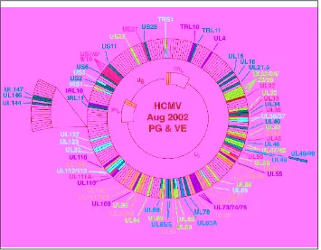
Figure 2C.4M‘Proteins of CMV which have been mapped to date. UL = unique long region; US = unique short region; TR = terminal repeat; IR = inverted repeat. The genome is linear within the virus but has been circularized for convenience. Open reading frames of known function are coloured according to the following code: orange = transactivators; pink = DNA replication; green = capsid and/or assembly; red = tegument; pale blue = envelope; dark green = immune evasion; dark blue = miscellaneous
Figure 2C.8M‘Schematic representation of the ways viruses can interfere with presentation of HLA-peptide complexes at the plasma membrane. Rib, ribosome; ER, endoplasmic reticulum; V, virus-encoded protein; PRO, proteosome; PM, plasma membrane; TAP, transporter associated with antigen presentation. Peptides derived from virus-infected cells are generated in the proteosome and actively transported by TAP into the lumen of the ER. A ribosome is shown producing a protein with a signal peptide, which folds in the ER to produce the HLA Class I chain. This should normally associate with peptide and be transported to the plasma membrane. Misfolded HLA molecules can be re-exported from the lumen of the ER back into the cytosol where they are degraded by the proteosome. Virus-encoded genes interfere with this process as follows: the proteins may be inherently insusceptible to proteosome digestion (EBNA of EBV) or may be modified to reduce their digestion (pp65 acts on MIE protein of HCMV). Proteins may block the function of TAP (ICP47 of HSV; US6 of HCMV). Proteins may bind mature class I molecules within the ER and so sequester them (E3-19K protein of adenoviruses; US3 protein of HCMV; m152 protein of MCMV). Two proteins of HCMV (US2 and US11) facilitate the re-export from the ER to the cytosol of mature HLA class I molecules. If all of these mechanisms are completely successful, the level of HLA display at the PM will be insufficient to prevent NK cells or macrophages recognising the cell as being abnormal and so destroying it. Proteins/peptides encoded within HCMV (UL18) or MCMV (m144) are presented at the plasma membrane to act as a decoy for NK cells by providing a negative signal. In addition, HCMV UL16 blocks transmission of a positive signal to another group of NK cells
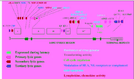
Figure 2F.5M‘Genome diagram of KSHV. Open boxes with Roman numbers denote groups of structural or metabolic genes which are conserved among ȍherpesviruses and also many other herpesviruses. The solid line represents the long unique (coding) region, open and filled
rectangles internal or terminal repeat regions. Solid circles represent originas of lytic relication [ori-(L), ori-(R)]. The position and transcriptional orientation of viral genes discussed in the text is indicated by pointed boxes. A red shading indicates genes known to be expressed in latently infected spindle cells and PEL cells (see text), light blue, dark blue and green shading refers to genes expressed in the different stages of the lytic cycle (see text). Colour coding of the names of individual viral genes refers to their presumed function, as indicated (see also text)
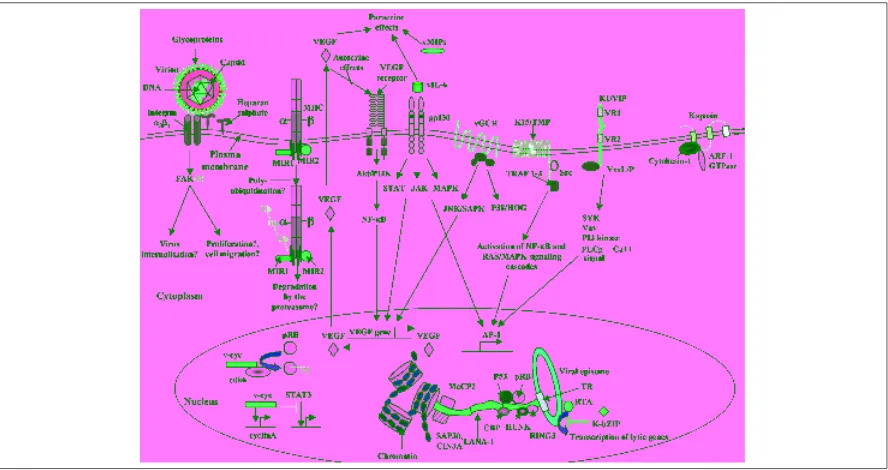
Figure 2F.7M‘Overview of some KSHV–encoded proteins that may contribute pathogenesis. Vital proteins are coloured in red, interacting cellular proteins in grey. See text for a detailed explanation of the pathways and receptors engaged by KSHV proteins
TE VI
xxii
Figure 21.3M‘World distribution of rabies. Rabies free areas are white; red indicates terrestrial rabies with or without bat rabies, and countries with only bat lyssaviruses are green
Figure 24.5M‘Children with characteristic slapped cheek appearance and reticular lacy rash of fifth disease
Figure 28.3M‘Recent Emergence and Reemergence of Human Viral Diseases, Examples
1
Diagnostic Approaches
Katie Jeffery
1and Deenan Pillay
21
John Radcliffe Hospital, Oxford, and
2
Royal Free and University College Medical School, London, UK
INTRODUCTION
If clinical virology in the 1980s was characterised by the widespread use of enzyme-linked immunosorbent assay (ELISA) technology, then there is no doubt that the 1990s will be seen as the time when molecular methods of virus detection entered routine diagnostic use. Following on from this development, the first few years of the twenty-first century will be seen as the period when real-time PCR and virus quantitation came of age, along with increasing automation of molecular diagnostics. Concurrently, the emphasis and priorities of diagnostic virology laboratories have shifted in response to: the availability of rapid diagnostic methods; the identification of new viruses, many of which are non- or poorly cultivatable; the increasing availability of effective antiviral agents; the emergence of antiviral resistance; the increasing number of immunocompromised patients, in whom opportunistic viral infections are life-threatening; and new cost pressures on pathology services.
This chapter provides an overview of diagnostic techniques against this background, and highlights those clinical scenarios of particular importance to virologists in a diagnostic setting.
TECHNIQUES—AN OVERVIEW
Virus Isolation
Many of the advances in clinical virology have come about because of the ability to propagate viruses in the
laboratory. Historically, viruses were propagated in laboratory animals and embryonated eggs, although most virus isolation techniques now rely on cultured cells. With appropriate specimens and optimal cell lines, this technique can be highly sensitive and specific, and a presumptive diagnosis made on the basis of a characteristic cytopathic effect (CPE), confirmed by immunostaining. The judicious use of two or three cell lines, such as a monkey kidney line, a human continuous cell line and a human fibroblast line, will allow the detection of the majority of cultivatable viruses of clinical importance, such as herpes simplex virus (HSV), varicella zoster virus (VZV), cytomegalovirus (CMV), enteroviruses, respiratory syncytial virus (RSV), adenovirus, parainfluenza viruses, influenza viruses and rhinoviruses. In addition, the ability to grow virus from a clinical specimen demonstrates the presence of viable virus (albeit viable within the chosen cell line)—this is not necessarily the case with detection of viral antigen or genome. For example, following initiation of antiviral therapy for genital herpes, HSV antigen can be detected from serial genital swabs for longer than by virus propagation in cell culture. This infers that antigen persists in the absence of viral replication and underlines the impor-tance of correct interpretation of laboratory results. Nevertheless, virus isolation has now been shown to be less sensitive than molecular amplification methods for this and other viruses (see later).
The advantages of virus isolation include: the ability to undertake further examination of the isolate, such as drug susceptibility assays (see later) or typing (Table 1.1); the provision of epidemiological information on
viruses of public health importance; and the culture and identification of previously unrecognised viruses, e.g. human metapneumovirus (van den Hoogenet al., 2001) and SARS-associated coronaviruses (Drostenet al., 2003). However, routine cell culture techniques available in most laboratories will not detect a number of clinically important viruses such as gastroenteritis viruses, hepatitis viruses, Epstein–Barr virus (EBV), Human herpesvirus 6,7and8(HHV-6, -7, -8), and/or human immunodeficiency virus (HIV). Other than HSV, for which most isolates will grow in human fibroblast cells within 3 days, the time for CPE (or, for example, haemadsorption) to develop for most clinical viral isolates is between 7 and 21 days. For this reason, a number of modifications to conventional cell culture have been reported, to provide more rapid results. These include centrifugation of specimens on to cell monolayers, often on cover slips, and immunostaining with viral protein-specific antibodies at 48–72 h follow-ing inoculation (Shell Vial Assay) (e.g. Stirk and Griffiths, 1988). Such techniques can also be under-taken in microtitre plates (O’Neillet al., 1996).
The role of conventional cell culture for routine diagnosis of viral infections is diminishing and is a subject of active debate within the virology community (Carman, 2001; Ogilvie, 2001). Many laboratories are discontinuing or downgrading virus isolation methods in favour of antigen or genome detection for the rapid diagnosis of key viral infections (usually those that are treatable, such as CMV and VZV). Nevertheless, it is important for large laboratories to maintain the ability to employ this methodology for the reasons given above. Where primary diagnosis is undertaken by cell culture, there will be increasing pressure to generate quicker results by use of the many rapid techniques that have been reported.
Antigen Detection
Immunofluorescence
One of the most effective rapid diagnostic tests is indirect or direct immunofluorescence (IF). Initially
undertaken with polyclonal antisera, and then sub-sequently with pools of monoclonal antibodies, this method uses either indicator-labelled antibody or a labelled antispecies antibody (indirect) to directly visualise viral antigens in clinical specimens. Usually, the label used is fluorescein. The indirect method is more sensitive, since more label can be bound to an infected cell. Results can be available with 1–2 h of specimen receipt. The most common use of this technique is for the diagnosis of respiratory viral infections whereby a panel of reagents are utilised to detect RSV, parainfluenza viruses, influenza A and B and adenovirus in multiple wells of a microscope slide. This technique is sensitive compared to cell culture, especially for the detection of RSV. The ideal specimen for such testing is a nasopharyngeal aspirate, most usually obtained from infants with suspected bronch-iolitis, for whom a rapid result is essential for correct clinical management and implementation of infection control measures. However, detection can also be made from a well-taken throat/nasal swab. There is increasing evidence that community or nosocomial acquired respiratory viruses lead to severe disease in immunocompromised patients (for review, see Ison and Hayden, 2002), and it is important that broncho-alveolar lavage specimens from such patients with respiratory disease are also tested for these viruses in addition to the more common pathogens, such as CMV. Respiratory virus antigens are expressed within the epithelial cells, and the success of the technique depends on an adequate collection of cells. A particular advantage of IF, compared to the commer-cial rapid antigen tests available for RSV and influenza, is that microscopic examination of the fixed cells can determine the presence of adequate cell numbers for analysis (Table 1.2). IF has been used widely for the direct detection of HSV and VZV in vesicle fluid, and has advantages over electron micro-scopy in both sensitivity and specificity. IF methods have also been used to detect more unusual viruses, such as Lassa fever (Wulff and Lange, 1975). An important limitation of IF is that well-trained micro-scopists are required for interpretation, which remains subjective.
Table 1.1 Virus isolation
Advantages Disadvantages
Sensitive Slow (conventional cell culture) ‘Catch-all’ Labour-intensive
Generates isolate for further study
Multiple cell lines required
Detects ‘viable’ virus Adaptation for rapid result
Table 1.2 Antigen detection by immunofluorescence
Advantages Disadvantages
Rapid Requires skilled staff Sensitive for some viruses
(e.g. RSV)
Variable sensitivity Dependent on high-quality
Detection and semi-quantitation of CMV antigen-containing cells in blood can also be undertaken by direct IF (CMV/pp65 antigenaemia assay). This technique involves separation of peripheral blood mononuclear cells (PBMCs) and fixing on a slide, followed by staining with a monoclonal antibody directed against the matrix protein pp65. The fre-quency of positive cells can predict CMV disease in the immunocompromised patient (van der Bijet al., 1989), and is used in a number of laboratories. However, it is labour-intensive, needs large numbers of PBMCs (making it unsuitable for all patient populations) and requires a rapid processing of blood specimens if a reduction in sensitivity of detection is to be avoided (Boeckh et al., 1994). Therefore, PCR is rapidly replacing antigenaemia as the method of choice for qualitative and quantitative detection of CMV.
ELISA/Latex Agglutination for Antigen Detection
Solid phase systems for antigen detection are now used widely. ELISAs are based on the capture of antigen in a clinical specimen to a solid phase via a capture antibody, and subsequent detection using an enzyme-linked specific antibody. Variation in capture and detector antibody species has increased the sensitivity of these assays, which are widely used for hepatitis B virus (HBV) surface antigen detection (HBsAg) and, more recently, for hepatitis C core antigen in donor blood-testing laboratories (Peterson et al., 2000). ELISA-based systems for the diagnosis of, for exam-ple, RSV, influenza and HSV may be appropriate in some contexts for point-of-care testing, although often at the expense of sensitivity when compared to IF and/ or virus culture.
Small latex particles coated with specific antibody can be agglutinated in the presence of antigen, which can then be observed with the naked eye. This rapid assay is used for rotavirus diagnosis, with an equiva-lent sensitivity to electron microscopy. Capture of antibody, rather than antigen, can also be undertaken, although latex assays for CMV and VZV antibodies may lack sensitivity and specificity compared to ELISA systems (see below).
Electron Microscopy
Electron microscopy (EM) is the only technique available for directly visualising viruses, and therefore
has many applications beyond purely diagnostic purposes. The major role of EM in a clinical setting is in the diagnosis of viral gastroenteritis, for which many of the aetiological agents are non-cultivatable, and analysis of skin lesions for herpes, pox and papillomaviruses.
Preparation of specimens and the technique of negative staining are straightforward and quick, and the method is a ‘catch-all’ approach to detecting viruses. Nevertheless, it has a limit of sensitivity of approximately 106viral particles per millilitre of fluid.
Vast numbers of virions are present during acute skin and gastrointestinal disease, and a diagnosis is easily made. This becomes more difficult later in the course of infection, when viral shedding is reduced below the level of detection. Sensitivity can be enhanced by antibody-induced clumping of virus (immune EM) or ultracentrifugation; however, it is unrealistic to under-take these methods routinely. The advantages and disadvantages of electron microscopy are summarised in Table 1.3.
The survival of EM within the routine clinical virology laboratory hinges on the availability of alternative, more sensitive methods of diagnosis. Many centres already use latex agglutination for rotavirus diagnosis, and polymerase chain reaction (PCR) is more sensitive for the detection of herpes-viruses in vesicular fluid (Beards et al., 1998). Currently, EM in diagnostic virology laboratories is used primarily for outbreak investigation. Now that PCR-based methods of Norovirus detection are established (Green et al., 1995), show increased sensitivity with respect to EM (O’Neill et al., 2001) and can be adapted for real-time PCR detection (Miller et al., 2002), the future of EM in clinical virology is in doubt. One of the first indications for electron microscopy was for the rapid diagnosis of smallpox; in the era of bioterrorism, EM will continue to play a role in specialist centres in the event of a bioterrorist attack, such as confirming VZV infection in cases of vesicular rash.
Table 1.3 Electron microscopy
Advantages Disadvantages
‘Catch-all’ Requires skilled staff Economical running costs Poor sensitivity Detects unculturable viruses Large capital outlay Adaptable, e.g. immunoelectron
Histology/Cytology
Direct microscopic examination of stained histology or cytology specimens can on occasion provide the first indication that a virus may be responsible for a pathological process; e.g. the intranuclear (early) or basophilic (late) inclusions seen in interstitial nephritis in renal transplant biopsies due to BK polyoma virus; changes in cervical cytology seen in association with human papillomavirus (HPV); and the nuclear inclu-sions seen in erythroid precursor cells in parvovirus B19 infection.
Serology
All viral infections generate a humoral response, and this can be used for diagnostic purposes. The classical pattern of response following an acute infection is illustrated in Figure 1.1. The functional nature of this response is extremely variable. In some instances, these antibodies are neutralising and can be assessed for this activity (e.g. polioviruses). However, many infections are controlled more effectively by T cell responses, and antibody detection is used as a surrogate of infection. Traditionally, methods of antibody detection did not distinguish between IgG and IgM responses, and diagnosis was based on seroconversion or a significant rise in antibody titre between acute and convalescent samples (10–14 days apart). The complement fixation test was used widely in this respect; however, assay insensitivity and the cross-reactivity of many antigens used within the assay limited its clinical usefulness. Most importantly, a diagnosis could only be made after the time of acute illness. Currently, the major use of this assay is for the diagnosis of ‘atypical’ pneumonia (Chlamydia psittaci/pneumoniae, Coxiella
or Mycoplasma), since there are few alternative serological methods. Other serological techniques include haemagglutination inhibition, latex agglutina-tion and immunofluorescence (used most widely for EBV diagnosis). Serum is the specimen of choice for most serological assays, but oral fluid can be used as a non-invasive alternative for the detection of a number of different antibodies, which may be useful for surveillance studies or in children (Perry et al., 1993; Parry et al., 1989). In patients with viral central nervous system infections, the cerebrospinal fluid (CSF) may be tested for virus antibodies, and the antibody ratio compared with serum to confirm intrathecal antibody synthesis.
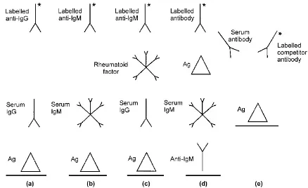
Increasingly, solid-phase ELISAs are used in diag-nostic laboratories. Recent technological advances, e.g. using synthetic peptides or recombinant antigens instead of whole viral lysates, and improvements in signal detection have led to more sensitive, specific and rapid methods for measuring virus specific antibody levels. The ELISA format is extremely versatile, and new assays can be designed quickly to cope with clinical demands, e.g. the investigation of new viruses, such as severe acute respiratory syndrome (SARS)-associated coronaviruses. Many of these assays are available commercially, and can be automated. They are essentially of three types (Figure 1.2):
. Indirect assays. Viral antigen is immobilised onto a solid phase, specific antibody in the patient serum sample binds to this antigen and, after a washing step, this antibody is detected by an enzyme-labelled antihuman immunoglobulin. In this way, either specific IgG or IgM can be detected, depending on the indicator immunoglobulin (Fig-ure 1.2a,b). Clearly, detection of IgM species is dependent on the prevailing level of IgG, such that a high level of specific IgG reduces the sensitivity of an IgM assay for the same virus. If rheumatoid factor is present in the clinical sample, it may lead to false-positive IgM results (Figure 1.2c).
. Capture assays. IgG or IgM species are captured onto the solid phase by antihuman immunoglobu-lin, followed by addition of antigen and then labelled antibody. With regard to IgM assays, this method reduces the potential interference of rheumatoid factor, and is used increasingly for a number of IgM species (Figure 1.2d).
. Competitive assays. In this case, a labelled antibody in the ELISA system competes for binding to immobilised antigen with antibody in the clinical sample. This assay improves both the specificity and sensitivity of the assay (Figure 1.2e).
Serological diagnosis of acute infection is best suited to situations in which detection of the virus itself is difficult, time-consuming or where virus excretion is likely to have ceased by the time of investigation, such as hepatitis A, rubella or parvovirus B19. There are situations, however, where IgM is produced over a prolonged period, or in response to reinfection, as is the case for rubella. In these cases, past infection can be distinguished from recent infection by antibody avidity tests. These are based on the principle that antibody responses mature over time, such that high-avidity antibodies predominate at a later stage. By using a chaotropic agent (e.g. urea) during the ELISA washing stage, low-affinity antibodies (representing recent infection) will be preferentially dissociated from
antigen, compared to higher-affinity antibodies
(Thomas and Morgan-Capner, 1991).
Immunoblot methods can be used for confirmation of HIV (Western blot) and hepatitis C virus (HCV) infection. These methods are based on the detection by antibodies within a serum sample of multiple epitopes blotted on to a membrane. Non-specific reactions within ELISAs are often clarified in these systems, since possible cross-reacting antibodies can be
identified by non-viral antigenic epitopes. Immunoblot assays are expensive and technically demanding.
Serology is also essential for the diagnosis and screening of persistent infections where antibodies are detectable in the presence of virus replication, such as HIV and HCV. The availability of sensitive assays allows widespread screening for immunity against, for example, HBV, rubella, VZV and hepatitis A.
Finally, for HIV infection, combined p24/antibody assays have been developed, for which a signal indicates the presence of either/both components. This is of advantage for a first-line screening assay, since p24 antigen is detectable prior to antibodies following primary infection, and thus leads to a shorter window period (Hashidaet al., 1996).
Despite these more recent advances in serological techniques, there remain some inherent limitations with this form of virological diagnosis (Table 1.4). It is highly dependent on the ability of the individual to mount appropriate immune responses to infection. Thus, serological methods have a limited role for diagnosing viral infections in the immunocompro-mised patient (Payaet al., 1989) and every effort must be made to detect the virus itself. Transfusion or
receipt of blood products may also lead to spurious serological results, e.g. leading to a false interpretation that seroconversion indicates acute infection. The major role of serology in transplant patients is in identifying immune status at baseline in order to ascribe a risk to primary infection, reinfection or reactivation during subsequent immunosuppression (see Table 1.10).
Interpretation of Serological Assays
Viral serology results should be interpreted carefully, taking into account the patient’s history and symp-toms. The diagnosis of a primary virus infection can be made by demonstrating seroconversion from a nega-tive to a posinega-tive specific IgG antibody response, or by detecting virus-specific IgM. A four-fold rise in IgG antibody titre between acute and convalescent samples can also be indicative of a primary infection (e.g. by complement fixation test). Detection of virus-specific IgG in a single sample, or no change in virus antibody titre between acute and convalescent phase sera, indicate exposure to the virus at some time in the past. Results of serological assays can be complicated by a number of factors; the age of the patient (the production of serum IgG or IgM antibodies can be absent or impaired in the immunocompromised, neonates and the elderly); receipt of blood products with passive antibody transfer; maternal transfer of IgG antibodies; and non-specific elevation of certain virus antibodies due to recent infection with other viruses. This last point is particularly common with herpesvirus infections, which have group-specific cross-reacting epitopes. IgM antibodies may persist for extended periods of time following primary infection, and may also be produced as a result of reactivation of latent infection, although not reliably so (e.g. CMV, EBV). The production of virus-specific
intrathecal antibody (requires demonstration of an intact blood–brain barrier) can confirm the diagnosis of viral CNS infection, e.g. subacute sclerosing panencephalitis. Serological assays may be comple-mented and confirmed by molecular assays, e.g. PCR for HCV RNA in the presence of hepatitis C antibody, and PCR for HIV, used in the diagnostic setting for the investigation of infants of HIV-seropositive mothers.
NUCLEIC ACID DETECTION
The area of most rapid development in diagnostic virology relates to molecular amplification assays to provide qualitative and quantitative results. PCR and other similar molecular assays have been applied to the diagnosis of virtually all human viruses. In general, the sensitivity of these assays far exceeds that of other virus detection systems, and subsequent interpretation of results in a clinical setting may be very difficult. A number of commercial kits and automated systems are now available, with the advantage of standardisation improving quality control and inter-laboratory varia-bility. These issues will be discussed following a brief review of the techniques available.
Polymerase Chain Reaction
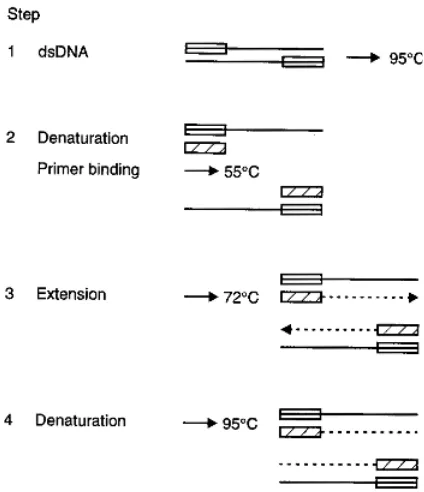
This technique uses a thermostable DNA polymerase to extend oligonucleotide primers complementary to the viral DNA genome target (Saiki et al., 1988). Consecutive cycles of denaturation, annealing and extension result in an exponential accumulation of target DNA. This is limited only by substrate (nucleotide) availability and possible competition between target genome and non-target amplicons for reaction components (Figure 1.3). RNA genomes require transcription to complementary DNA (reverse
Table 1.4 Serology
Advantages Disadvantages
Specific IgG assays good indicator of prior infection Retrospective (e.g. rising CFT titres) CFTs insensitive, especially Capture IgM assays good indicator of recent infection to assess previous infection
Allows retrospective diagnosis if no acute clinical specimen Cross-reactivity and interference
obtained Insensitive for diagnosing some congenital infections (e.g. CMV) Rapid Not appropriate for immunocompromised
Automated Spurious results possible following receipt of blood products Diagnosis of unculturable or poorly culturable viruses
Can utilise non-invasive clinical samples, e.g. saliva, urine
transcription) prior to the PCR reaction. Undertaking a second PCR round on the first round amplicon can increase the overall sensitivity of detection (nested PCR). This uses a different set of PCR primers internal to the first set, and therefore can act as a confirmation of the correct amplicon produced by the first round reaction.
Primers
The correct choice of primers is an important determinant of the success of any PCR. Clearly, the nucleic acid sequence of at least a part of the viral genome needs to be known, and primers must target a well-conserved region. This can be done using multiple alignment programmes; however, the final success of the PCR depends on the availability of sequence data from a range of different viral isolates. Since unusual viral variants may not be detected by an established PCR technique, it is important for the clinical virologist to have knowledge of the primer targets in order to interpret results correctly. This issue is equally as important for commercial assays as it is for ‘in-house’ assays, as has been demonstrated regarding suboptimal HIV subtype detection by a commercial quantitative PCR assay (Arnold et al., 1995). The appropriateness of primers also requires continual re-evaluation in the light of new selective pressures on
viral evolution, such as antiviral drug resistance. Other important aspects of primer design include the avoidance of secondary structure, or complementarity between primers (leading to so-called primer–dimer amplification artefacts). Computer programs used to design primer sequences address these aspects.
Preparation of Clinical Specimen
Viral gene detection methods do not require the maintenance of viral infectivity within the clinical specimen. Thus, specimens can be transported and stored in the refrigerator or freezer prior to analysis, with more flexibility than that required for virus isolation. This is a major advantage over traditional methods of virus detection. However, of particular concern is the susceptibility of RNA to nucleases, which are present in all biological material. In addition, certain specimen types, e.g. intraocular fluids (Wiedbrauket al., 1995) and urine (Cherneskyet al., 1997), are intrinsically more susceptible to false negative results. Thus, specimens for qualitative and, especially, quantitative PCR require careful prepara-tion and assays need to be evaluated for individual specimen types and patient groups. The anticoagulant heparin is contraindicated for blood samples because it inhibits the PCR reaction. Currently, it is recom-mended that ethylenediaminetetraacetic acid (EDTA) blood for HIV RNA quantitation is separated as soon as possible, after which the plasma can be stored frozen until analysis. If multiple tests are to be undertaken on one sample, it should be aliquoted on receipt to avoid multiple freeze–thawing. A number of different nucleic acid extraction methods are available, and their use depends on the nature of the clinical specimen and whether the target is RNA or DNA.
Detection of Product
The PCR product of any specific reaction has a known size, and can therefore be detected on an agarose gel in comparison to a molecular weight ladder. However, more than one band may be seen, or the band may not be in precisely the correct position. For this reason, specific detection of the product by hybridisation with a nucleic acid probe is to be encouraged. This can be undertaken within a microtitre plate format, with a colorimetric end-point, and read in a standard spectro-photometer, e.g. Gor et al.(1996). Many commercial PCR assays employ this system. The addition of such a
step enhances further the specificity of the assay, and may also improve sensitivity.
Multiplex PCR
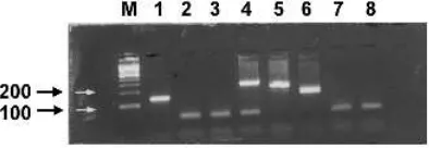
Since more than one viral target is often sought in one specimen, efforts have been made to combine multiple sets of primers, against different targets, within one PCR reaction (Jefferyet al., 1997). Each set of primers requires specific conditions for optimal amplification of the relevant target, and the development of a multiplex system requires a detailed evaluation of these conditions to ensure that the efficiency of amplification for any one target is not compromised. Identification of the specific product in this system may be based on different sized amplicons, or use of different probes. Figure 1.4 illustrates a multiplex PCR for HSV, VZV and CMV with agarose gel-based detection.
Quantitation
Conventional PCR is inherently a qualitative assay. Initial attempts to produce quantitative information involved the simultaneous analysis of samples with known target genome copy number, and comparing the intensity of bands on an agarose gel with that of the test specimen. However, the efficiency of amplifica-tion within any one PCR reacamplifica-tion is exquisitely sensitive to changes in condition, or indeed inhibitory factors within the clinical specimen, as above. It is therefore important that internal standards (within the same PCR reaction) are used for quantitative compe-titive (qcPCR) assays. These control sequences should mimic the target genome as closely as possible, yet be detectable as a distinct entity on final analysis. This can
involve the incorporation of restriction enzyme sites, whereby the control amplicon, but not target sequence, can be cleaved subsequently (Fox et al., 1992), or merely a control sequence of different size (Piataket al., 1993). Commercial assays often use a jumbled sequence as a control, with subsequent use of probes against both control and target sequences. In all cases, since the number of input control genomes is known, simple proportions can be applied to the signals to generate a quantitative value for the clinical specimen (Figure 1.5a). These assays are inherently variable, due to both laboratory and biological variation. Many commercial qcPCR assays are associated with a variation of 0.5 log10, and this must be considered in the clinical interpretation of results (Saag et al., 1996). The dynamic range of these assays is determined by the linearity of the reaction. Variability is more likely at the extremes of this range, and this variability determines the lowest value (lower limit) at which the user can be confident that the value given approx-imates to the truth. The lower limit of sensitivity for quantitative HIV PCR can be increased by initial ultracentrifugation of plasma to concentrate virus.
Figure 1.4 A 2% agarose gel of ethidium bromide-stained products from a multiplex PCR for HSV, VZV and CMV. Lanes: M, size markers (the 100 and 200 bp markers are indicated on the left); 1, isolate of HSV; 2, 3, 7 and 8 are negative specimens; 4, CMV control; 5, VZV control; 6, HSV control
Real-time PCR
The type of PCR reaction discussed above depends on end-point detection of product, with the aim of maximising the amplification reaction. More sensitive detection methods allow the kinetics of the amplifica-tion to be measured, and require fewer cycles of amplification for product to be detected.
Real-time PCR systems allow the reactions to be undertaken within a closed system, and fluorescence generated by the assay is measured without further manipulation. These systems produce very rapid temperature cycling times and, together with removal of post-PCR detection procedures, PCR reactions can be completed within minutes. Many of the signalling technologies rely on energy transfer between a fluoro-phore and a proximal quencher molecule (fluorescence resonance energy transfer). In the ‘TaqMan’ system, a specific probe binds to the relevant amplicon, and subsequent hydrolysis of this probe produces an increase in fluorescence (Morris et al., 1996). The ‘LightCycler’ system allows product detection by the incorporation of an intercalating dye into double-stranded DNA, with an increase in fluorescence as product accumulates (Wittweret al., 1997). Specificity of this reaction for the correct product (rather than artefacts) is provided by analysing a decrease in fluorescence at the melting temperature specific for that product. These commercial systems are under constant review, with new systems and applications for
the diagnostic laboratory coming on-line (e.g. the ‘iCycler iQ real-time PCR detection system’). A number of probe systems are also available to generate sequence-specific fluorescence signals (reviewed in Mackayet al., 2002).
The major advantage of real-time PCR is that it is inherently suitable for quantitative PCR, based on the number of temperature cycles required for a threshold fluorescence signal to be reached, usually in compar-ison with an external standard curve. An example of real-time detection of a calibration series for the detection of hepatitis C is shown in Figure 1.6. The dynamic range of real-time PCR of at least 8 log10 copies of template surmounts the problems encoun-tered by many qcPCR reactions, of an inability to quantitate high virus loads while maintaining sensitiv-ity at the lower end of the assay. In addition, intra- and inter-assay variability is reduced in comparison with qcPCR (Locatelliet al., 2000; Abeet al., 1999).
The disadvantages of real-time PCR in comparison with conventional PCR include an inability to monitor the size of the amplicon or to perform a nested PCR reaction without opening the system, incompatibility of some systems with some fluorescent chemistries, and currently limited capability with multiplexed reactions because of the limited number of fluorophores avail-able. Recent advances in the design of hairpin primers, hairpin and nuclease oligoprobes and novel combina-tions of fluorophores will allow discrimination of an increasing number of targets in single rapid reactions
Figure 1.6 Real-time PCR detection of a hepatitis C calibration series from 10 million IU/ml down to 10 IU/ml in 1 log steps.
xaxis=cycle number;yaxis=log fluorescence. The sample with the highest virus load (107IU/ml) crosses the baseline at cycle 18,
in future (Mackay et al., 2002; Vet et al., 1999). In addition, the start-up costs of real-time PCR may be prohibitive. Despite these difficulties, real-time PCR is now used routinely in many diagnostic virology laboratories, for both qualitative and quantitative applications. As with conventional PCR, real-time PCR has proved to be cost-effective in high-through-put laboratories in comparison with traditional culture-based methods of viral diagnosis.
PCR Contamination and Control Reactions
PCR is highly susceptible to contamination from amplified products generated in a previous reaction, from target sequences cloned in plasmid vectors and from other positive clinical specimens. By contrast, a false negative result can arise from inadequate nucleic acid extraction from a sample and inhibitory factors in the PCR reaction. Similarly, the sensitivity of the assay may be reduced but not completely inhibited. Relevant controls within each PCR run are essential for a correct interpretation of a positive or negative result, and these are highlighted in Table 1.5. The limit of sensitivity for any one assay must be assessed. This can be undertaken by serial dilutions of a tissue culture fluid of known median tissue culture infective dose (TCID50), purified viral genome, tissue culture fluid with known virion concentration (by EM) or plasmid containing the target genome. Many laboratories are reluctant to introduce plasmids into the molecular biology area because of the risk of widespread contamination.
There are two specific procedures designed to reduce PCR contamination. First, extraneous DNA within PCR reagents can be inactivated by subjecting ‘clean’ PCR
reagents to ultraviolet irradiation. This introduces thymidine dimers into the DNA chain, rendering it unamplifiable. More effective is the substitution of dUTP for dTTP in the PCR reaction (Longo et al., 1990); this does not affect specific product detection. In subsequent PCR reactions, the addition of uracil DNA glycosylase prevents DNA polymerisation of any uracil-containing DNA, but has no effect on thymidine-containing DNA template. Thus, any contaminating DNA from a previous reaction is not amplified.
Physical Organisation of the Laboratory
The physical requirements for undertaking ‘in-house’ PCR reactions are demanding (Victor et al., 1993). A ‘clean room’ is required, in which preparation and aliquoting of reagents occurs. This must be isolated from any possible contamination with viral nucleic acid. A separate area is also required for nucleic acid extraction, although this can be undertaken in a diagnostic area. A dedicated PCR room is required for setting up reactions and siting of thermal cyclers. Finally, another room is required for post-PCR analysis, such as gel running and genome detection.
Dedicated laboratory coats and equipment are
required for each of these areas, and strict adherence to protocol by all staff is essential (Table 1.6).
Clearly, the provision of such a dedicated set of rooms for molecular biology is a challenge for busy diagnostic virology laboratories. Nevertheless, it is of paramount importance that diagnostic PCR reactions are undertaken with minimal risk of contamination, and every effort must be made to provide the relevant space if such assays are to enter the routine diagnostic armamentarium. Some of the newer automated commercial assays incorporate some of the above steps within a self-contained machine. However, it is unwise to use such assays outside of a molecular biology environment in which staff are well trained in this type of work.
Table 1.5 PCR—recommended controls
Negative controls
. Extraction control —to control for contamination during extraction
—mimic clinical sample
. Reagent control —to control for contamination of reagents
—use solvent in which extracted nucleic acid is suspended
Positive controls
. Extraction control —use positive clinical specimen
. Control genome —to control for PCR efficiency, specifically to assess sensitivity
. Alternate target —to control for inhibition of reaction
Table 1.6 PCR—physical separation
1. Preparation of reagents —‘clean’ room (no nucleic acid) —separate room
2. Nucleic acid extraction
3. Amplification reactions (in cases of nested reactions, second round PCR should be further separated
Quality Control
Molecular biology is expensive compared to more traditional virological methods, making it difficult for each laboratory to undertake comprehensive evalua-tions of each PCR assay. For this reason, there is an urgent requirement for standardised methodologies and, at least in this respect, the availability of commercial assays is welcome. There is also a need for external quality control (QC) schemes, since major clinical and therapeutic decisions are made on the basis of molecular assay results, many of which have been developed ‘in house’ (Valentine-Thon, 2002). QC programmes such as Quality Control for Molecular Diagnostics (www.qcmd.com) provide QC schemes for blood-borne viruses and other pathogens, such as CMV and enterovirus; however, such programmes are still under development in terms of repertoire, and participation can represent a significant expense for diagnostic laboratories.
Other Amplification Systems
Other amplification systems include the ligase chain reaction (LCR) which, as with PCR, requires a thermal cycler. Technical innovations in molecular amplifica-tion allow techniques such as nucleic acid sequence-based amplification (NASBA), transcription mediated amplification (TMA), strand displacement amplifica-tion (SDA) and branched chain DNA (bDNA) to detect minute quantities of nucleic acid without the use of a specialised thermal cycler.
Ligase Chain Reaction
LCR involves hybridisation of two oligonucleotide probes at adjacent positions on a strand of target DNA which are joined subsequently by a thermostable ligase. The reaction also takes place on the comple-mentary strand, so multiple rounds of denaturation, annealing and ligation lead to an exponential ampli-fication of the viral DNA target (Hsuihet al., 1996). RNA targets require prior reverse transcription.
Nucleic Acid Sequence-based Amplification
This technique uses RNA as a target, utilising three enzyme activities simultaneously: reverse transcriptase (RT), RNase H, and a DNA-dependent RNA
polymerase (Guatelli et al., 1990). A DNA primer incorporating the T7 promoter hybridises to the target RNA and is extended by RT. RNase degrades the RNA strand, and the RT utilising a second primer produces double-stranded DNA. T7 polymerase then forms multiple copies of RNA from this DNA template. This method is suited to the detection of RNA viruses, or mRNA transcripts of DNA viruses. In addition, it can be modified to a quantitative assay using internal controls (Figure 1.5b). Different detec-tion formats for the amplified RNA product have been developed, including electrochemiluminescence and molecular beacon detection technologies, and adapted for rapid detection of many viruses, e.g. West Nile and St Louis encephalitis (Lanciotti and Kerst, 2001). Transcription-mediated amplification (TMA) tech-niques are very similar. Detection of the amplicon is commonly performed with detection systems such as the Hybridization Protection detection system, which uses a chemiluminescent signal, as in Gen-Probe# systems. TMA is widely used to detect HIV and HCV nucleic acid sequences.
Strand Displacement Amplification
Strand displacement amplification technology has been established in a fully automated system known as BDProbeTec#. In SDA technology, an oligonucleotide primer containing a restriction enzyme site binds to its complementary (target) nucleic acid. An exonuclease-deficient DNA polymerase (exo-) is used in the presence of dGTP, dATP, dUTP and a dCTP contain-ing an a-thiol group (dCTP aS) to produce double-stranded DNA containing a restriction enzyme site. Upon binding, the restriction enzyme nicks the strand without cutting the complementary thiolated strand. The exo-DNA polymerase recognises the nick and extends the strand from the site, displacing the previously created strand. The recognition site is repeatedly nicked and restored by the restriction enzyme and exo-DNA polymerase with continuous displace-ment of DNA strands containing the target segdisplace-ment. The process becomes exponential with the addition of an antisense primer containing the appropriate recognition site.
Branched Chain DNA
since the 1980s. These are based on the hybridisation of a labelled oligonucleotide probe to a unique complementary piece of viral genome, and can be undertaken either on a solid phase or in situ. These short probes are 20–30 bases in length and can be RNA (riboprobe) or DNA. Many solid-phase assays are of the dot–blot or slot–blot type, and have been widely used for HPV diagnosis and typing. The bDNA assay is a modification of probe assays. This method uses a signal amplification system rather than amplify-ing target genome. Samplify-ingle-stranded genome (RNA or DNA) is hybridised to an assortment of hybrid probes, which are captured in turn onto a solid phase by further complementary sequences. Branched DNA amplifier molecules then mediate signal amplification via enzyme-labelled probes with a chemiluminescent output. This method can also provide quantitative results (Dewar et al., 1994; van Gemen et al., 1993) (Figure 1.5c).
Clinical Use of Molecular Techniques
The application of qualitative and quantitative mo-lecular analysis to human viral infections has provided new insights into the natural history of human viral infections, such as HIV, HBV, HCV and the herpes-viruses. This includes the nature of viral persistence and latency, viral replication and turnover rate, and an understanding of the response to antiviral therapies. It follows that molecular diagnostic assays do not merely offer an increase in sensitivity over alternative methods; rather, their correct interpretation demands an under-standing of our transformed knowledge. These issues are discussed further below (Table 1.7).
Diagnosis of Infection
Infection is defined by the presence of virus in a clinical specimen. The infection may be asymptomatic or symptomatic (disease). However the key determinant for correct diagnosis is the sensitivity of the assay, with a goal of detecting viral genome if it is present at any level. A sensitive qualitative assay is relevant, for instance, in the diagnosis of HIV in infants (proviral DNA in PBMCs) (Lyall et al., 2001) or HCV (serum RNA) infection (Zeuzemet al., 1994). Before introdu-cing such an assay into routine use, the sensitivity and specificity of the new test must be established, according to the formulae in Table 1.8. Note that, in
Table 1.7 Clinical use of molecular amplification techniques
. Diagnosis of infection
. Diagnosis of disease
. Virus genotyping
. Prediction of disease/staging of infection
. Monitoring antiviral therapy
. Prediction of transmission
. Confirming transmission events
. Epidemiology of infection
Table 1.8 Evaluation of a new diagnostic assay
Parameter Description Formula
Sensitivity Proportion of true positives correctly identified by test
positive results true positives
Specificity Proportion of true negatives correctly identified by test
negative results true negatives
Positive predictive value
Proportion of patients with positive test results who are correctly diagnosed
sensitivityprevalence
sensitivityprevalenceþ ð1specificityÞ ð1prevalenceÞ
Negative predictive value
Proportion of patients with negative test results who are correctly diagnosed
sensitivity ð1prevalenceÞ
ð1sensitivityÞ prevalenceþspecificity ð1prevalenceÞ
Likelihood ratio Indicates how much a given diagnostic test result will raise or lower the pretest probability of the target disorder
sensitivity