review article
Medical Progress
Whipple’s Disease
Florence Fenollar, M.D., Ph.D., Xavier Puéchal, M.D., Ph.D., and Didier Raoult, M.D., Ph.D.
From Unité des Rickettsies, IFR 48, Cen-tre National de la Recherche Scientifique UMR 6020, and Université de la Méditer-ranée, Faculté de Médecine — both in Marseille, France (F.F., D.R.); and the Department of Rheumatology, Le Mans General Hospital, Le Mans, France (X.P.). Address reprint requests to Dr. Raoult at Université de la Méditerranée, Faculté de Médecine, 27 Blvd. Jean Moulin, 13385 Marseille CEDEX 5, France, or at didier. [email protected].
Drs. Fenollar and Puéchal contributed equally to this article.
N Engl J Med 2007;356:55-66.
Copyright © 2007 Massachusetts Medical Society.
T
he discovery of whipple’s disease and its causative bacterium,Tropheryma whipplei, is a prime example of how modern technologies have contributed to medical knowledge. Although Whipple’s disease was first described in 1907,1 the first successful culture of T. whipplei was performed nearly a century later, in 2000. This accomplishment led to a new level of understanding of the disease.
During the 20th century, knowledge of this chronic disease slowly accumulated (Table 1).2-6 At the dawn of the 21st century, two major events — molecular ampli-fication of the 16S ribosomal RNA (rRNA) of T. whipplei by polymerase-chain-reaction (PCR) assay and cell culture of the organism — greatly improved our understand-ing of Whipple’s disease.7-9 Initially, the organism was named Tropheryma whippelii, from the Greek trophi (food) and eryma (barrier), because of the malabsorption fre-quently observed in the disease.8 The successful isolation and serial culture of the bacterium9 were followed by the sequencing of its genome11,12 and made it possible to define the organism’s antibiotic susceptibility.13,14 The name was subsequently changed slightly to Tropheryma whipplei.10
Whipple’s disease is rare, though there is no valid estimate of its actual preva-lence. Only about 1000 cases have been reported to date.15 In postmortem studies, the frequency of the disease is less than 0.1%.16 Although it occurs in people of all ages throughout the world, the typical patient is a middle-aged white man.17
Whipple’s disease is characterized by two stages — a prodromal stage and a much later steady-state stage. The prodromal stage is marked by protean symptoms, along with chronic nonspecific findings, mainly arthralgia and arthritis. The steady-state stage is typified by weight loss, diarrhea, or both, and occasionally there are other manifestations, since many organs can be involved.17 The average time between the prodromal and the steady-state stages is 6 years. If a patient has received immuno-suppressive therapy, such as treatment with corticosteroids or tumor necrosis factor antagonists, a more rapid clinical progression may occur.18,19 For example, diarrhea has been reported to develop shortly after the initiation of immunosuppressive therapy for chronic arthritis in patients with Whipple’s disease.19
Thus, there are several presentations linked to T. whipplei infection: histologic lesions in the gastrointestinal tract in association with diverse clinical manifestations (classic Whipple’s disease), endocarditis with negative blood cultures, and isolated neurologic infection.
Without treatment, Whipple’s disease is ulti-mately fatal. Even with a specific antibiotic regi-men, clinical relapse occurs in 2 to 33% of cases
after an average of 5 years; relapse is usually char-acterized by neurologic involvement.26
Classic Whipple’s Disease
The most common gastrointestinal symptom of classic Whipple’s disease is weight loss, often as-sociated with diarrhea.15,21,24,25 Occult bleeding from the intestinal mucosa is observed in 20 to 30% of patients. Abdominal pain may be present. Hepatosplenomegaly and, occasionally, hepatitis may occur.15 Ascites has been reported in about 5% of patients.15
Joint involvement has been reported in 65 to 90% of patients with classic Whipple’s dis-ease.17,21,24,25 The presenting symptom in most patients with joint involvement is intermittent migratory arthralgia, arthritis, or both.17,21,23,25 Polyarthritis is most common, but oligoarthritis may occur. Although joint involvement alone is uncommon, Whipple’s disease should be consid-ered in the differential diagnosis in any middle-aged man with intermittent episodes of unex-plained polyarthritis or oligoarthritis of the large joints, even in the absence of digestive symp-toms.17,27 Less frequent in Whipple’s disease is chronic seronegative polyarthritis, which can be destructive and is often mistaken for rheumatoid arthritis.17 On rare occasions, spondyloarthropa-thy, hypertrophic osteoarthropaspondyloarthropa-thy, and infection of a knee prosthesis have been described in pa-tients with classic Whipple’s disease.28 Skeletal muscle myalgia and cramps in skeletal muscle may be present.15
Neurologic involvement has been reported in
Table 1. Milestones in the History of Whipple’s Disease and Tropheryma whipplei.
Date Investigators Advance
1907 Whipple1
First description of the disease 1947 Oliver-Pascual et al.2
First diagnosis before the death of a patient 1949 Black-Schaffer3
Development of periodic acid–Schiff staining for diagnosis
1952 Paulley4
First reported efficacy of antibiotic treatment 1961 Chears and Ashworth,5
Yardley and Hendrix6
Detection of bacteria in macrophages by electron microscopy
1991 Wilson et al.7
Partial sequencing of 16S rRNA of an unknown bacterium 1992 Relman et al.8
Confirmation and extension of the 16S rRNA sequence; first naming of the bacterium: T. whippelii
2000 Raoult et al.9
First cultivation of the Whipple bacillus 2001 La Scola et al.10
First phenotypic characterization of the Whipple bacillus; renaming of the bacterium: T. whipplei
2003 Bentley et al.,11
Raoult et al.12
Full sequencing of two genomes from two different strains of T. whipplei
Table 2. Demographic and Clinical Features of Classic Whipple’s Disease.*
Feature Patients with Whipple’s Disease
no./total no. (%)
Male sex 770/886 (87)
Arthralgia or arthritis 244/335 (73)
Diarrhea 272/335 (81)
Weight loss 223/240 (93)
Fever 128/335 (38)
Adenopathy 174/335 (52)
Melanoderma 99/240 (41)
Neurologic signs† 33/99 (33)
Ocular signs† 6/99 (6)
Pleural effusion 26/190 (14)
* Data are from reports on seven case series, all published since 1960, by Chears et al.,22
Enzinger and Helwig,16
Kelly and Weisiger,23
Maizel et al.,24
Dobbins,15
Fleming et al.,25
and Durand et al.21
Total numbers refer to the total num-ber of patients evaluated for Whipple’s disease. The ages of the patients at diagnosis ranged from 1 to 83 years. † Supranuclear ophthalmoplegia is included as a
6 to 63% of patients with classic Whipple’s dis-ease.15,18,29 However, in a small autopsy series, central nervous system lesions were described in 10 of 11 patients (91%).16 The neurologic
mani-festations of classic Whipple’s disease are diverse and can resemble those of almost any neurologic condition (Table 3).18,29 Cognitive changes are common, affecting 71% of patients with
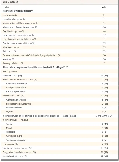
neuro-Table 3. Clinical Features of Neurologic Whipple’s Disease and Blood Culture–Negative Endocarditis Associated with T. whipplei.
Feature Value
Neurologic Whipple’s disease29
No. of patients 84
Cognitive change — % 71
Supranuclear ophthalmoplegia — % 51
Altered level of consciousness — % 50
Psychiatric signs — % 44
Upper motor neuron signs — % 37
Hypothalamic manifestations — % 31
Cranial nerve abnormalities — % 25
Myoclonus — % 25
Seizures — % 23
Oculomasticatory, or oculofacialskeletal, myorhythmia — % 20
Ataxia — % 20
Sensory deficits — % 12
Blood culture–negative endocarditis associated with T. whipplei30-38
No. of patients 17
Male sex — no. (%) 14 (82)
Previous valvular disease — no. (%) 7 (41)
Acute rheumatic fever 3 (18)
Bicuspid aortic valve 2 (12)
Aortic bioprosthesis 2 (12)
Antecedent — no. (%) 12 (71)
Arthralgia or arthritis 8 (47)
Seronegative polyarthritis 2 (12)
Psoriatic arthritis 1 (6)
Myalgia 1 (6)
Interval between onset of symptoms and definite diagnosis — range (mean) 2 mo–20 yr (5 yr)
Involved valves — no. (%)
Aortic 8 (47)
Mitral 4 (24)
Tricuspid 1 (6)
Aortic and mitral 3 (18)
Aortic and tricuspid 1 (6)
Fever — no. (%) 2 (12)
Cardiac vegetations — no. (%) 13 (76)
Congestive heart failure — no. (%) 10 (59)
logic signs, and may even extend to dementia.29 Psychiatric symptoms such as depression and personality changes are observed in roughly half the patients who have neurologic involvement. Similarly, half have supranuclear ophthalmoplegia at presentation.29 Myoclonus is observed in one quarter of patients with neurologic involvement. Hypothalamic involvement, evidenced by polydip-sia, hyperphagia, a change in libido, and changes in the sleep–wake cycle, is present in less than one third of patients with neurologic signs. Movement abnormalities of the eye muscles, termed oculo-masticatory, or oculofacioskeletal, myorhythmia, are considered pathognomonic for Whipple’s dis-ease.29
The prognosis for patients with central nervous system infection remains poor. More than 25% of such patients die within 4 years, and the same proportion of patients have major sequelae.39 Asymp tomatic neurologic involvement in classic Whipple’s disease has been demonstrated through detection of DNA from T. whipplei in cerebrospinal fluid by means of a PCR assay.40 Ocular involve-ment, excluding ophthalmoplegia, occurs in up to 11% of patients with classic Whipple’s disease.15,24,41 Anterior or posterior uveitis, usually chronic and bilateral at diagnosis, is the most frequent ocular manifestation.
Cardiac involvement has been reported in a wide range of patients with classic Whipple’s dis-ease (17 to 55%).21,42 However, two older autopsy studies showed nearly invariable involvement of the pericardium, myocardium, or endocardium; PAS-positive macrophages were found in 79% of reported cases.16,43 Pericarditis occurs in more than half of people with Whipple’s disease.24 Myo-carditis occurs far less often and is sometimes first evident with the onset of heart failure or with sudden death. Pulmonary involvement occurs in an estimated 30 to 40% of patients with classic Whipple’s disease,15 and pleural effusion, pulmo-nary infiltration, or granulomatous mediastinal adenopathy was often described in the earliest reported cases.15
Noncaseating epithelioid- and giant-cell gran-ulomas, most often lymph-node grangran-ulomas, have been found in 9% of people with classic Whipple’s disease.15 Involvement of the abdominal, espe-cially the mesenteric, lymph nodes is not uncom-mon, but peripheral lymphadenopathy is rare. Cutaneous manifestations vary.15,44 Melanoderma is a classic finding, but like other researchers,21
we have found that it is rarely observed these days, since Whipple’s disease is recognized earlier in its course. Kidney involvement, which is only occasionally described, typically occurs late in the course of the disease.15 Other manifestations, such as hypothyroidism, epididymitis, and orchitis, have occasionally been reported in cases of classic Whipple’s disease.15,45,46
Endocarditis Associated with T. whipplei
T. whipplei may be associated with blood culture– negative endocarditis. This was initially observed by chance in one patient in a study by Golden-berger et al. in which cardiac valves obtained from 18 patients with endocarditis were screened with a broad-range PCR strategy that targeted the 16S rRNA sequence of T. whipplei.47 Four addi-tional cases were reported two years later.30 To date, 17 cases of blood culture–negative endocar-ditis associated with T. whipplei (Table 3) have been described,30-38 most of which have involved native cardiac valves in men with an average age of ap-proximately 60 years. Arthralgia or arthritis, often preceding the diagnosis of blood culture–nega-tive endocarditis by some years, has been the pre-dominant extracardiac symptom in these cases.30 Clinical signs of infection appear to be rare.30
Physicians often use the Duke criteria to diag-nose endocarditis,42 but in patients with blood culture–negative endocarditis, two of the criteria — fever and a history of valvulopathy — are absent, making it difficult to diagnose endocar-ditis associated with T. whipplei.48 To date, this manifestation of Whipple’s disease has generally been confirmed by a PCR assay of DNA taken from surgically obtained cardiac valves, although in one case, an assay of intestinal tissue speci-mens was positive.30
Isolated Neurologic Manifestations
lymphadenopa-thy (2). PCR assays of intestinal tissue specimens were positive in 4 of the 32 patients. The pre-dominant symptoms included cognitive impair-ment, ophthalmoplegia, ataxia, and upper motor neuron disorder. Of the 30 patients for whom follow-up data were available, 18 (60%) had im-provement, and 10 died (33%); in 1 patient, the disease stabilized. Whether earlier detection and treatment would have improved the outcome is unknown, though arguably likely.
Other Presentations
Cases of Whipple’s disease with isolated arthri-tis,59-61 spondylodiskitis,62 and uveitis63 in the ab-sence of clinical or histologic evidence of diges-tive involvement have also been described. In these cases, the diagnosis was established with PCR assays of synovial fluid or tissue,59,61 specimens from disk puncture biopsy,62 or aqueous humor 63 or with electron microscopical examination and PAS staining of synovial tissue.60
Asymptomatic Carriers
There is controversy regarding the prevalence of T. whipplei in duodenal-biopsy specimens, saliva, stool, and blood from healthy persons.64 Some PCR studies have detected the organism in people without evident Whipple’s disease. For example, in one small study in which a PCR assay for T. whipplei was performed on blood samples from apparently healthy donors, 1 of 174 samples was positive.65 In two other studies, T. whipplei DNA was detected in saliva from 19%66 and 35%67 of healthy subjects. PCR assays have also detected DNA from T. whipplei in patients with disorders other than Whipple’s disease; positive findings have been reported in duodenal-biopsy samples (in 5% of patients), gastric secretions (12%), and stool (11%).66,68,69 However, neither our labora-tory70 nor that of Dr. David Relman at Stanford University71 has identified T. whipplei DNA in sam-ples from duodenal biopsies in control subjects. Among patients without Whipple’s disease, we have detected T. whipplei using a PCR assay on DNA isolated from saliva in 4 of 620 patients (0.6%) and from stool in 2 of 133 patients (1.5%) (un-published data).
T H E O R G A N I S M
T. whipplei appears to be present in the general environment, though neither its source nor its
transmission is well established. Studies using PCR have demonstrated T. whipplei DNA in sewage plant effluent72 as well as in human stool.73 Fur-thermore, an association between Whipple’s dis-ease and Giardia lamblia infection has been report-ed.74 Since the protozoan G. lamblia is present in the environment, it is plausible that both micro-organisms occupy the same ecologic niche.74 In-deed, it has been suggested that T. whipplei might be acquired through fecal–oral transmission.75
The complete genome of two strains of the bacteria has been sequenced.11,12T. whipplei pos-sesses a very small circular chromosome (less than 1 megabase), as reported for other intracellular bacteria. Organisms with adaptive strategies in-volving host dependence are generally associated with genome reduction, and genome annotation in T. whipplei has revealed that the biosynthetic pathways for 16 amino acids are missing or im-paired, suggesting a requirement for external nu-trients. Recombination of regions encoding for surface proteins has been detected, possibly asso-ciated with the production of many diverse mem-brane proteins, which may enable the bacterium to evade host immunity.11
T. whipplei has been isolated from mammalian cell cultures.9 With this approach, 18 novel iso-lates (7 from cerebrospinal fluid, 4 from blood, 2 from cardiac valves, 2 from lymph nodes, 1 from duodenal tissue, 1 from synovial fluid, and 1 from skeletal muscle) have been established in serial cultures.11,76,77 According to genomic analyses, it is also possible to culture T. whipplei without mammalian cells, simply by adding the missing amino acids to the culture medium.78 Using this strategy, we have recently isolated and established two strains of T. whipplei from cerebrospinal fluid, two from blood, one from synovial fluid, one from a lymph node, one from a cardiac valve, one from skeletal muscle, and one from stool.79
PA T H O PH Y S I O L O G Y O F W H I PPL E ’ S D I S E A S E
antigen among those with the disease. However, no causal association with any specific genetic fac-tor has been demonstrated, and some studies do not support the existence of genetic risk factors.81
Massive infiltration of infected tissues by mac-rophages on microscopy typifies Whipple’s dis-ease.82 After treatment, bacteria disappear, yet macrophages persist. T. whipplei multiplies in mac-rophages but not in monocytes from healthy sub-jects.83 In contrast, in patients with Whipple’s disease, T. whipplei multiplies in both monocytes and macrophages.83 Replication of T. whipplei in macrophages is associated with apoptosis of the host cell,83 which may be crucial for bacterial dis-semination and is also correlated with expression and release of interleukin-16.84 Antibodies neu-tralizing interleukin-16 inhibit the growth of T. whipplei in macrophages.83 Serum interleukin-16 levels and markers of apoptosis correlate with the activity of Whipple’s disease, decreasing to normal levels on successful treatment.83
Humoral responses do not appear to be impli-cated in Whipple’s disease.80 Several studies have demonstrated defective macrophage function in patients with the disease. Although macrophages from affected patients phagocytose bacteria nor-mally, they appear to be unable to degrade bac-terial antigens efficiently.15 Experimental data suggest that this inability to degrade bacterial antigens is related to inadequate production of interleukin-12,85 which may lead to diminished interferon-γ production by T cells and defective macrophage activation. A decrease in interleukin-12 production might then prevent the develop-ment of an effective type 1 helper T-cell immune response and would favor a shift toward a type 2 helper T-cell response. In support of this hypoth-esis, the gene expression profile of macrophages in intestinal lesions from one patient with classic Whipple’s disease indicated that genes encoding CCL18 and interleukin-10 were uniquely up-regu-lated in intestinal lesions.86 A similar pattern in up-regulated genes has been associated with mac-rophage 2, also known as alternatively activated macrophages, reflecting a predominance of type 2 helper T cells in the local immune response.
C L I N I C A L D I A G N O S I S
Blood Studies
Several nonspecific findings may together suggest the diagnosis of Whipple’s disease. For example,
before treatment there may be elevated levels of acute-phase reactants, anemia, leukocytosis, thrombocytosis, and laboratory evidence of mal-absorption.21,25 Thrombocytopenia is present on occasion.44 Eosinophilia has also been reported.42
Endoscopy
Pale yellow, shaggy mucosa alternating with erod-ed, erythematous, or mildly friable mucosa has been described on endoscopic examination of the postbulbar region of the duodenum and jejunum in patients with classic Whipple’s disease.64
Other Diagnostic Tools
Electron microscopy may detect the distinctive tri-laminar cell wall of T. whipplei; laboratories with experience in detecting T. whipplei are best at iden-tifying this feature.75 However, the classic tool for diagnosing Whipple’s disease is PAS staining of small-bowel–biopsy specimens, which on light-microscopical examination shows magenta-stained inclusions within macrophages of the lamina pro-pria (Fig. 1A). Several biopsy samples should be studied, because the lesions can be focal and sparse.
Depending on clinical manifestations, other tissues might be biopsied and stained with PAS.64,75,81 However, the PAS-positive inclusions within cells are nonspecific.64,75 For example, PAS-positive cells are also seen in patients with Mycobacterium avium complex.75 Ziehl–Neelsen staining, which is positive for patients infected with M. avium complex and negative for those with Whipple’s disease, may be used to differentiate between these two infections. Noncaseous gran-ulomas composed of epithelioid cells, which are PAS-negative in 40% of cases, may be present in the lymphatic tissue, gastrointestinal tract, bone marrow, kidneys, synovial tissue, liver, or lungs in patients with Whipple’s disease.64,75,81
own serum is used (rather than antibodies devel-oped in the laboratory). With this technique, the organism was detected in heart-valve samples from five patients with blood culture–negative endocarditis.90
As noted above, PCR can be used to detect T. whipplei in samples from a variety of tissue types and body fluids.91 As with all PCR assays, it is critical to avoid contamination of the DNA sample and to include positive and negative con-trols to validate the test. Initially, PCR assays targeting the 16S rRNA gene and 16S–23S inter-genic regions of the T. whipplei gene were used.8,81 More recently, a quantitative real-time PCR assay targeting this intergenic region was developed70 that offers the advantages of a reduced detection time and a lower risk of sample contamination. Now, on the basis of genome analysis, a new quantitative real-time PCR assay has been devel-oped that targets repeated sequences of T. whipplei, with substantially greater sensitivity than the earlier PCR assays and the same specificity.92
When amplified product is detected, the iden-tification of T. whipplei should be confirmed by sequencing or by using fluorescence-labeled oligo-nucleotide hybridization probes in a real-time PCR assay. Discrepancies between laboratories suggest that results obtained with “homemade” (not standardized) PCR should be interpreted with caution. The many positive PCR results from people without Whipple’s disease have been ob-tained primarily with the use of nested or semi-nested techniques, which carry a high risk of contamination.66,68,69,93,94 Nonetheless, it is im-portant to pay attention to a positive PCR assay, as suggested by the death of a patient in whom one of three PCR tests was positive but whose duodenal biopsy specimens were negative on PAS staining 95; the diagnosis of Whipple’s disease was thought to be ruled out, yet the autopsy revealed Whipple’s disease. Cultivation of T. whipplei from various samples can be achieved, but this tech-nique is not generally available.9,14,41,76,77,87
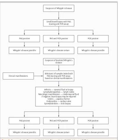
Our strategy for diagnosing Whipple’s disease uses the results of PAS staining and PCR in par-allel (Fig. 2). However, another group has recently proposed histologic examination of a small-bowel– biopsy specimen as the first step, with PCR per-formed only if the histologic findings are nega-tive.96 The main limitation of this approach is that the specificity of both histologic assessment and PCR is less than optimal.
T R E A T M E N T
Whipple’s disease was invariably fatal before the advent of antibiotics. However, current
recommen-A
B
C
Figure 1. Detection of Whipple’s Disease by PAS Stain-ing and Immunohistochemical Analysis.
Small-bowel biopsy with PAS staining and PCR assay Suspicion of Whipple’s disease
PAS and PCR positive PAS positive
Whipple’s disease possible
PCR positive
Whipple’s disease certain
Selections of samples tested with PAS staining and PCR assay based on clinical manifestations Clinical manifestations
Arthritis — synovial fluid or biopsy Lymphadenopathies — lymph nodes Neurologic manifestation — cerebrospinal fluid;
if negative, brain biopsy may be required Uveitis — aqueous humor Endocarditis — cardiac valve Spondylodiskitis — disk biopsy
Suspicion of localized Whipple’s disease
Whipple’s disease possible
PAS and PCR positive PAS positive
Whipple’s disease possible
PCR positive
Whipple’s disease certain Whipple’s disease possible
Figure 2. Strategy for the Diagnosis of Whipple’s Disease Using PAS Staining and PCR Assay.
dations are not based on therapeutic trials or the susceptibility of T. whipplei to various antimicro-bial agents. Tetracycline has long been prescribed as a first-line treatment, but the frequency of re-currence after treatment with this agent has been high (28% on average).21,25,26 Thus, the standard for antibiotic therapy currently favors antibiotics that are capable of crossing the blood–brain bar-rier, such as trimethoprim–sulfamethoxazole. The recommended treatment is oral administration of 160 mg of trimethoprim and 800 mg of sulfa-methoxazole twice per day for 1 to 2 years, usually preceded by parenteral administration of strepto-mycin (1 g per day) together with penicillin G (1.2 million U per day) or ceftriaxone (2 g daily) for 2 weeks. However, lack of a clinical response has been reported with this strategy, and recur-rence is also possible (Table 4).21,25,26,39,97-99
Patients with a neurologic recurrence of Whip-ple’s disease have a poor prognosis.39 Interferon gamma has been proposed for treatment of re-current central nervous system disease, and one report noted that a positive effect was still present at least 1 year after interferon gamma therapy had been stopped.100 The susceptibility of T.
whip-plei to various antimicrobial agents has been tested with the use of both cell and axenic cultures.12,13 Many antibiotics, including doxycycline and sulfa-methoxazole, are active in vitro, but trimethoprim is not, as predicted from genomic analysis,13,14,101 since T. whipplei lacks the coding sequence for di-hydrofolate reductase, which trimethoprim tar-gets.101 In cell culture, cephalosporins (including ceftriaxone) and fluoroquinolones are not active.13 In axenic medium, ceftriaxone and levofloxacin are active.14 Vacuole acidification has been shown to be critical to the survival of T. whipplei in phago-somes, since agents that increase the intravacuo-lar pH decrease bacterial viability.102 A regimen based on this observation — doxycycline (200 mg per day) and an alkalinizing agent, hydroxychloro-quine (200 mg three times per day) — has been effective in vitro. This combination has, thus far, been the only successful bactericidal regimen against T. whipplei in vitro.13,102 Whether this regi-men will work in a general clinical setting is un-known, though it has been successful in four of our patients: two with classic Whipple’s disease and two with blood culture–negative endocardi-tis (unpublished data).
On the basis of previous work,102 we suggest using a regimen of doxycycline and hydroxychlo-roquine to eradicate the intracellular organisms in patients with Whipple’s disease who do not have neurologic involvement (as indicated by a negative PCR assay on cerebrospinal fluid and the absence of neurologic signs). In patients with neurologic involvement, we suggest adding a high dose of sulfamethoxazole or sulfadiazine to the regimen described above. There is no established marker that can be used to determine how long treatment should be continued. By analogy with other chronic infections,103,104 it would seem rea-sonable to use this regimen for at least 12 to 18 months. Clinical trials are needed to confirm our approach and to establish whether these personal suggestions are effective.
DI R E C T IONS F OR F U T U R E R E SE A R C H
The recent cultivation of T. whipplei, along with the complete sequencing of its genome, should pro-vide new opportunities for investigating, under-standing, and treating Whipple’s disease. The res-ervoir of T. whipplei remains to be established, and transmission mechanisms remain to be elucidat-ed. The significance of possible asymptomatic carriers must be clearly addressed. Isolates of T. whipplei should be routinely genotyped to iden-tify associations among clinical forms, different strains, and geographic origin. Although PCR has expanded the recognized clinical spectrum of the disease, many facets remain elusive. In the future, the development of an assay for detection of spe-cific antibodies in the serum may help with diag-nosis of the disease. Improvement in diagnostic approaches is of paramount importance for
reli-Table 4. Initial Treatment and Subsequent Relapse in Whipple’s Disease.*
Antibiotic
No. of Relapses/ No. of Patients Treated (%)
Tetracycline 43/133 (32)
Trimethoprim–sulfamethoxazole 1/46 (2)
Penicillin and streptomycin 2/6 (33)
Other 12/64 (19)
Total 58/249 (23)
* Data are from six reports on case series, published since 1985, by Keinath et al.,26
Fleming et al.,25
Bai et al.,97
Geboes et al.,98
Feurle and Marth,99
able detection. Improved detection will in turn lead to decreases in the morbidity, and perhaps the mortality, associated with the disease, which is treatable when diagnosed early but may have fatal consequences when the diagnosis is delayed. Prospective trials are needed to evaluate therapy.
Drs. Raoult and Fenollar are among the inventors named on a patent held by the Université de la Méditerranée that does not involve commercialized products. No other potential conflict of interest relevant to this article was reported.
We thank Drs. Melanie Ihrig, Sally Cutler, Jean-Louis Mège, Hubert Lepidi, and Benoît Desnues for helpful comments re-garding the manuscript.
References
Whipple GH. A hitherto undescribed disease characterized anatomically by de-posits of fat and fatty acids in the intesti-nal and mesenteric lymphatic tissues. Bull Johns Hopkins Hosp 1907;18:382-91.
Oliver-Pascual E, Galan J, Oliver-Pas-cual A, Castillo E. Un caso de lipodystro-fia intestinal con lesiones ganglionares, mesentericas de granulomatosis lipof agica (Enfermedad de Whipple). Rev Esp Enferm Apar Dig 1947;6:213-26.
Black-Schaffer B. The tinctorial dem-onstration of a glycoprotein in Whipple’s disease. Proc Soc Exp Biol Med 1949;72: 225-7.
Paulley JW. A case of Whipple’s dis-ease (intestinal lipodystrophy). Gastroen-terology 1952;22:128-33.
Chears WC Jr, Ashworth CT. Electron microscopic study of the intestinal mu-cosa in Whipple’s disease: demonstration of encapsulated bacilliform bodies in the lesion. Gastroenterology 1961;41:129-38.
Yardley JH, Hendrix TR. Combined electron and light microscopy in Whipple’s disease: demonstration of “bacillary bod-ies” in the intestine. Bull Johns Hopkins Hosp 1961;109:80-98.
Wilson KH, Blitchington R, Frothing-ham R, Wilson JA. Phylogeny of the Whip-ple’s disease-associated bacterium. Lancet 1991;338:474-5.
Relman DA, Schmidt TM, MacDermott RP, Falkow S. Identification of the uncul-tured bacillus of Whipple’s disease. N Engl J Med 1992;327:293-301.
Raoult D, Birg M, La Scola B, et al. Cultivation of the bacillus of Whipple’s disease. N Engl J Med 2000;342:620-5. [Erratum, N Engl J Med 2000;342:1538.]
La Scola B, Fenollar F, Fournier PE, Altwegg M, Mallet MN, Raoult D. Descrip-tion of Tropheryma whipplei gen. nov., sp. nov., the Whipple’s disease bacillus. Int J Syst Evol Microbiol 2001;51:1471-9.
Bentley SD, Maiwald M, Murphy LD, et al. Sequencing and analysis of the genome of the Whipple’s disease bacterium Tro-pheryma whipplei. Lancet 2003;361:637-44. Raoult D, Ogata H, Audic S, et al. Tro-pheryma whipplei Twist: a human pathogen-ic Actinobacteria with a reduced genome. Genome Res 2003;13:1800-9.
Boulos A, Rolain JM, Raoult D. Anti-biotic susceptibility of Tropheryma whipplei in MRC5 cells. Antimicrob Agents Chemo-ther 2004;48:747-52. Raoult D. Molecular evaluation of antibi-otic susceptibility of Tropheryma whipplei in axenic medium. J Antimicrob Chemo-ther 2005;55:178-81.
Dobbins WO III. Whipple’s disease. Springfield, IL: Thomas, 1987.
Enzinger FM, Helwig EB. Whipple’s disease: a review of the literature and report fifteen patients. Virchows Arch Pathol Anat Physiol Klin Med 1963;336: 238-69.
Puéchal X. Whipple disease and arthri-tis. Curr Opin Rheumatol 2001;13:74-9.
Gerard A, Sarrot-Reynauld F, Liozon E, et al. Neurologic presentation of Whipple disease: report of 12 cases and review of the literature. Medicine (Baltimore) 2002; 81:443-57.
Mahnel R, Kalt A, Ring S, Stallmach A, Strober W, Marth T. Immunosuppressive therapy in Whipple’s disease patients is associated with the appearance of gastro-intestinal manifestations. Am J Gastroen-terol 2005;100:1167-73.
Misbah SA, Mapstone NP. Whipple’s disease revisited. J Clin Pathol 2000;53: 750-5.
Durand DV, Lecomte C, Cathébras P, Rousset H, Godeau P. Whipple disease: clinical review of 52 cases. Medicine (Bal-timore) 1997;76:170-84.
Chears WC Jr, Hargrove MD Jr, Verner JV Jr, Smith AG, Ruffin JM. Whipple’s dis-ease: a review of twelve patients from one service. Am J Med 1961;30:226-34.
Kelly JJ III, Weisiger BB. The arthritis of Whipple’s disease. Arthritis Rheum 1963;6:615-32.
Maizel H, Ruffin JM, Dobbins WO III. Whipple’s disease: a review of 19 patients from one hospital and a review of the lit-erature since 1950. Medicine (Baltimore) 1993;72:343-55.
Fleming JL, Wiesner RH, Shorter RG. Whipple’s disease: clinical, biochemical, and histopathologic features and assess-ment of treatassess-ment in 29 patients. Mayo Clin Proc 1988;63:539-51.
Keinath RD, Merrell DE, Vlietstra R, Dobbins WO III. Antibiotic treatment and relapse in Whipple’s disease: long term follow-up of 88 patients. Gastroenterology 1985;88:1867-73.
Puéchal X, Schaeverbeke T, Sibilia J, Saraux A, Poveda JD. Polymerase chain reaction testing for Tropheryma whippelii in 14.
unexplained isolated cases of arthritis. Arthritis Rheum 2002;46:1130-2.
Frésard A, Guglielminotti C, Berthe-lot P, et al. Prosthetic joint infection caused by Tropheryma whippelii (Whipple’s bacillus). Clin Infect Dis 1996;22:575-6.
Louis ED, Lynch T, Kaufmann P, Fahn S, Odel J. Diagnostic guidelines in central nervous system Whipple’s disease. Ann Neurol 1996;40:561-8.
Gubler JG, Kuster M, Dutly F, et al. Whipple endocarditis without overt gastro-intestinal disease: report of four cases. Ann Intern Med 1999;131:112-6.
Geissdorfer W, Wittmann I, Seitz G, et al. A case of aortic valve disease associ-ated with Tropheryma whippelii infection in the absence of other signs of Whipple’s disease. Infection 2001;29:44-7.
Naegeli B, Bannwart F, Bertel O. An uncommon cause of recurrent strokes: Tropheryma whippelii endocarditis. Stroke 2000;31:2002-3.
Schneider T, Salamon-Looijen M, von Herbay A, et al. Whipple’s disease with aortic regurgitation requiring aortic valve replacement. Infection 1998;26:178-80.
Aiouaz H, Celard M, Puget M, et al. Whipple’s disease endocarditis: report of 5 cases and review of the literature. Rev Med Interne 2005;26:784-90. (In French.)
Dreier J, Szabados F, von Herbay A, Kroger T, Kleesiek K. Tropheryma whipplei infection of an acellular porcine heart valve bioprosthesis in a patient who did not have intestinal Whipple’s disease. J Clin Microbiol 2004;42:4487-93.
Richardson DC, Burrows LL, Koritho-ski B, et al. Tropheryma whippelii as a cause of afebrile culture-negative endocarditis: the evolving spectrum of Whipple’s dis-ease. J Infect 2003;47:170-3.
Smith MA. Whipple endocarditis with-out gastrointestinal disease. Ann Intern Med 2000;132:595.
Lepidi H, Fenollar F, Dumler JS, et al. Cardiac valves in patients with Whipple endocarditis: microbiological, molecular, quantitative histologic, and immunohisto-chemical studies of 5 patients. J Infect Dis 2004;190:935-45.
Schnider PJ, Reisinger EC, Gerschlager W, et al. Long-term follow-up in cerebral Whipple’s disease. Eur J Gastroenterol Hepatol 1996;8:899-903.
and monitoring by cytology and poly-merase chain reaction analysis of cerebro-spinal fluid. Gastroenterology 1997;113: 434-41.
Drancourt M, Raoult D, Lepidi H, et al. Culture of Tropheryma whippelii from the vitreous fluid of a patient presenting with unilateral uveitis. Ann Intern Med 2003;139:1046-7.
Fenollar F, Lepidi H, Raoult D. Whip-ple’s endocarditis: review of the literature and comparisons with Q fever, Bartonella infection, and blood culture-positive endo-carditis. Clin Infect Dis 2001;33:1309-16.
McAllister HA Jr, Fenoglio JJ Jr. Car-diac involvement in Whipple’s disease. Circulation 1975;52:152-6.
Misbah SA, Ozols B, Franks A, Map-stone N. Whipple’s disease without mal-absorption: new atypical features. QJM 1997;90:765-72.
Dearment MC, Woodward TA, Menke DM, Brazis PW, Bancroft LW, Persellin ST. Whipple’s disease with destructive arthri-tis, abdominal lymphadenopathy, and cen-tral nervous system involvement. J Rheu-matol 2003;30:1347-50.
Tran HA. Reversible hypothyroidism and Whipple’s disease. BMC Endocr Dis-ord 2006;6:3.
Goldenberger D, Kunzli A, Vogt P, Zbinden R, Altwegg M. Molecular diagno-sis of bacterial endocarditis by broad-range PCR amplification and direct sequencing. J Clin Microbiol 1997;35:2733-9.
Raoult D. Afebrile blood culture-neg-ative endocarditis. Ann Intern Med 1999; 131:144-6.
Messori A, Di Bella P, Polonara G, et al. An unusual spinal presentation of Whipple disease. AJNR Am J Neuroradiol 2001;22:1004-8.
Lohr M, Stenzel W, Plum G, Gross WP, Deckert M, Klug N. Whipple disease confined to the central nervous system presenting as a solitary frontal tumor: case report. J Neurosurg 2004;101:336-9.
Peters G, du Plessis DG, Humphrey PR. Cerebral Whipple’s disease with a stroke-like presentation and cerebrovascu-lar pathology. J Neurol Neurosurg Psychia-try 2002;73:336-9.
Lee AG. Whipple disease with supra-nuclear ophthalmoplegia diagnosed by polymerase chain reaction of cerebrospinal fluid. J Neuroophthalmol 2002;22:18-21.
Posada IJ, Ferreiro-Sieiro A, Lopez-Valdes E, Cabello A, Bermejo-Pareja F. Whipple’s disease confined to the brain: a clinical case with pathological confirma-tion at necropsy. Rev Neurol 2004;38:196-8. (In Spanish.)
Papadopoulou M, Rentzos M, Nicolaou C, Ioannidou V, Ioannidis A, Chatzipana-giotou S. Cerebral Whipple’s disease diag-nosed using PCR: the first case reported from Greece. Mol Diagn 2003;7:209-11.
Akar Z, Tanriover N, Tuzgen S, et al.
Intracerebral Whipple disease: unusual lo-cation and bone destruction. J Neurosurg 2002;97:988-91.
Lynch T, Odel J, Fredericks DN, et al. Polymerase chain reaction-based detection of Tropheryma whippelii in central nervous system Whipple’s disease. Ann Neurol 1997;42:120-4.
Schröter A, Brinkhoff J, Günthner-Lengsfeld T, et al. Whipple’s disease pre-senting as an isolated lesion of the cervi-cal spinal cord. Eur J Neurol 2005;12: 276-9.
Coria F, Cuadrado N, Velasco C, et al. Whipple’s disease with isolated central nervous system symptomatology diag-nosed by molecular identification of Tropheryma whippellii in peripheral blood. Neurologia 2000;15:173-6.
O’Duffy JD, Griffing WL, Li CY, Abdel-malek MF, Persing DH. Whipple’s arthri-tis: direct detection of Tropheryma whippelii in synovial f luid and tissue. Arthritis Rheum 1999;42:812-7.
Farr M, Hollywell CA, Morris CJ, Struthers GR, Bacon PA, Walton KW. Whipple’s disease diagnosed at hip arthro-plasty. Ann Rheum Dis 1984;43:526-9.
Bruhlmann P, Michel BA, Altwegg M. Diagnosis and therapy monitoring of Whipple’s arthritis by polymerase chain reaction. Rheumatology (Oxford) 2000;39: 1427-8.
Weber U, Morf MH, Gubler JG, Alt-wegg M, Maibach RC. Spondylodiscitis as the first manifestation of Whipple’s disease — a removal worker with chronic low back pain. Clin Rheumatol 2003;22: 443-6.
Rickman LS, Freeman WR, Green WR, et al. Uveitis caused by Tropheryma whip-pelii (Whipple’s bacillus). N Engl J Med 1995;332:363-6.
Marth T, Raoult D. Whipple’s disease. Lancet 2003;361:239-46.
Misbah SA, Stirzaker D, Ozols B, Franks A, Mapstone NP. Anonymous sur-vey of blood donors by polymerase chain reaction for Tropheryma whippelii. QJM 1999; 92:61.
Ehrbar HU, Bauerfeind P, Dutly F, Koelz HR, Altwegg M. PCR-positive tests for Tropheryma whippelii in patients with-out Whipple’s disease. Lancet 1999;353: 2214.
Street S, Donoghue HD, Neild GH. Tropheryma whippelii DNA in saliva of healthy people. Lancet 1999;354:1178-9.
Maibach RC, Dutly F, Altwegg M. De-tection of Tropheryma whipplei DNA in feces by PCR using a target capture method. J Clin Microbiol 2002;40:2466-71.
Dutly F, Hinrikson HP, Seidel T, Mor-genegg S, Altwegg M, Bauerfeind P. Tro-pheryma whippelii DNA in saliva of patients without Whipple’s disease. Infection 2000; 28:219-22.
Fenollar F, Fournier PE, Raoult D, 56.
Gerolami R, Lepidi H, Poyart C. Quantita-tive detection of Tropheryma whipplei DNA by real-time PCR. J Clin Microbiol 2002; 40:1119-20.
Maiwald M, von Herbay A, Persing DH, et al. Tropheryma whippelii DNA is rare in the intestinal mucosa of patients without other evidence of Whipple disease. Ann Intern Med 2001;134:115-9.
Maiwald M, Schuhmacher F, Ditton HJ, von Herbay A. Environmental occur-rence of the Whipple’s disease bacterium (Tropheryma whippelii). Appl Environ Micro-biol 1998;64:760-2.
Gross M, Jung C, Zoller WG. Detec-tion of Tropheryma whippelii (Whipple’s disease) in faeces. Ital J Gastroenterol Hepatol 1999;31:70-2.
Fenollar F, Lepidi H, Gerolami R, Drancourt M, Raoult D. Whipple disease associated with giardiasis. J Infect Dis 2003;188:828-34.
Fenollar F, Raoult D. Whipple’s dis-ease. Clin Diagn Lab Immunol 2001;8:1-8. Fenollar F, Birg ML, Gauduchon V, Raoult D. Culture of Tropheryma whipplei from human samples: a 3-year experience (1999 to 2002). J Clin Microbiol 2003;41: 3816-22.
Maiwald M, von Herbay A, Fredricks DN, Ouverney CC, Kosek JC, Relman DA. Cultivation of Tropheryma whipplei from cerebrospinal fluid. J Infect Dis 2003;188: 801-8.
Renesto P, Crapoulet N, Ogata H, et al. Genome-based design of a cell-free cul-ture medium for Tropheryma whipplei. Lan-cet 2003;362:447-9.
Raoult D, Fenollar F, Birg M-L. Cul-ture of T. whipplei from the stool of a pa-tient with Whipple’s disease. N Engl J Med 2006;355:1503-5.
Marth T, Strober W. Whipple’s disease. Semin Gastrointest Dis 1996;7:41-8.
Dutly F, Altwegg M. Whipple’s disease and “Tropheryma whippelii.” Clin Microbiol Rev 2001;14:561-83.
Lepidi H, Fenollar F, Gerolami R, et al. Whipple’s disease: immunospecific and quantitative immunohistochemical study of intestinal biopsy specimens. Hum Pathol 2003;34:589-96.
Desnues B, Raoult D, Mege JL. IL-16 is critical for Tropheryma whipplei replication in Whipple’s disease. J Immunol 2005;175: 4575-82.
Elssner A, Doseff AI, Duncan M, Ko-tur M, Wewers MD. IL-16 is constitutively present in peripheral blood monocytes and spontaneously released during apoptosis. J Immunol 2004;172:7721-5.
Marth T, Neurath M, Cuccherini BA, Strober W. Defects of monocyte interleu-kin 12 production and humoral immunity in Whipple’s disease. Gastroenterology 1997;113:442-8.
ing cells exhibit a transcriptional pattern of M2/alternatively activated macrophages. J Infect Dis 2005;192:1642-6.
Raoult D, La Scola B, Lecocq P, Lepidi H, Fournier PE. Culture and immunologi-cal detection of Tropheryma whippelii from the duodenum of a patient with Whipple disease. JAMA 2001;285:1039-43.
Baisden BL, Lepidi H, Raoult D, Ar-gani P, Yardley JH, Dumler JS. Diagnosis of Whipple disease by immunohistochemi-cal analysis: a sensitive and specific meth-od for the detection of Tropheryma whipplei (the Whipple bacillus) in paraffin-embed-ded tissue. Am J Clin Pathol 2002;118: 742-8.
Dumler JS, Baisden BL, Yardley JH, Raoult D. Immunodetection of Tropheryma whipplei in intestinal tissues from Dr. Whipple’s 1907 patient. N Engl J Med 2003; 348:1411-2.
Lepidi H, Coulibaly B, Casalta JP, Raoult D. Autoimmunohistochemistry: a new method for the histologic diagnosis of infective endocarditis. J Infect Dis 2006;193:1711-7.
Fenollar F, Raoult D. Molecular tech-niques in Whipple’s disease. Expert Rev Mol Diagn 2001;1:299-309.
Fenollar F, Fournier PE, Robert C, 87.
88.
89.
90.
91.
92.
Raoult D. Use of genome selected repeat-ed sequences increases the sensitivity of PCR detection of Tropheryma whipplei. J Clin Microbiol 2004;42:401-3.
Apfalter P, Reischl U, Hammerschlag MR. In-house nucleic acid amplification assays in research: how much quality con-trol is needed before one can rely upon the results? J Clin Microbiol 2005;43:5835-41.
Amsler L, Bauernfeind P, Nigg C, Mai-bach RC, Steffen R, Altwegg M. Preva-lence of Tropheryma whipplei DNA in patients with various gastrointestinal diseases and in healthy controls. Infection 2003;31:81-5.
Müller SA, Vogt P, Altwegg M, See-bach JD. Deadly carousel or difficult in-terpretation of new diagnostic tools for Whipple’s disease: case report and review of the literature. Infection 2005;33:39-42.
Olmos M, Smecuol E, Maurino E, Bai JC. Decision analysis: an aid to the diag-nosis of Whipple’s disease. Aliment Phar-macol Ther 2006;23:833-40.
Bai JC, Crosetti EE, Maurino EC, Mar-tinez CA, Sambuelli A, Boerr LA. Short-term antibiotic treatment in Whipple’s dis-ease. J Clin Gastroenterol 1991;13:303-7.
Geboes K, Ectors N, Heidbuchel HP, Rutgeerts PJ, Desmet VJ, Vantrappen GR. Whipple’s disease: the value of upper gas-93.
94.
95.
96.
97.
98.
trointestinal endoscopy for the diagnosis and follow-up. Acta Gastroenterol Belg 1992;55:209-19.
Feurle GE, Marth T. An evaluation of antimicrobial treatment for Whipple’s dis-ease: tetracycline versus trimethoprim-sulfamethoxazole. Dig Dis Sci 1994;39: 1642-8.
Schneider T, Stallmach A, von Herbay A, Marth T, Strober W, Zeitz M. Treat-ment of refractory Whipple disease with interferon-γ. Ann Intern Med 1998;129: 875-7.
Cannon WR. Whipple’s disease, ge-nomics, and drug therapy. Lancet 2003;361: 1916.
Ghigo E, Capo C, Aurouze M, et al. Survival of Tropheryma whipplei, the agent of Whipple’s disease, requires phagosome acidification. Infect Immun 2002;70:1501-6.
Maurin M, Raoult D. Q fever. Clin Microbiol Rev 1999;12:518-53.
Scollard DM, Adams LB, Gillis TP, Krahenbuhl JL, Truman RW, Williams DL. The continuing challenges of leprosy. Clin Microbiol Rev 2006;19:338-81.
Copyright © 2007 Massachusetts Medical Society.
99.
100.
101.
102.
103.
104.
FULLTEXTOFALLJOURNALARTICLESONTHEWORLDWIDEWEB