Oriental Pharmacy and Experimental Medicine 2003 3(3), 151-156
Induction of apoptosis by protein fraction isolated from the leaves of
Mirabilis
jalapa
L on HeLa and Raji cell-line
Zullies Ikawati
1,*, Sudjadi
1, Widyaningsih Elly
1, Dyah Puspitasari
1and Sismindari
1,21Faculty of Pharmacy, Gadjah Mada University, Jogjakarta, Indonesia; 2Life Science Laboratory, Gadjah Mada
University, Jogjakarta, Indonesia
SUMMARY
The leaves of Mirabilis jalapa L contains protein fraction presumed ribosome-inactivating protein (RIP). RIP is a group of protein that has RNA N-glycosidase activity that is capable to inhibit protein synthesis. Protein fraction of the plant was shown to be cytotoxic on HeLa cell-line, however, the mechanism by which the protein kill the cells is not identified yet, whether trough apoptosis, necrosis, or other mechanism. This research aim to study the mechanism of cell death caused by the protein fraction isolated from the leaves of this plant on HeLa and Raji cell-line, as representative of different kind of cancer cells. Results showed that protein fraction isolated from the leaves of Mirabilis jalapa L was more cytotoxic to HeLa cell-line (LC50: 0.65 mg/ml) than to Raji cell-line (1.815 mg/ml) on 48 hours incubation time. Moreover, it was demonstrated that the death of HeLa cells caused by the protein fraction was due to induction of apoptosis, while on Raji cell-line was due to non-apoptosis way, presumably via necrosis.
Key words: Ribosome-inactivating protein (RIP); Leaves of Mirabilis jalapa L; Cytotoxicity;
Apoptosis; HeLa and Raji cell-line
INTRODUCTION
Cancer is the second largest cause of death in both men and women in many countries, after cardiovascular disease (Anonymous, 1998). To date, many anticancer drugs have been developed. However, resistance to anticancer drug was discovered. Therefore, the development of new and safe drugs have become necessary by the pharmaceutical industry. Natural products have served as a major source of drugs for centuries, and about half of the pharmaceuticals in use today are derived from natural products (Clark, 1996). This is especially obvious in the case of antitumor drugs, as exemplified by paclitaxel, vincristine, vinorelbin, teniposide and various water-soluble analogs of camptothecin (Pezzuto, 1997). Therefore it is promising to discover new potential anticancer drug from natural product.
Indonesia is rich of natural biodiversity, and thousands plants are used in folk medicine. One of them is Mirabilis jalapa L., which is known as four oíclock plant. This plant has been utilized for traditional medicine in many countries for years including Indonesia. Leaves and roots of the plant were reported to contain an antiviral protein, which was active against the mechanical transmission of certain plant viruses (Kubo et al., 1990). This protein belongs to ribosome-inactivating proteins (RIPs) that are widely distributed in higher plants, and hold promise for agricultural and pharmaceutical application.
RIP is a group of protein that capable to inactivate ribosome by modifying the 28S rRNA through its N-glycosidase activity, which is manifested by cleavage of the N-glycosidic bond at a specific adenine. The adenine at position 4324 of rat liver 28S rRNA and corresponding adenine on other eukaryotic or prokaryotic models are the target sites (Stirpe et al., 1992). Through this mechanism, the binding of elongation factor 2 is prevented, Correspondence: Zullies Ikawati, PhD, Faculty of Pharmacy,
with the consequent arrest of protein synthesis. RIPs have many biological activities, one of them is the capability to cleave supercoiled double stranded DNA become nick-circular and linear form. This property may be used to identify the presence of RIP in a protein mixture from a plant (Ling et al., 1994).
In addition to its antiviral property, protein fraction isolated from Mirabilis jalapa L was found to have cytotoxic activity on HeLa cell line (Sismindari et al., 2001). However, the mechanism by which the protein kills the cells was not defined yet, whether through apoptosis or necrosis or other mechanism. Apoptosis, a kind of cellular death characterized by the early activation of endogenous proteases, cell shrinkage, membrane blebbing, and DNA fragmentation, is commonly observed following treatments with various anticancer agent in vitro and in vivo (Saikumar et al., 1999), and it is the death cells expected after the treatment of anticancer drugs. In this study, we investigated the cytotoxic effect of protein fraction isolated from Mirabilis jalapa L in HeLa and Raji cells, which represent different kind of cancer cells, and observed whether the protein fraction could induce apoptosis. We also confirmed whether the protein belongs to RIP. We found that the protein fraction could kill the cells through different mechanism.
MATERIALS AND METHODS
Plant material
The fresh leaves of Mirabilis jalapa L (Nyctagynaceae) were collected from Yogyakarta area in January 2002. A voucher specimen is deposited in the Laboratory of Life Science Gadjah Mada University.
Cell culture
The HeLa and Raji cell line were obtained from the stock of Life Science Laboratory Gadjah Mada University. The cells were maintained in RPMI 1640 (SIGMA) supplemented with 10% fetal bovine serum (Gibco), 1% penicillin-streptomycin (Gibco) and 0.5% fungizon (Gibco). The cells culture were incubated at 37oC in a humidified atmosphere of 5% CO2.
Isolation of RIP-containing protein from Mirabilis jalapa L
Leaves of Mirabilis jalapa L (30 gram) were ground
to a powder and stored at -20oC until use. The protein extracts were prepared by homogenizing the ground tissue with extraction buffer (5 mM sodium phospate, pH 7.2, with 140 mM NaCl) in 4oC. The solution was subsequently centrifuged for 15 min at 1,500 g in 4oC. The supernatant was then added with saturated ammonium sulfate solution under constant stirring and followed by centrifugation for 10 min at 1,500 g. Pellet was solubilised in minimal volume of 5 mM sodium phospate buffer pH 6.5, and were dialyzed with two changes of 5 mM NaPO4 buffer pH 6.5 in 4
o
C for overnight. The dialysate was centrifuged at 8,500 g for 10 min in 4oC, and the supernatant was collected as protein sample. Protein concentration was determined using UV spectrophotometry as total protein (Layne, 1957).
Cleavage of super-coiled DNA by the protein extracts
To confirm the presence of RIP in the protein extracts, we assayed the capability of the protein to cleave a supercoiled double stranded DNA. In this experiment, pUC18 was isolated from Eschericia coli DH5a cultured in LB medium containing ampicillin 150 mg/ml at 37oC. After reaching the stationary growth phase, total DNA plasmid was purified by the modified alkaline lysis procedure (Sambrook et al., 1989). One µg of pUC18 was incubated with various amounts of protein extracts to final volume of 10 µl containing 50 mM Tris-HCl, 10 mM MgCl2, 100 mM NaCl, pH 8.0, at 30
o
C for 1 hour. At the end of the reaction, 4 µl of loading buffer were added. Electrophoresis was carried out on a 1% agarose gel in 0.5×TBE (Tris Borat EDTA) buffer. DNA bands were visualized by staining with ethidium bromide (Sismindari et al., 2000).
Cytotoxicity assay
The exponentially growing cells (5×10⁴ cells/ml) were seeded in 96-well microculture plate with a serial dilution of protein extracts in a volume of 100
µl. The blank was treated with medium containing equivalent concentration of DMSO. After 48 hr incubation, the number of viable cells was ascertained with direct observation for HeLa cells, and with Trypan Blue exclusion method for Raji cells. LC50 value was calculated using
Detection of DNA fragmentation
HeLa or Raji cells at a density of 106 cells/ml were treated with 2.5 mg/ml of protein extracts isolated from leaves of Mirabilis jalapa L for 48 hours and then collected by centrifugation at 325×g for 5 min. The cell pellets were resuspended in PBS buffer and lysed in 700 µl of TE buffer containing 0.1% SDS and proteinase K (10 µg/ml) for overnight at 37oC. After two successive extractions with phenol: chloroform: isoamyl alcohol (25:24:1) and chloroform: isoamyl alcohol (24:1) respectively, the aqueous layer was transferred to a fresh centrifuge tube. The DNA was ethanol precipitated and resuspended in TE buffer and containing RNAse (100 µg/ml). The DNA fragmentation was visualized by gel agarose electrophoresis using 2% agarose gel in 0.5×TBE buffer.
Apoptosis assay by deadend colorimetric apoptosis detection system
Apoptosis assay in HeLa cells was done with DeadEnd Colorimetric Apoptosis Detection System (Promega) developed from TUNEL assay. Briefly, cells (control and treatment) were incubated with biotinylated-12-dUTP in the presence of terminal deoxynucleotidyl transferase (TdT) enzyme to label 3’-OH ends of fragmented DNA. All apoptotic cells were detected using enzyme-peroxidase staining.
RESULTS
Cleavage of supercoiled DNA by the protein extract
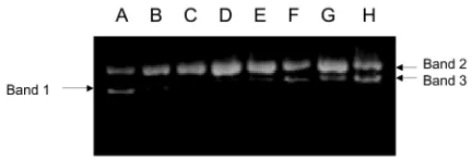
Protein extracts isolated from the leaves of Mirabilis jalapa L in the concentration of 2 µg were found to cleave the supercoiled DNA of pUC18 to be nicked circular form (Fig. 1a lane 3). Even in the higher
concentration, i.e. 3 µg, the nicked circular was cleaved to be linear form (Fig. 1), which moved between supercoiled DNA and nicked circular form. The cleavage of supercoiled DNA activity seemed to be concentration-dependent, as shown by gradual fainting of supercoiled DNA and gradual appearing of nicked and linear DNA band in the agarose gel, parallel with the increasing amount of protein (Fig. 1). As the cleavage of supercoiled DNA is one property of RIP (Ling et al., 1994), these results indicate that the protein extracts might contain RIP.
Cytotoxic activity
The presumed RIP-containing proteins demonstrated cytotoxic activity in HeLa and Raji cell line with different level, respectively, as shown in Fig. 2. After 48 hours incubation, LC50 of the protein fraction on HeLa cell line was 0.65 mg/ml, while on Raji cell-line was 1.815 mg/ml.
Induction of apoptosis
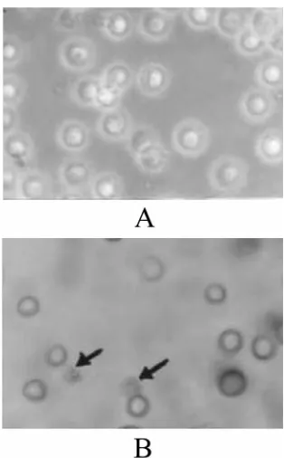
In order to confirm the mechanism of death cells caused by the extracts, apoptosis assay was done. Apoptosis assay was carried out with two methods, i.e. agarose gel electrophoreses to detect DNA fragmentation and staining method developed from TUNEL assay. As indicated on Fig. 3, it was found that protein extracts broke-down the DNA of HeLa cells into fragments (see Fig. 3a). The phenomenon, however, was not found in Raji cells (see Fig. 3b). The apoptotic death of HeLa cells was also shown by staining method using DeadEnd Colorimetric Apoptosis Detection System (Fig. 4a and b).
Fig. 1. Effect of protein fraction on the cleavage of DNA plasmid (pUC18). Lane A: pUC 18 control; lane B - H: pUC18 treated with protein fraction with concentration of 0.5, 1.0, 2.0, 6.0, 10.0, 12.0, and 14.0 mg/ml, respectively. Band 1: supercoiled form of DNA; band 2: nick-circular form of DNA, band 3: linear form of DNA.
The cells was shrinked and stained dark brown indicating that the cells undergo apoptosis process.
DISCUSSION
The results of our research show that the leaves of
Mirabilis jalapa L contain protein fraction that presumed to belongs to RIP. The presence of RIP is indicated by the capability of the protein extracts to cleave supercoiled DNA to be linear and nick-circular form. This property is one of the characteristics of RIP beside the N-glycosidase activity.
Several proteins have been found in genus Mirabilis. One of them is Mirabilis Antiviral Protein (MAP) isolated form the root of Mirabilis jalapa (Kataoka et al., 1990). MAP, a type I RIP, was purified to homogeneity and was revealed to be lysine rich and basic (pI 9.8), with molecular weight close to 24.2 kDa as determined by sodium dodecyl sulfate-polyacrilamide gel electrophoresis (SDS-PAGE) (Takanami et al., 1990). MAP was shown to inhibit protein synthesis in Eschericia coli as well as in eukaryotes (Habuka et al., 1991) and to possess repellant properties against aphid and white flies (Verma and Kymar, 1979). Others that are isolated from the roots of Mirabilis expansa are ME1 (27.5 kDa) and ME2 (27 kDa), and shows high similarity to the MAP (Vivanco et al., 1999). The protein fraction in this study has not been investigated and identified further, however, from the purification we found a single protein with the molecular weight close to 30 kDa (Sudjadi et al., 2003). Other kind of RIP is expected to be found in Mirabilis jalapa L planted in Indonesia as we are investigating now. A study concerning this matter is now being carried out in our laboratory.
Our protein fraction showed cytotoxic effect on HeLa and Raji cell line with different level of potency. The protein was found to be more cytotoxic to HeLa than to Raji cell line. After 48 hours incubation, the LC50 of the protein was 0.65 mg/ml
for HeLa and 1.815 mg/ml for Raji cell. On the same concentration (the highest concentration tested: 2.50 mg/ml), the protein caused 100% cell death in HeLa cells, but only less than 65% on Raji cells. HeLa and Raji cells represent different kind of cancer cell, which enable us to have description of the effect of the protein fraction on different cell system. HeLa cells are the classic example of an immortalized cell line. These are human epithelial cells from a fatal cervical carcinoma transformed by human papillomavirus 18 (HPV18). While Raji cells are lymphoblast-like cells which was established by R.J.V. Pulvertaft in 1963 from a Burkitt lymphoma
Fig. 3. Induction of DNA fragmentation in HeLa cells (A) but not in Raji cells(B). The cells were treated with 2.5 mg/ml protein fraction isolated from Mirabilis jalapa for 24 hours at 37oC and were then harvested for DNA isolation. Characteristic apoptotic DNA ladder were observed in HeLa cells but not in Raji cells.
of the left maxilla of an 11-year-old Negro male. It seems that the mechanism of cell death in both cells is different each other, as consequence of different level of cytotoxicity of the protein fraction.
To address this question, we study the effect of the protein on induction of apoptosis, one of the mechanism of cell death. From this study, we found that the protein fraction caused DNA fragmentation in HeLa cells, indicating that the cells undergo apoptosis process (Fig. 3A). While in Raji cells, there was no evidence of DNA fragmentation after 48 hour incubation with the protein, indicating that there was no apoptosis. The resistance of Raji cells against induction of apoptosis by the protein fraction may correlate with the lower cytotoxic effect of the protein on those cells compared to HeLa cells. These results are in line with Kawabata (1999) reporting that Raji cells were resistant to apoptosis induction, due to a defect in the apoptotic pathway in the cytoplasm downstream of caspase-3. The death of Raji cells found on the cytotoxicity study is presumably due to necrosis as indicated by Tripan Blue exclusion. The necrotic cells were recognized by swelling and nuclear flocculation, and have no ability to exclude the dyes.
The mechanisms by which the protein fraction, presumed RIP, are able to induce apoptosis in HeLa cells are still not well understood. Apoptotic cell death is initiated by a cascade of events mainly through two different mechanisms involving distinct initiator caspases. One of these mechanisms is receptor-dependent and is directly related to the activation of caspase-8 and possibly caspase-10. The other mechanism, in response to genotoxic agents, anti-cancer drugs, etc., provokes alterations in the mitochondria, leading to the induction of a pathway that activates caspase-9. We did not yet investigate whether the protein fraction initiates the activation of any caspase. But some authors have suggested that any toxin that produces approximately 90% inhibition of protein biosynthesis may induce apoptosis (Kochi et al., 1993). In fact, it has also been reported that cleavage of the large ribosomal subunit 28S rRNA is a common feature in several cases of apoptosis-mediated cell death (Houge et al., 1995). As mentioned before, the capability to cleave of the N-glycosidic bond of the 28S rRNA is a property of RIP. Therefore, if the
protein fraction is really RIP, it is possible that the protein fraction from Mirabilis jalapa induces apoptosis in HeLa cells by its N-glycosidase activity. However, the molecular mechanism by which the RIP induce apoptosis through its N-glycosidase activity, whether dependent or independently from caspase activation, specially caspase-3, is still need to be elucidated.
ACKNOWLEDGEMENTS
This work was supported by grants from Quality of Undergraduate Education (QUE) Program Batch II Faculty of Pharmacy Gadjah Mada University.
REFERENCES
Anonymous. (1998) Cancer Facts and Figures, American Cancer Society, Atlanta.
Clark M. (1996) Natural products as a resource for new drugs. Pharm. Res. 13, 1133-1144.
Habuka N, Miyano M, Kataoka J, Noma M. (1991) Eschericia coli ribosome is inactivated by Mirabilis antiviral protein which cleaves the N-glycosidic bond at A2660 of 23 S ribosomal RNA. J. Mol. Biol.
221, 737-743.
Houge G, Robaye B, Eikhom TS, Goldstein J, Mellgren G, Gjertsen BT, lanotte M, Doskeland SO. (1995) Fine mapping of 28S rRNA sites specifically cleaved in cells undergoing apoptosis. Mol. Cell Biol. 15, 2051-2062.
Kawabata Y, Hirokawa M, Kitabayashi A, Horiuchi T, Kuroki J, Miura, AB. (1999) Defective Apoptotic Signal Transduction Pathway Downstream of Caspase-3 in Human B-Lymphoma Cells: A Novel Mechanism of Nuclear Apoptosis Resistance. Blood
94, 3523-3530.
Kochi SK, Collier RJ. (1993) DNA fragmentation and cytolysis in U937 cells treated with diphtheria toxin or other inhibitors of protein synthesis. Exp. Cell Res. 208, 296-302.
Kubo S, Ikeda T, Imaizumi S, Takanami Y, Mikami Y. (1990) A potent plant virus inhibitor found in Mirabilis jalapa L. Ann. Phytopathol. Soc. Jpn. 56, 481-487. Layne E. (1957) Spectrophotometric and turbidimetric
methods for measuring proteins. In: Methods of Enzymology, edited by Colowick and Kaplan, p. 477, Academic Press, New York.
Olmo N, Turnay J, Buitrago GH, Silanes IL, Gavilanes JG, Lizarbe MA. (2001) Cytotoxic mechanism of the ribotoxin a-sarcin: induction of cell death via apoptosis. Eur. J. Biochem. 268, 2113-2123.
Pezzuto JM. (1997) Plant-derived anticancer agents. Biochem. Pharmacol. 53, 121-133.
Saikumar P, Dong Z, Mikhailov V, Denton M, Weinberg JM, Venkatachalam MA. (1999) Apoptosis: definition, mechanism, and relevance to disease. Am. J. Med.
107, 489-506.
Sambrook J, Fritsch EF, Maniatis T. (1989) Molecular cloning, A Laboratory Manual, 2nd edition, Cold Spring Harbor Laboratory Press, USA.
Sismindari, Lord JM. (2000) Ribosome-inactivating RNA N-glycosidase activity of Mirabilis jalapa L, Morinda citrifolia L, and Carica papaya L. Indon. J. Biotech. 342-345. Sismindari, Mubarika S, Sudjadi. (2001) Selective cytotoxic effects on cancer cell-lines of a protein fraction isolated from Annona squamosa and
containing ribosome-inactivating proteins, Indon. J. Biotech. 459-463.
Sudjadi, Sismindari, Herawati T, Prasetyawati AT. (2003) Purification of Ribosome-inactivating Protein from Mirabilis jalapa leaves by CM Sepharose CL-6B and Sephacryl S-300 HR columns, Indon. J. Pharm. in press.
Stirpe F, Barbieri R, Batteli MG, Soria M, Lappi DA. (1992) RIP from Plants: Present status and future prospect, Review. Biotechnology 10, 105-109. Takanami Y, Kuwata S, Ikeda T, Kubo S. (1990)
Purification and characterization of the anti-plant viral protein from Mirabilis jalapa L. Ann. Phytopathol. Soc. Jpn. 40, 228-287.