Virtual Screening on Neuraminidase Inhibitors activity of
plant- derived natural products by using Pharmacophore
Modelling and Docking
Muchtaridi1§, Habibah. A. Wahab1,2,C
1 Laboratory of Pharmaceutical Design and Simulation, School of Pharmaceutical Sciences, University Sains Malasysia, 11800, Penang, Malaysia.
2 Malaysian Institute of Pharmaceuticals and Nutraceuticals, Ministry of Science, Technology and Innovation, Block A, SAINS@USM, 11900, Penang, Malaysia.
§ Permanent office : Pharmaceutical chemistry Laboratorium of Faculty of Pharmacy, Universitas Padjadjaran, Jl. KM 21,5 Bandung-Sumedang, Jatinangor, Indonesia
CEmail: [email protected]
ABSTRACT
Neuraminidase (NA) plays an important role in the replication and release of new avian influenza virion. Due to this event, NA had been considered as a valid target in drug design against influenza virus. The aim of this study is to identify the new neuraminidase inhibitors, using pharmacophore modeling and docking based virtual screening from natural compounds of Malaysia. A variety of natural compounds from plant sources of Malaysia which collected in NADI database has been screened to possess substantial neuraminidase inhibitors properties. Pharmacophore model was developed using Catalyst software embedded in Discovery Studio 2.1 by using sialic acid derivatives which act as N1 inhibitors. The pharmacophore hypothesis selected had five features (one hydrogen bond donor (D), one negative ionizable (N), one positive ionizable (P), and two bond donor (D), one negative ionizable (N), one positive ionizable (P), and two hydrophobic moiety (Hy), also included two excluded volume. Best pharmacophore was validated by Hyporefine in DS 2.1 which has a lowest total cost value (92.055), the highest cost difference (107.807), the lowest RMSD (1.197), and the best correlation coefficient (0.944651)..The X-ray crystal structure of Neuraminidase N1 (1F8B) was used in the docking studies, using Autodock 3.0.5 software, and the free energy of binding was used to rank the hits, whereas oseltamivir and DANA have been used as the ligands in controlled docking. In silico screening had been carried out on the compounds of our laboratories database (NADI). The mapping of NADI database natural compounds shows that 46 compounds of 2350 NADI compounds map to three feature of common pharmacophore training set. MSC458 (Morinda citrifolia) have the highest fit value with free energy docking -9.48 kcal/mol (Ki 1.12x 10⁻⁷) into neuraminidase, whereas MSC605 have the most negative free energy of bind (-12.10) and Ki 1.35x 10⁻⁹.
Keywords: Neuraminidase, Pharmacophore, Docking, Virtual Screening
1. INTRODUCTION
Influenza virus has impacted the world since long time ago. It infects nearly 20% of the human population of the world [1]. This virus consists of a membrane-enveloped, segmented, negative-strand RNA viruses, and two glycoproteins on the surface of the hemagglutinin (HA) and neuraminidase (NA). HA and NA have a major role in viral replication. HA is a trimer macromolecules responsible for attachment to the surface of the cell receptors that are associated with terminal sialic acid [2] whereas NA is an important viral enzyme that play a role in virus proliferation and infectivity. Therefore, blocking its activity generates antivirus effects, making it attractive to target this viral glycoprotein for the design and development of anti influenza drugs. In influenza virus, it was found that the enzyme active site of viral NA of both virus A and B is highly conserved in amino acid sequence variation.
In the recent time, the analog NA inhibitors was developed from chemical reagent starting as lead compound, but little case which using by natural plants bioactive compounds as starting materials for be developed lead compounds of NA Inhibitors, because Malaysia or Indonesia has great potential to develop her abundant natural resources to increase the market based on herbal products.
NADI (Natural Based Drug Discovery) [3] is a database of Malaysian medicinal plants which aims to be a one-stop centre for in silico drug discovery from natural products. NADI was developed with the aim to assist the research in performing drug discovery at Universiti Sains Malaysia. It provides structural information on 3000 different compounds along with the information of the botanical sources of plants species that could be used in virtual screening.
2. COMPUTATIONAL DETAILS
Materials
Database of Bioactive Compounds in NADI (USM)
The bioactive compounds of diversity Laboratories collection in NADI, was used as a test set in this virtual screening. The database, at present, contains 3000 compounds of Malaysia natural product which geometry have been built and optimized using MOPAC semi-empirical programme.
Training Set : 24 representative structures obtained from the literature (Table 1) was taken for training set in this study.
Software for Virtual Screening : Hyperchem 7.0. (Hyperchem, Inc), Autodock 3.0.5 (Molecular Graphics Laboratory, The Scripps Research Institute), Discovery Studio 2.5. (Accelryl Inc.).
Methods
Generation of Conformation Library of Bioactive Compounds: For the training and test sets molecules, conformational models representing their available conformational space were calculated. All molecules were built using the 2D and 3D sketcher of Hyperchem 7.0, and optimized using MM2 in Hyperchem 7.0. A conformational set was generated for each molecule using the poling algorithm and the best energy option, based on CHARMm force field from Discovery Studio 2.5 [4]. The molecules associated with their conformational models were
������ ���� ��� ������������� ����� ����� ��� ����� ���� ������ �� ������ ��� ���������
conformation of each molecule. Generation of Pharmacophore Hypothesis: All the pharmacophore modeling calculations were carried out by using the Discovery Studio 2.1 software package (Accelrys, San Diego, USA). The HipHop modules within Catalyst in D.S 2.1 were used for the constructions of qualitative and quantitative models, respectively. The features considered were H-bond donor (D), hydrophobic (Hy), H-bond acceptor (A), and positive ionizable (P), and negative ionizable (N), and also excluded volume included (E). Validation of Pharmacophore Models: Based on the information of the qualitative models, the quantitative pharmacophore models were created by Hypogen within Catalyst in DS 2.1 packages. 255 Conformers was chosen to be minimized as best conformation, and 20 kcal/mol was set as energy threshold as global energy minimum for conformation searching [5], this protocol is available in DS 2.1 packages. The best pharmacophore models was validated according to Deng et al. [6] in terms of cost functions and other statistical parameters which were calculated by HypoRefine module during hypothesis generation. A good pharmacophore model should have a high correlation coefficient, lowest total cost and RMSD values, and the total cost should be close to the fixed cost and away from the null cost. The best pharmacophore model was further validated
�� ���� ��� ������ ��� ��������� ������������� ���� [7]. Pharmacophore-Based Virtual
Screening of NADI database: Out of the 3000 molecules contained in NADI database, 2350 molecules were filtered as drug-like molecules which were then converted into separate Catalyst molecules were filtered as drug-like molecules which were then converted into separate Catalyst libraries. Using the Ligand Pharmacophore Mapping protocol, the ������������������������� ���� ��� ������ ������� ������� ��� ������� ������� ��������were set to zero, and one [8].
Molecular Docking: Linux operating system version Fedora 6 Redhat on dual core processor was used, to screen the potential bioactive compounds from NADI database. Ligands dataset was already available in pdb file. The neuraminidase protein of subtype N1 binding with DANA complex (PDB code : 1F8B)[9] and oseltamivir (2HUO) [10] was used as the target. Docking simulations were performed with AutoDock [11]. The AutoDockTools (ADT) script was used to convert the ligand PDB to the pdbq format by adding Gasteiger charges, checking polar hydrogens and assigning ligand flexibility. In addition, the ADT was also performed to prepare the protein targets for the simulations. Using ADT interface, the Kollman charges were added for the macromolecule and a grid box of 60 x 60 x 60 points, with a spacing of 0.375 Å, centered on the binding site for the co-crystallized ligand (26.507; 17.972; 57.828) was setup for AutoGrid and AutoDock calculations.
3. RESULTS AND DISCUSSION
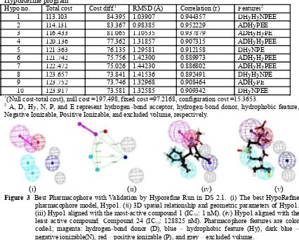
Table 1. Statistical parameters of the top 10 hypotheses of Neuraminidase inhibitors generated by HypoRefine program
Hypo no. Total cost Cost diff.1 RMSD (Å) Correlation (r) Features2
1. 113.103 84.395 1.03907 0.944357 DHDHyHyNPEE
2. 114.131 83.367 0.98385 0.952229 ADHyPEE
3 116.433
3. 116.433 81.065 1.1053581.065 1.10535 0.9378790.937879 ADH H PEADHyHyPEPEPE
4. 120.136 77.362 1.31857 0.907315 ADHyHyPEE
5. 121.363 76.135 1.29581 0.912158 DHDHyNPE
6. 121.742 75.756 1.42300 0.889973 ADHyHyPEE
7. 122.472 75.026 1.44230 0.886802 ADHyHyPEE
8. 123.657 73.841 1.41536 0.892491 DHDHyHyNPE
9. 123.752 73.746 1.32968 0.908464 ADHyPEPE
10
10. 123.917 73.581 1.32585 0.909342 DHDHyNPEE
1(Null cost-total cost), null cost =197.498, fixed cost =97.2168, configuration cost =15.3653.
2A, D, Hy, N, P, and E represent hydrogen -bond acceptor, hydrogen-bond donor, hydrophobic feature,
Negative Ionizable, Positive Ionizable, and excluded volume, respectively.
(i) (iiii) (iviv) (v)
Figure 3. Best Pharmacophore with Validation by Hyporefine Run in DS 2.1. (i) The best HypoRefine pharmacophore model, Hypo1. (iiii) 3D spatial relationship and geometric parameters of Hypo1. (iii) Hypo1 aligned with the most-active compound 1 (IC5050: 1 nM). (iviv) Hypo1 aligned with the
least active compound, Compound 24 (IC5050: 128825 nM). Pharmacophore features are color
coded; magenta: hydrogen-bond donor (D), blue � hydrophobic feature (Hy), dark blue � negative ionizable(N), red �positive ionizable (P), and grey �excluded volume.
Validation of Pharmacophore Model 1. Fischer Randomization test
2. Test set
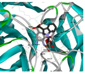
Figure 5. Conformation of MSC605 (black carbon) and OSTM X ray (pink carbon) bonded N1, surface visualizased with DS 2.1.
In addition, MSC927 was found that binding interaction into N1 (1F8B) [9] have similarity with DANA X-ray, although have fit value (mapping) and free energy docking less than better if be compared with Oseltamivir (2HUO)[10] and DANA X-ray (1F8B)[9]. MSC927 also have mapping similar with OSTM, but MSC927 ������� ��� �������� ��������� ��������
Figure 6b and 6c show CGA and OSTM interaction at N1.
(i) (ii) Figure 6.
Figure 6. (i) Conformation of MSC927 (green) and OSTM X ray (orange) bonded N1, surface visualizased with DS 2.1. Non-polar hydrogen atom at ligand did not seems in order to clear. B. (ii) Pocket area of site active of neuraminidase A (green : NI, red : strong hydrophobic, purple : PI, blue : weak hydrophobic) with visualizased VMD 1.8.5 by Linux
4. CONCLUSION
The best quantitative pharmacophore model, Hypo1 showed the lowest total cost value (92.055), the highest cost difference (107.807), the lowest RMSD (0.966317), and the best correlation coefficient (0.941732) compared to other models. Hypo1 contains four features: one hydrogen-bond donor (D), one Hydrophobic aliphatic moiety (Hy), one negatively ionizable (N), and one positive ionizable (P). There were 45 compounds of 3000 NADI database that showed high positive ionizable (P). There were 45 compounds of 3000 NADI database that showed high predictive affinity when matched into ������� HBD, Hy, NI and PI by optimizing the minimum
REFERENCES
1. Moscona, A., Oseltamivir resistance--disabling our influenza defenses. N Engl J Med, 2005. 353(25): p. 2633-6.
2. Colman, P.M., Influenza virus neuraminidase: structure, antibodies, and inhibitors. Protein Sci, 1994. 3(10): p. 1687-96.
3. Wahab, H.A., et al., Nature based drug discovery (NADI) & Its Application to Novel Neuraminidase Inhibitors Identification by virtual screening, pharmacophore modelling and mapping of Malaysian Medicinal Plants. , in Drug Design and Discovery for Developing Countries. , E. Megnassan, L. Owono Owono, and S. Miertus, Editors. 2009, ICS-UNIDO,Trieste, Italy.: Trieste, Italy. 4. Brooks, B.R., et al., CHARMM: A Program for Macromolecular Energy,
Minimization, and Dynamics Calculations. J Comput Chem, 1983. 4(2): p. 187-217.
5. Wang, H.Y., et al., Pharmacophore modeling and virtual screening for designing potential PLK1 inhibitors. Bioorg Med Chem Lett, 2008. 18(18): p. 4972-7. 6. Deng, X.Q., et al., Pharmacophore modelling and virtual screening for
identification of new Aurora-A kinase inhibitors. Chem Biol Drug Des, 2008. 71(6): p. 533-9.
7. Abu Hammad, A.M. and M.O. Taha, Pharmacophore modeling, quantitative structure-activity relationship analysis, and shape-complemented in silico screening allow access to novel influenza neuraminidase inhibitors. J Chem Inf Model, 2009. 49(4): p. 978-96.
8. Adane, L., D.S. Patel, and P.V. Bharatam, Shape- and chemical feature-based 3D-pharmacophore model generation and virtual screening: identification of 3D-pharmacophore model generation and virtual screening: identification of potential leads for P. falciparum DHFR enzyme inhibition. Chem Biol Drug Des, 2010. 75(1): p. 115-26.
9. Smith, B.J., et al., Analysis of inhibitor binding in influenza virus neuraminidase. Protein Sci, 2001. 10(4): p. 689-96.
10. Russell, R.J., et al., The structure of H5N1 avian influenza neuraminidase suggests new opportunities for drug design. Nature, 2006. 443(7107): p. 45-9. 11. Morris, G.M., R. Huey, and A.J. Olson, Using AutoDock for ligand-receptor
docking. Curr Protoc Bioinformatics, 2008. Chapter 8: p. Unit 8 14.
12. Kim, C.U., et al., Structure-activity relationship studies of novel carbocyclic influenza neuraminidase inhibitors. J Med Chem, 1998. 41(14): p. 2451-60. 13. Williams, M., et al., Structure-activity relationships of carbocyclic influenza
neuraminidase inhibitors Bioorganic & Medicinal Chemistry Letters, 1997. 7(14): p. 1837
14. Yi, X., Z. Guo, and F.M. C hu, Study on molecular mechanism and 3D-QSAR of influenza neuraminidase inhibitors. Bioorg Med Chem, 2003. 11(7): p. 1465-74. 15. Wang, G.T., et al., Design, synthesis, and structural analysis of inhibitors of
influenza neuraminidase containing a 2,3-disubstituted tetrahydrofuran-5-carboxylic acid core. Bioorg Med Chem Lett, 2005. 15(1): p. 125-8.