The effect of combination of triamcinolone
acetonide and methotrexate on
keloid-fibroblast activity in dermis equivalent
Endra Yustin E. S., Fajar Waskito, Yohanes Widodo Wirohadidjojo
Department of Dermatovenereology, Faculty of Medicine, Gadjah Mada University/ DR. Sardjito Hospital Yogyakarta
ABSTRACT
Endra Yustin E. S., Fajar Waskito, Yohanes Widodo Wirohadidjojo - The effect of combination of triamcinolone acetonide and methotrexate on the keloid-fibroblast activity in dermis equivalent
Background: Triamcinolone acetonid (TA) intralesion has been a standard treatment for keloids for many years, due to its effect in inhibiting collagen synthesis and fibroblast proliferation. However, until now the clinical result is unsatisfactory. Keloid flattening is slow and sometimes adverse reactions may occur. Methotrexate (MTX) is a chemotherapeutic agent having an antiproliferating effect which act as an antifolic acid. Because of this effect, MTX is potential to be used in combination with TA for the treatment in keloid. Fibroblast populated collagen lattice (FPCL) was a dermal equivalent usually used for fibroblast activity measurement.
Objective: To understand the inhibition of fibroblast keloid activities of MTX in vitro on FPCL contraction, compared to TA and MTX plus TA.
Methods: This research used simple parallel multigroups experimental study design, and conducted on third passage keloid fibroblast culture, which was isolated from one patient. Fibroblast was cultivated in collagen type 1 from rat tail (FPCL). Keloid fibroblasts was classified into 16 groups, and treated with 5, 10, 20 mM TA, 1.75, 3.5, 7 mM MTX, combination of TA and MTX, and a control negative. FPCL contraction indicating activities of fibroblast was measured using Scion Image software. Mean of FPCL contraction was analyzed using one way analysis of variance (ANOVA).
Results: All treatments could inhibit FPCL contraction until day 2 (p<0.05). The highest inhibition of FPCL was found in combination of TA 20 mM + MTX 7 mM (p<0.05). The treatment that could inhibit FPCL contraction until day 3 was only group MTX 3.5 mM + TA 20 mM. This result indicated that a combination of TA and MTX was stronger in inhibiting keloid fibroblast activities compared with TA and MTX alone.
Key words: keloid fibroblast - growth factors - triamcinolone acetonid - methotrexate - FPCL contraction
ABSTRAK
Endra Yustin E. S., Fajar Waskito, Yohanes Widodo Wirohadidjojo - Efek kombinasi triamsinolon asetonid dan metotreksat terhadap aktivitas fibroblas keloid pada ekivalen jangat
Latar Belakang. Injeksi triamsinolon asetonid (TA) intralesi telah merupakan standar terapi keloid selama beberapa tahun, karena dapat menghambat sintesis kolagen dan proliferasi fibroblas. Namun demikian, sampai dewasa ini hasil klinisnya tidak memuaskan. Penipisan keloid terjadi sangat lamban dan kadang-kadang timbul reaksi yang tidak diinginkan. Metotreksat (MTX) digunakan sebagai agen kemoterapi yang memiliki efek antiproliferasi yang bekerja sebagai anti-asam folat. Oleh karena efek tersebut, MTX berpotensi sebagai kombinasi dengan TA pada terapi keloid. Fibroblast populated collagen lattice (FPCL) yang dikenal sebagai ekivalen kulit jangat sering dipakai untuk menilai aktivitas fibroblas.
Tujuan: Mengetahui daya hambat aktivitas fibroblas keloid secara in vitro terhadap kontraksi FPCL antara kombinasi TA dan MTX dalam berbagai dosis dengan berbagai dosis TA dan MTX secara tunggal.
Metode: Penelitian ini menggunakan rancangan penelitian eksperimental sederhana paralel multigrup, yang memakai kultur fibroblas keloid passage 3 dari satu penderita keloid. Fibroblas dibiakkan dalam kolagen tipe 1 dari buntut tikus (FPCL). Fibroblas keloid dibagi menjadi 16 grup dan diterapi dengan TA 5, 10, 20 mM, MTX 1,75, 3,5, 7 mM, kombinasi TA dan MTX, dan kontrol negatif tanpa perlakuan. Kontraksi FPCL sebagai indikasi aktivitas fibroblas keloid dinilai dengan Scion Image software. Rerata luas FPCL diuji dengan memakai analisis varian satu jalan (ANOVA).
Hasil: Kelompok MTX dan TA tunggal ataupun kombinasi pada semua dosis dapat menghambat FPCL pada hari pertama dan kedua (p<0,05), hambatan terbesar dijumpai pada kombinasi MTX 7 mM + TA 20 mM (p<0,05). Terapi yang dapat menghambat kontraksi sampai hari ke-3 hanya dijumpai pada kelompok MTX 3.5 mM + TA 20 mM. Hasil penelitian ini menunjukkan bahwa kombinasi TA + MTX lebih kuat menghambat
aktivitas fibroblas keloid dibanding TA and MTX yang diberikan tersendiri.
INTRODUCTION
Keloids are benign hyperproliferative growth of dermal collagen that is usually resulted from excessive tissue response to skin trauma,1 expands
beyond the boundaries of the original injury as it heals and does not regress spontaneously.2,3 Keloids
are often pruritic, painful, causes cosmetic disfigurement and skin contracture.4 Keloids may
be caused by minor skin wound such as acne, piercing, vaccination, or even a mosquito bite or post-surgery, especially on presternal region, superior back of the trunk and posterior region of the neck.5 Keloid scars mostly affected persons
between 10 and 30 years of age, in Blacks and Asians race, with predisposition in individuals with human leukocyte antigen (HLA) B 14, BW 16 and blood group A.5
The pathology of keloids remains poorly understood. In vitro researches had shown that keloid scars were considered to be a result of abnormal wound healing, due to overproliferation of fibroblast followed by low activities of collagenase that caused overexcessive collagen in the wound.3 Fibroblast proliferation was stimulated
by growth factors and cytokines, such as vascular endothelial growth factor (VEGF),6 transforming
growth factor beta (TGF-b), tumor necrosis factor alfa (TNF-α), and interleukin 6 (IL-6).7 The most
important factor is TGF-b8, followed by VEGF.6
Keloid scars exclusively affected Homo-sapiens, and that is why no experiments were possible in animal models. The experiment for keloids usually used in vitro model that investigated the organization of collagen fibers by fibroblast in fibroblast populated collagen lattice (FPCL) contraction model. It was composed of cultured
fibroblast suspended in collagen lattice isolated from rat tail. This model was a dermal equivalent, which was usually used for fibroblast activities measurement.9,10
Keloid therapy was fraught with varying degrees of success. There was no one modality that was always successful. Various methods had been developed to treat keloid, from pharmacologic treatments such as triamcinolone acetonide (TA),11
tretinoin,12 tacrolimus,13 5-flourourasil (5-FU),14
tamoxifen,15 to surgery.1 All of these modalities had
unsatisfactory results, the keloid flattening was followed by high rates of recurrences.1,3 The most
commonly used therapeutics for keloid was intralesion TA injection as a single modality or combined with others.1,5 A study showed that 10
mM TA caused a significant decrease in the production of TGF-b keloid fibroblast2 with side
effect like severe pain, atrophy, hypopigmentation or Cushing’s syndrome after several injection.16
Methotrexate (MTX) is a chemotherapeutic agent having an anti-proliferating effect which act as a folic acid antagonist that usually used to treat breast cancer,17 acute lymphoblastic leukaemia,18
psoriasis,19 rheumatoid arthritis,20 and
kerato-achantoma tumor as a intralesional treatment with no local or systemic adverse effects from the therapy.21,22
As an immunosuppressant, MTX might cause suppression of TNF-α, IL-6,23 TGF-b,24 and also
had anti-angiogenesis effect,17 that decreases the
level of VEGF.25 Muzaffar26 and Tonkin27 reported
FIGURE.1. Average contraction percentage of FPCL with treatment (TA) and control
METHODS
Keloid dermal fibroblast primary cultures
Primary cell lines of keloid dermal fibroblasts were established from operating room specimens. The keloid tissue was taken from deltoid region. A standard explant method was used to establish the primary fibroblast cultures from the operative specimens. This explant method was performed using sterile technique under a laminary flow hood. After all of the subcutaneous tissue was removed with a sterile scalpel blade, the gross specimens were minced into small fragments in a petri dish. The specimens underwent antimicrobial treatment with Dulbecco’s phosphate-buffered saline (PBS) solution with 5% penicillin/streptomycin/ amphotericin (PSA). Minced fragments measuring approximately 1 mm were then placed into tissue culture flasks containing 5 ml of explant tissue culture media (40% fetal calf serum in Dulbecco’s modified eagle medium with 1% PSA). These flasks were then stored in a humidified incubator containing a 5% CO2 atmosphere at 37°C. The explant-containing flasks were examined daily under light microscopy for the outgrowth of fibroblasts from the tissue fragments. Until light microscopy demonstrated such fibroblast outgrowth, explant tissue culture media was changed daily. Once fibroblast outgrowth approached confluence, the tissue fragments were removed. The fibroblasts were then trypsinized with 0.05% trypsin and subcultured into culture flasks containing 10 ml of primary culture media (10% fetal calf serum in Dulbecco’s modified eagle medium with 1% PSA). All flasks were stored in a humidified incubator containing a 5% CO2 atmosphere at 37 °C. Primary culture media was changed twice a week. Successive cultures were passed at confluence. Experiments were performed using passage 3 keloid fibroblasts.
Preparation of fibroblast-populated collagen lattices
Collagen lattices were prepared from type 1 collagen extracted from rat tail tendons in 0.1% (vD v) acetic acid. A working solution was constituted by diluting the collagen to 2 mg/ml on the day of the experiment. Keloid fibroblast was trypsinized, resuspended in medium and counted
using a haemocytometer. A collagen–keloid fibroblast suspension was prepared in DMEM and divided into 16 groups, then treated with 5, 10, 20 µM TA, 1,75, 3.5, 7 µM MTX, combination of TA and MTX, and a control negative. Fibroblast-populated collagen lattices with treatments were seeded into the wells of 24-well plates at density of 6 × 10⁴ cells/ml. Plates were kept in an incubator with 5% CO2 at 37°C.
Macroscopic evaluation of the gel contraction
The area of collagen gel populated with fibroblasts was photographed with a digital camera (Olympus SLR E.500) at day 0, 1, 2 and 3 after the experiment had begun, the gel area was calculated with Scion Image software. The percentage of gel contraction in each interval studied was calculated with the following formula:
Percentage of contraction =
100 ) (
1 2 1
A
A A
where: A1 is initial gel area and A2 is the area at the observed intervals.
Statistical Analysis
The gel contraction data were analyzed using ANOVA and were expressed as mean percentage of contraction and standard deviation. Posthoc test used Tukey HSD with p<0.05.
RESULTS
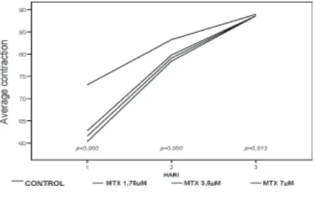
FIGURE 2. Average contraction percentage of FPCL with treatment (MTX) and control
FIGURE 3. The best average contraction percentage of FPCL with treatment (MTX, TA, combination of MTX
and TA) and control.
FIGURE 1 and 2 show that all treatments with various doses of TA and MTX given separately caused a statistically significant (p<0.05) inhibition on FPCL contraction until day 2, compared to control. The inhibition of FPCL contraction was in dose-dependent manner, a higher dose of TA or MTX may paradoxically increase inhibition of FPCL contraction.
From the combination of TA and MTX groups (data were not shown), we found a similar result with treatment group which given separately. All various doses combination of TA and MTX caused a statistically significant (p<0.05) inhibition on FPCL contraction until day 2, compared to control. A higher dose of combination treatment might increase the inhibition of FPCL contraction. The best inhibition of FPCL contraction from each combination of TA and MTX was found in all various doses of MTX with 20 µM TA.
FIGURE 3 shows that all treatments could inhibit FPCL contraction until day 2 (p<0.05). The highest inhibition of FPCL was found in combination of TA 20 µM + MTX 7 µM (p<0.05). The treatment that could inhibit FPCL contraction until day 3 was only group MTX 3.5 µM + TA 20 µM combination (post-hoc test). This result indicated that a combination of TA and MTX would be stronger in inhibiting keloid fibroblast activities than TA and MTX given separately.
DISCUSSION
Experiments with cells derived from keloid tissue revealed a number of abnormalities in cellular functions, such as in proliferation, apoptosis, or expression of growth factors.3 The most important
factor is TGF-α,9,10,28 followed by VEGF,6,29,30
IL6,7,30 and TNF-α.31
Keloid explants embedded in collagen lattice were morphologically similar with in vivo wound healing, it could be used for maintaining long-term viable keloid dermal explants until 6 weeks as a tool for investigating the pathogenesis of keloid scar formation.32 FPCL were used for investigating
TGF-ß,9,10 FGF,10 5-FU,14 chitin, and chitosan on keloid.33
This study (FIGURE1-3) showed the differences of average contraction percentage of FPCL with treatment and control on day 1 until 3. This result indicated a viable fibroblast keloid and the consistency of FPCL contraction due to the treatment.
Triamcinolone acetonide (TA) could alter matrix extracellular due to fibroblast proliferation and TGF-ß. Cruz and Korchin found that 10 µM TA caused statistically significant growth inhibition of keloid and fetus fibroblasts.34 Carrol2 found that 10 and
20 µM TA caused a statistically significant decrease in the level of TGF-ß keloid fibroblast on day 1. Exposure with 5 µM TA did not cause a statistically significant decrease in the level of TGF-ß. A higher dose of TA may increase cellular proliferation.2
FPCL and inhibition of FPCL were not only caused by TGF- ß. The inhibition of FPCL contraction was in dose-dependent manner, a higher dose of TA or MTX might paradoxically increase inhibition of FPCL contraction.
Methotrexate (MTX) had been shown to suppress TNF-α due to apoptosis.35 Neurath23
found that 1-10 µg/ml MTX on murine caused suppression TNF-a and IL-6. Sigmundsdottir36
found that 1-10 µM MTX markedly reduced the expression of vascular E-selectin and ICAM 1 in psoriatic skin, and no further inhibition was observed after 10 µM MTX. This result was similar with our study. FIGURE 2 shows that 1.75, 3.5 and 7 µM MTX caused a statistically significant (p<0.05) inhibiton on FPCL contraction until day 2, compared with control it was due to the inhibition of fibroblast proliferation, TNF-α, or may be the other factors, on dose-dependent.
The study of combination MTX with other chemotherapy or immunosuppresant for cancer or rheumatoid arthritis had been conducted. The target mechanism was VEGF, TGF ß1 and TNF-a. Low-dose MTX with cyclophosphamide caused significant decreased level of VEGF in breast cancer,17 and with dexamethasone in rheumatoid
artrithis.37 Combination of 5-FU and
cyclophospha-mide caused significant decreased plasma TGF-ß1 concentration in breast cancer.24 Muzaffar26 and
Tonkin27 reported a success in treating keloid with
oral low dose MTX combined with reconstruction syndactily with no recurrences. In our study, all treatments with various doses of MTX and TA given separately or in combination could inhibit FPCL contraction until day 2 (p<0.05), except in group MTX 3.5 µM + TA 20 mM combination that could inhibit FPCL contraction until day 3 (p<0.05) (post-hoc test). That combination might be an ideal dose to get synergism effect. The highest inhibition of FPCL was found in combination of TA 20 µM + MTX 7 µM (p<0.05). This result indicated that combination of TA and MTX would inhibit further the keloid fibroblast activities than TA or MTX given separately. This result might be based on synergistic or additive effects of the drugs.
To summarize, we had demonstrated that the inhibition of FPCL contraction was dose-dependent, a higher dose of TA or MTX might paradoxically
increase the inhibition of FPCL contraction. The combination of TA and MTX would inhibit further keloid fibroblast activities than TA or MTX given separately; strongly suggesting based on synergistic or additive effects of the drugs. These data suggested a new therapeutic approach for keloid.
REFERENCES
1. Kelly AP. Medical and surgical therapies for keloids. Dermatol Therap, 2004; 17: 212-18.
2. Carroll LA, Hanasono MH, Miculec AA, Kita M, Koch RJ. Triamcinolone stimulates bFGF production and inhibits TGF-β1 production by human dermal fibroblasts. Dermatol Surg 2002; 28: 704-709. 3. Maneros AG, Krieg T. Keloids-clinical, pathogenesis,
and treatment options. JDDG, 2004; 2: 905-13. 4. Lee SS, Yosipovitch G, Chan YH, Goh CL. Pruritus,
pain, and small nerve fiber function in keloids: a control study. J Am Acad Dermatol, 2004; 51: 1002-1006. 5. Shejbal D, Badecovic V, Ivkic M, Kalogjera L, Aleric Z,
Drvis P. Strategies in the treatment of keloid and hypertrophic scars. Acta Clin Croatia, 2004; 43: 417-22.
6. Le AD, Zhang Q, Wu Y, Messadi DV, Akhondzadeh A, Nguyen AL, Aghaloo TL, Kelly AP, Bertolami CN. Elevated vascular endothelial growth factor in keloids: relevance to tissue fibrosis. Cells Tissues Organs. 2004;176(1-3):87-94.
7. Tosa M, Ghazizadeh M, Shimizu H, Hirai T, Hyakusoku H, Kawanami O. Global gene expression analysis of keloid fibroblast in response to electron beam irradiation reveals in the involvement of interleukin-6 pathway. J Invest Dermatol, 2005;124: 704-13.
8. Diegelmann RF, Evans MC. Wound healing: an overview of acute, fibrotic and delayed healing. Frontiers in Bioscience, 2004; 9: 282-89.
9. Mukhopadhyay A, Tan EKJ, Khoo YTA, Chan SY, Lim IJ, Phan TT. Conditioned medium from keloid keratinocyte/keloid fibroblast coculture induce contraction of fibroblast-populated collagen lattices. Br J Dermatol. 2005; 152: 639-45.
10. Kamamoto F, Paggiaro AO, Rodas A, Herson MR, Mathor MB, Ferreira. A wound contraction experimental model for studying keloid and wound-healing modulators. Intern Soc Artific Organs, 2003; 27: 701-705.
11. Shaffer JJ, Taylor SC, Bolden FC. Keloidal scars: a review with a critical look at therapeutic options. J Am Acad Dermatol, 2002; 46: 63-97.
12. Uchida G, Yoshimura K, Kitano Y, Okazaki M, Harii K. Tretinoin reverses up regulation of matrix metalloproteinase-13 in human keloid-derived fibroblast. Exp Dermatol, 2003; 12: 35-42.
gli-1 oncogene in keloids. J Am Acad Dermatol. 2001;45(5):707-11.
14. Levinson H, Liu W, Peled Z. 5-fluorouracil inhibits keloid fibroblast proliferation and keloid fibroblast populated collagen lattice contraction. J Burns & Surg Wound Care, 2002; 1: 1-10.
15. Haczynski J, Tarkowski R, Jarzabek K, Wolczynski S, Magoffin DA, Czarnocki KJ, Ziegert M, Jakowicki J, Jakimiuk AJ. Differential effects of estradiol, raloxifene and tamoxifen on estrogen receptor expression in cultured human skin fibroblasts. Int J Mol Med. 2004;13(6): 903-8.
16. Kumar S, Sigh RJ, Reed AM, Lteif AN. Cushing’s syndrome after intra-articular and intradermal administration of triamcinolone acetonide in three pediatric patients. Pediatrics, 2008; 6:1820-24. 17. Colleoni M, Rocca A, Sandri MT, Zorzino L, Masci G,
Nolè F, Peruzzotti G, Robertson C, Orlando L, Cinieri S, de BF, Viale G, Goldhirsch A. Low-dose oral metho-trexate and cyclophosphamide in metastatic breast cancer: antitumor activity and correlation with vascular endothelial growth factor levels. Ann Oncol. 2002;13(10):1689-90. .
18. Estlin EJ. Novel targets for therapy in paediatric oncology. Current Drug Targets-Immune, Endocrine & Metabol Disorders. 2002; 2:141-150.
19. Haustein UF, Rytter M. Methotrexate in psoriasis: 26 years’ experience with low-dose long-term treatment. Eur Acad Dermatol Venereol. 2000; 14: 382-88. 20. Kramer I, Wibulwas A, Croft D, Genot E, Rheumatoid
arthritis: targetting in proliferative fibroblasts. Progr Cell Cycle Res. 2003; 5: 59-70.
21. Hurst LT, Gan BS. 1995. Intralesional methotrexate in keratoacanthoma of the nose. Br J Plastic Surg. 48: 243-46.
22. Annest NM, VanBeek MJ, Arpey CJ, Whitaker DC. Intralesional methotrexate treatment for keratoachantoma tumors: a retrospective study and review of the literature. J Am Acad Dermatol. 2007; 56: 989-93.
23. Neurath MF, Hildner K, Becker C, Schlaak JF, Barbulescu K, Germann T, Schmitt E, Schirmacher P, Haralambous S, Pasparakis M, Meyer Zum Büschenfelde KH, Kollias G, Märker-Hermann E. Methotrexate specifically modulates cytokine production by T cells and macrophages in murine collagen-induced arthritis (CIA): a mechanism for methotrexate-mediated immunosuppression. Clin Exp Immunol. 1999;115(1):42-55.
24. Kajdaniuk D, Marek B, Swietochowska E, Ostrowska Z, Glogowska-Szelag J, Kos-Kudla B, Ciesielska-Kopacz N, Wielkoszyski T. Plasma transforming growth factor beta1 in breast cancer patients treated with CMF chemotherapy. J Clin Pharm Ther. 2000;25(4):291-4. 25. Lennernäs B, Albertsson P, Lennernäs H, Norrby K.
Chemotherapy and antiangiogenesis. Drug-spesific, dose-related effects. Acta Oncol. 2003; 42: 294-303.
26. Muzaffar AR, Rafols F, Masson I, Ezaki M, Carter PR. Keloid formation after syndactyly reconstruction: associated conditions, prevalence, and preliminary report of a treatment method. J Hand Sur. 2004; 29: 201 208. 27. Tokin MA, Willis KR, Lawson RD. Keloid formation resulting in acquired syndactyly of an initially normal web space following syndactyly release of an adjacent web space. J Hand Surg. 2008; 1: 29-31.
28. Bock O, Yu H, Zitron S, Bayat A, Ferguson MWJ, Mrowietz U. Studies of transforming growth factors beta 1-3 and their receptors I and II in fibroblast of keloids and hypertrophic scars. Act Dermatol Venereol. 2005; 85: 216-20.
29. Gira AM, Brown LF, Woshington CV, Cohen C, Arbiser JL. Keloids demonstrate high-level epidermal expression of vascular endothelial growth factor. J Am Acad Dermatol. 2004; 50: 850-53.
30. Giugliano G, Pasquali D, Notaro A, Brongo S, Nicoletti G, D’Andrea F, Bellastella A, Sinisi AA. Verapamil inhibits interleukin-6 and vascular endothelial growth factor production in primary cultures of keloid fibroblasts. Br J Plast Surg. 2003;56(8):804-09. . 31. Berman B, Villa AM, Ramirez CC. Novel opportunities
in the treatment and prevention of scarring. J Cutan Med Surg. 2005: 32-36.
32. Duong HS, Zhang Q, Kobi A, Le A, Messadi DV. Assesment of morphological and immunohistological alterations in long-term keloid skin explants. Cells Tissues Organs. 2005; 181: 89-102.
33. Howling GI, Dettmar PW, Goddard PA, Hampson FC, Dornish M, Wood EJ. The effect of chitin and chitosan on fibroblast populated collagen lattice contraction. Biotechnol App Biochemist. 2002; 36: 247-53. 34. Cruz and Korchin. cit Carroll LA, Hanasono M, Miculec
AA, Kita M, Koch RJ. 2002. Triamcinolone stimulates bFGF production and inhibits TGF- α1 production by human dermal fibroblasts. Dermatol Surg. 1994; 28: 704-709.
35. Lathounder SD, Gerards AH, Dijkmans BAC, Aarden LA. Two inhibitors of DNA-synthesis lead to inhibition of cytokine production via different mechanism. Nucleosides, Nucleotides & Nucleic Acids. 2004; 23: 1089-1100.
36. Sigmundsdottir H, Johnston A, Gudjonsson JE, Bjarnason B, Valdimarsson H. Methotrexate markedly reduces the expression of vascular E-selectin, cutaneous lymphocyte-associated antigen and the numbers of mononuclear leucocytes in psoriatic skin. Exp Dermatol. 2004;13(7):426-34.