3
CHAPTER 2
LITERATURE REVIEW
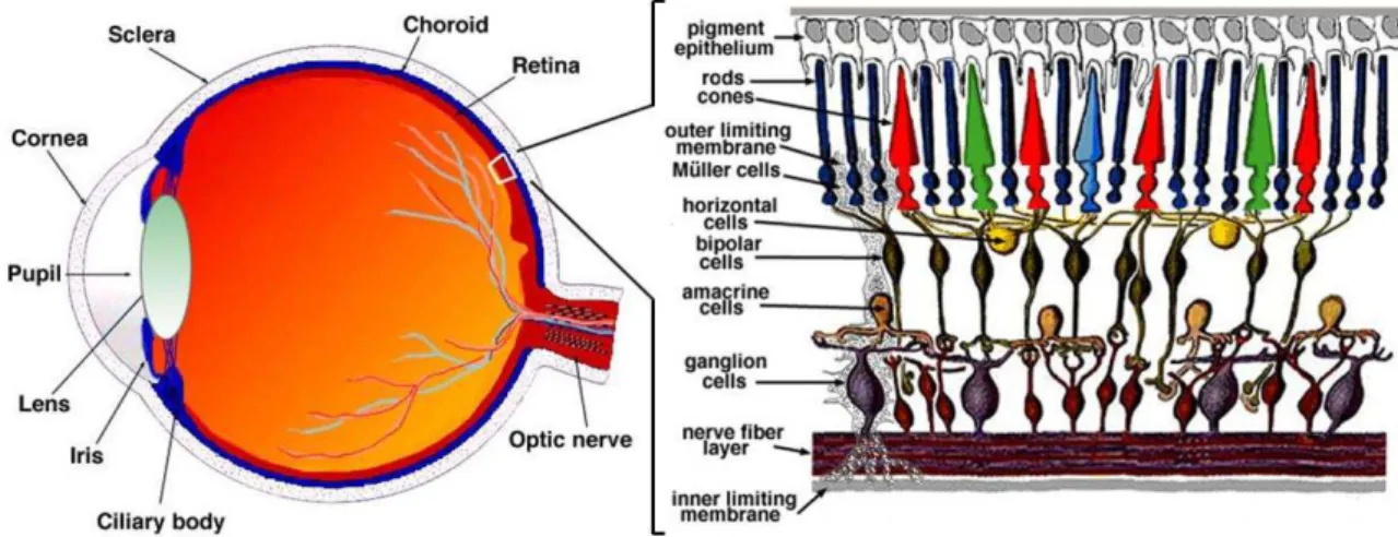
Eye is the organ responsible for the vision, which is considered one of the most important sense of human being. Retina is the inner most layer at the posterior part of the
eyeball which is a clear, thin, and photosensitive tissue. This structure converts light into electrical signals which is enabled by specialized cells, the photoreceptors, in a process termed phototransduction. There are two types of photoreceptors cells: rods and cones, which are located in humans in the peripheral and central retina, respectively. Rod cells are involved in the dim light vision while cone cells are associated with color detailed vision. In addition to the photoreceptors, retinal pigmented epithelium cells also play an important role in maintaining retinal function. (Figure 1) Losing either one or both of these photoreceptors as well as retinal pigment epithelium lead to a condition called retinal dystrophies.10
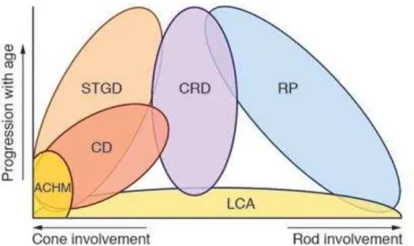
There are many types of retinal dystrophies, affecting only one cell type or both and different progression with age. These disorder phenotype range from achromatopsia, Stargardt’s disease, cone dystrophy, cone-rod dystrophy, retinitis pigmentosa, to Leber congenital amaurosis. (Figure 2) Amongst others, Leber congenital amaurosis is one of the most severe and it accounts for ~20% of children in the special school for the blind.3
4
Figure 2. Classification of IRDs according to age progression and cell involvement. There are two kind of
photoreceptor cells involvement: rod and cone. Rod is responsible for dim light and peripheral vision. Cone is responsible for color and central vision. STGD: Stargardt disease, CD: cone dystrophy, ACHM: achromatopsia, CRD: cone-rod dystrophy, RP: retinitis pigmentosa, LCA: Leber congenital amaurosis.10 (Adapted from: den Hollander, et al. 2010)
2.1. Leber Congenital Amaurosis
Leber congenital amaurosis (LCA; OMIM 204000) is the most severe retinal dystrophy with the early onset in the first year of life. It was described for the first time by the German doctor, Theodore Leber, in 1869. He found that the disease was characterized by unresponsive pupil, wandering nystagmus, and a fundus that initially appears normal and then turn to be typical for retinitis pigmentosa in early childhood. He introduced this disease as the congenital form of retinitis pigmentosa.11 Pinckers thought that the disease Leber found actually was more or less the same as what we call now as neuronal ceroid lipofuscinosis.12 This disease accounts for ~5% of all inherited retinopathies with a prevalence of
approximately 1:50.000.13.14
2.1.1. Clinical Characteristics
The three main features of LCA are congenital onset of visual loss, amaurotic pupils; no response in light reflex, and nystagmus.11 Franceschetti and Dieterle mentioned the importance of elecroretinogram in retinal degeneration.15 Absent electroretinogram became one of the diagnostic criteria other than bilateral congenital blindness.16 Foxman and colleagues suggested that elevtroretinogram (ERG) should be examined before the age of one year.17
5
(75%) of visual acuity of the patients remained stable while visual deterioration and even improvement were observed in 15% and 10% patients, respectively.18-20
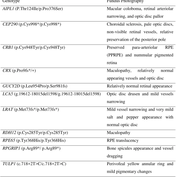
Table 1. Fundus photography results of LCA patients with known mutations in
causative genes.1
Genotype Fundus Photography
AIPL1 (P.Thr124Ile/p.Pro376Ser) Macular coloboma, retinal arteriolar
narrowing, and optic disc pallor
CEP290 (p.Cys998*/p.Cys998*) Choroidal sclerosis, pale optic discs,
non-visible retinal vessels, relative preservation of the posterior pole
CRB1 (p.Cys948Tyr/p.Cys948Tyr) Preserved para-arteriolar RPE (PPRPE) and nummular pigmented retina
CRX (p.Pro9fs*/+) Maculopathy, relatively normal
appearing vessels and optic disc
GUCY2D (p.Leu954Pro/p.Ser981fs) Relatively normal retinal appearance
LCA5 (g.19612-18015del1598/g.19612-18015del1598) Optic disc drusen and mild vessels narrowing
LRAT (p.Met73fs*/p.Met73fs*) Mild vessel narrowing and very mild salt and pepper appearance with normal optic disc
RDH12 (p.Cys285Tyr/p.Cys285Tyr) Maculopathy
RPE65 (p.Tyr368His/p.Tyr368His) RPE translucency
RPGRIP1 (p.Arg89*/ p.Arg89*) Bone spicules appearance and vessel
dragging
TULP1 (c.718+2T>C/c.718+2T>C) Perivofeal yellow annular ring and
mild pigmentary changes
All LCA patients in this table have recessive mutations, except for the patient with CRX mutation.
6
correlation between preserved para-arteriolar RPE and CRB1 LCA-causing mutation. However, it is still difficult to have a convincing genotype-phenotype correlation in IRDs.1,21
ERG is essential to assess the visual function of LCA patients. ERG serves to measure the function of cone and rod cells. The electrical signal a-wave comes from the photoreceptor and the b-wave comes from bipolar and Müller cells. ERG test shows nonresponsive signals
in LCA patients while a-wave and b-wave are detectable in normal persons. This is especially important to diagnose LCA and also to figure out the genotype-phenotype correlation in LCA. Some studies showed that LCA patients carrying mutations in AIPL1 have a rod ERG impairment, in GUCY2D cone ERG impairment, and in RPGRIP1 both cone and rod impairment. Carriers with CRB1 heterozygous mutation may develop regional retinal dysfunction that can be determined using multi-focal ERG.1
Clinical characteristics are also important in order to understand, characterize, and predict the prognosis of the disease. While fundus photography provides a wide variety of retinal appearances, full-field ERGs never give positive results after treatment in previous study.22 Pupillometry and nystagmus assessment were used as an objective measurement in clinical trial of LCA patients. Still, subjective measurements such as best corrected visual acuity, Goldmann visual-field examination, and mobility testing can be very useful.9
2.1.2 Genetic Causes and Heritability of Leber Congenital Amaurosis
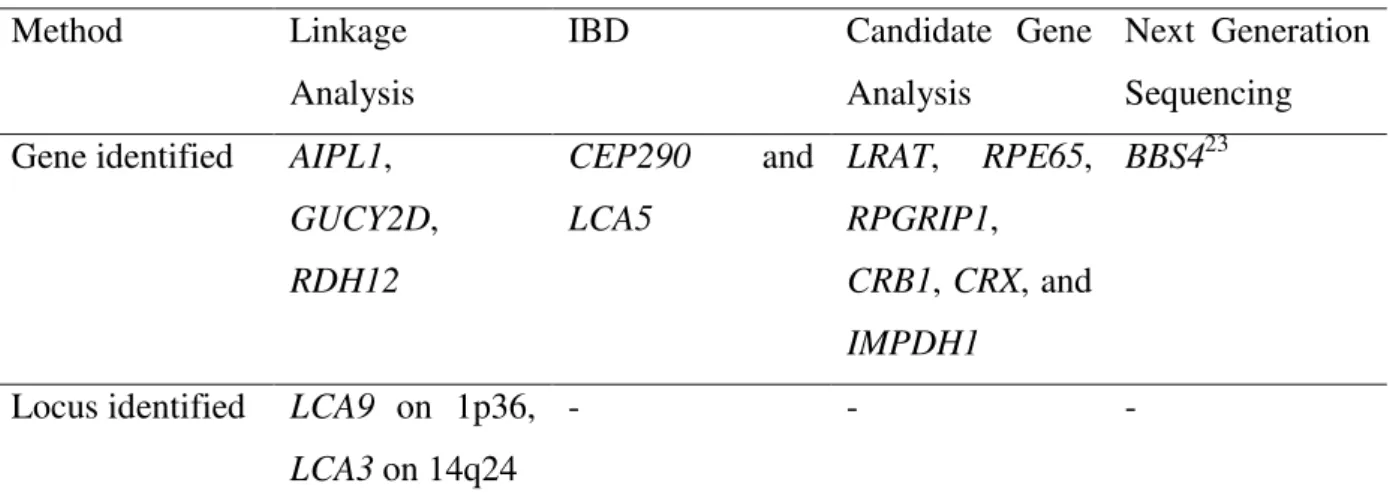
There are different approaches to identify the genetic causes of LCA such as linkage analysis, identity-by-descent (IBD) mapping, candidate gene analysis and whole exome sequencing (Table 2).
Table 2. Mutation identification strategy.1
Method Linkage
7
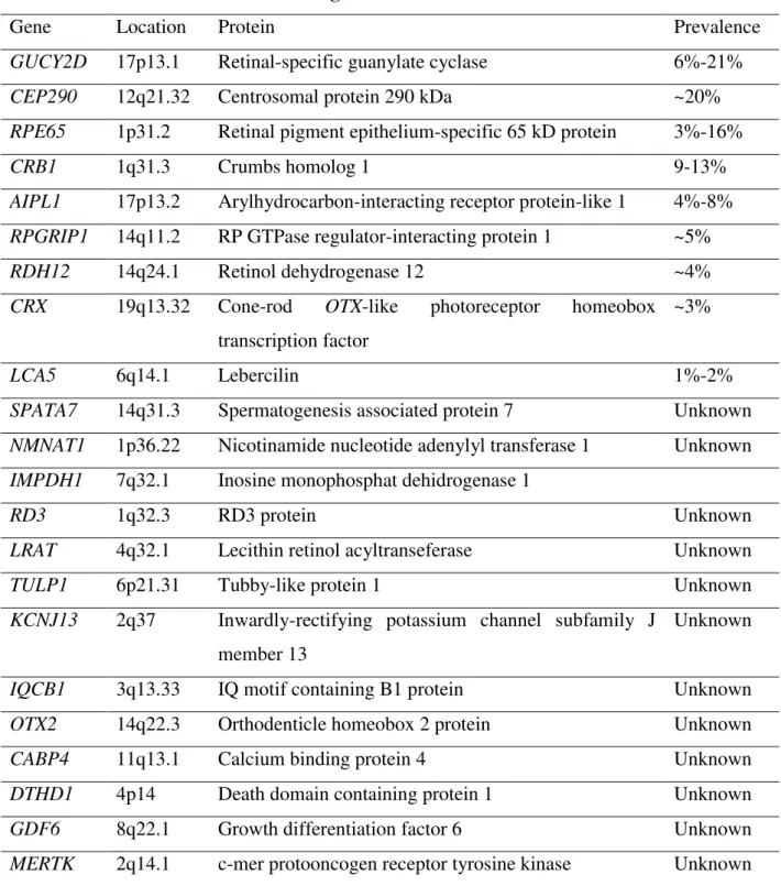
Table 3. Prevalence of LCA causative genes.6
Gene Location Protein Prevalence
GUCY2D 17p13.1 Retinal-specific guanylate cyclase 6%-21%
CEP290 12q21.32 Centrosomal protein 290 kDa ~20%
RPE65 1p31.2 Retinal pigment epithelium-specific 65 kD protein 3%-16%
CRB1 1q31.3 Crumbs homolog 1 9-13%
AIPL1 17p13.2 Arylhydrocarbon-interacting receptor protein-like 1 4%-8%
RPGRIP1 14q11.2 RP GTPase regulator-interacting protein 1 ~5%
RDH12 14q24.1 Retinol dehydrogenase 12 ~4%
CRX 19q13.32 Cone-rod OTX-like photoreceptor homeobox
transcription factor
~3%
LCA5 6q14.1 Lebercilin 1%-2%
SPATA7 14q31.3 Spermatogenesis associated protein 7 Unknown
NMNAT1 1p36.22 Nicotinamide nucleotide adenylyl transferase 1 Unknown
IMPDH1 7q32.1 Inosine monophosphat dehidrogenase 1
RD3 1q32.3 RD3 protein Unknown
LRAT 4q32.1 Lecithin retinol acyltranseferase Unknown
TULP1 6p21.31 Tubby-like protein 1 Unknown
KCNJ13 2q37 Inwardly-rectifying potassium channel subfamily J
member 13
Unknown
IQCB1 3q13.33 IQ motif containing B1 protein Unknown
OTX2 14q22.3 Orthodenticle homeobox 2 protein Unknown
CABP4 11q13.1 Calcium binding protein 4 Unknown
DTHD1 4p14 Death domain containing protein 1 Unknown
GDF6 8q22.1 Growth differentiation factor 6 Unknown
MERTK 2q14.1 c-mer protooncogen receptor tyrosine kinase Unknown
So far, 22 genes have been identified to cause LCA. Around 70% was caused by mutation in GUCY2D, CEP290, RPE65, CRB1, and AIPL1.
8
restricted to retina, therefore, genes that are expressed specifically in retina or have important function, can be presumed as candidate genes for LCA.1
Thus far, there are up to 22 genes involved in this disease (Table 3).6 From those genes, CEP290, GUCY2D, CRB1, IMPDH1, and RPE65 are the most common mutated genes, with a prevalence being 15%, 11.7%, 9.9%, 8.3%, and 6%, respectively in previous
studies on Caucasian populations. Since there is no comprehensive data about Asian population, it is important to accomplish the study. Inheritance pattern of LCA is mainly autosomal recessive, apart from CRX, IMPDH1, OTX which can be inherited in an autosomal dominant trait. These genes can be clustered into several group based on the mechanism in which they are involved: phototransduction, retinoid cycle, photoreceptor structure and development, connecting cilium transport system, guanine synthesis, outer segment phagocytosis.1
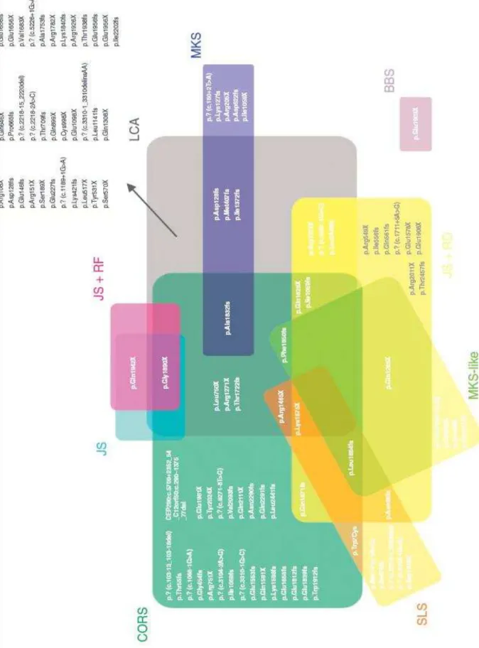
2.1.3 CEP290
CEP290 is one of the most mutated genes in LCA (~20%) in Caucasian populations. This gene encodes the centrosomal protein of 290 kDa composed of 2472 amino acids. This
protein is localized in the connecting cilium of the retinal photoreceptor cells, which may play a role in the transport system between the inner and outer segment of the photoreceptors. This transport system is very important because the proteins, which required for phototransduction, are synthesized in the inner segment of the photoreceptor cells. These proteins have to be transported to the outer segment, in order to conduct their correct function. In addition, CEP290 is expressed in almost all of the body cells localizing to the centrosomes or the basal body of the cilium. Cilium is a microtubule-related organelle which acts as the antenna of the cells, transferring sensory information from the extracellular surroundings. The importance of this organelle is represented by numerous disease associated with mutation in the ciliary genes, termed ciliopathies.24,25
9
Figure 3. Mutation spectrum in CEP290. CORS: Cerebello-oculo-renal syndrome, JS: Joubert syndrome,
10
2.2. Therapeutic Strategies
Therapeutic strategies for inherited retinal disorders comprise gene augmentation therapy and antisense oligonucleotide-based therapy. Nucleic acid sequence, either DNA or RNA, is used for both of them rather than protein or other molecules. The therapeutic agent is delivered to the target cells and allows restoration of gene function by acting as a replacement
(gene augmentation) or in case of splicing mutation, redirecting the correct splicing using antisense oligonucleotide.27,28
2.2.1 Gene Augmentation Therapy
Gene augmentation therapy aims to insert a full-length cDNA of a gene, to restore the defect caused by the mutation. As the full-length cDNA is insufficient to enter the cell itself, the presence of a vector is required. There are several vectors available for gene augmentation therapy, including viral and non-viral vectors (Table 4).27,29
Clinical trials for RPE65-associated LCA have already been conducted using adeno-associated virus (AAV) vector containing the full length RPE65 cDNA. This approach was proved to be safe and effective at least up to 3 years post-therapy.30
2.2.1.1 Lentiviral Vector
Lentivirus (LV) is a single-strand RNA retrovirus enable to infect both dividing and non-dividing cells and can be integrated in the chromosome of host cells. This ability gives a benefit of a long-term expression in dividing cells. However, random insertion of LV in the gene and gene spare long interspersed nuclear elements (LINE) can cause insertional
mutagenesis which can lead to other genetic diseases such as cancer. In addition, expression levels can be reduced because of moderate immune response. The cargo capacity of this virus is up to 10 kb, large enough for most of retinal dystrophy genes.31-33
11
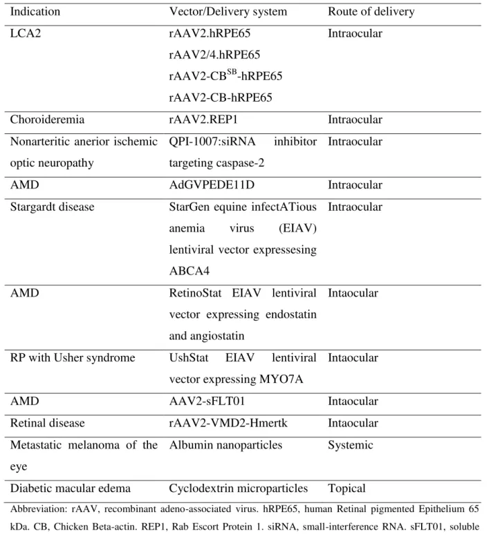
Table 4. Vectors used in clinical trial for various retinal diseases. (Adapted from: Rowe-Rendleman, et al.2014)
Indication Vector/Delivery system Route of delivery
LCA2 rAAV2.hRPE65
Stargardt disease StarGen equine infectATious anemia virus (EIAV)
lentiviral vector expressesing ABCA4
Intraocular
AMD RetinoStat EIAV lentiviral
vector expressing endostatin and angiostatin
Intaocular
RP with Usher syndrome UshStat EIAV lentiviral vector expressing MYO7A
Intaocular
AMD AAV2-sFLT01 Intaocular
Retinal disease rAAV2-VMD2-Hmertk Intaocular
Metastatic melanoma of the eye
Albumin nanoparticles Systemic
Diabetic macular edema Cyclodextrin microparticles Topical
Abbreviation: rAAV, recombinant adeno-associated virus. hRPE65, human Retinal pigmented Epithelium 65 kDa. CB, Chicken Beta-actin. REP1, Rab Escort Protein 1. siRNA, small-interference RNA. sFLT01, soluble Fms-like tyrosine kinase. MYO7A, Myosin VIIA. VMD2, Vitelliform Macular Dystrophy 2. hMERTK, human c-mer Proto-Oncogen Tyrosine Kinase. AMD, Age-related Macular Degeneration. RP, retinitis pigmentosa.
12
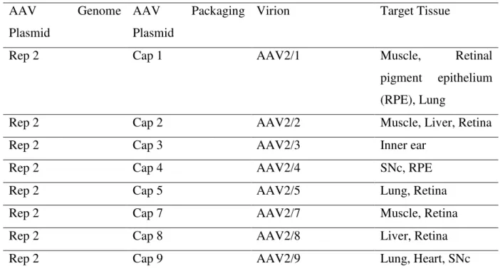
and packing them separately in two different AAV (dual AAV vectors). Dual AAV vectors can be occurred as AAV has the ability to form intermolecular concatemers in the nuclei of targeted cells.31,36
Table 5. AAV serotype and target tissue. (Adapted from: Surace , 2008).35
AAV Genome
Abbreviations: Rep, Replication. Cap, Capsid. AAV packaging plasmid can be used to target different organs.
2.2.1.3 Nanoparticles
13
2.2.2.1 AON Sequences
Theoretically, AON sequences are specifically designed to induce a biological effect, such as interfering expression of interest gene via RNase H-dependent mechanism or manipulating aberrant transcript caused by splicing mutation. Basically, two groups of AON are known: RNase H-dependent oligonucleotides, which degrade mRNA, and sterick-blocker
oligonucleotides, which interfere with the splicing machinery. Both of them need to be specific, but the action mechanism of AON itself is complex and poorly understood. Several predictive tools have been established, mostly based on the secondary structures of local sequences and thermodynamic properties of AONs. One of them is ESEfinder (http://rulai.cshl.edu/cgibin/tools/ESE3/esefinder.cgi?process=home), which has been used in determining AON sequences as therapeutic approach for CEP290-associated LCA.39,40
2.2.2.2 Chemical Structure of AON
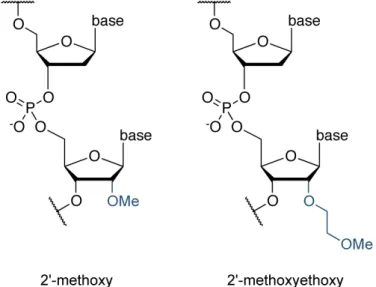
Phosphorothioate is the most widely used antisense oligonucleotide backbone, which is more soluble than methylphosphonates, the first synthesized oligonucleotides backbones. Modification are also important in the 2’-position of ribose by an O-alkyl group to enhance the stability of AONs inside the cell without decreasing the potency, such as 2’-methoxy (2’ -OMe) and 2’-methoxyethoxy (2’-MOE) (Figure 4). The stability of these modifications probably due to the structural protection provided by the alkyl group.40However, convincing differences between these two chemical modifications are poorly understood yet.41
Figure 4. Chemical structure of AON. Adapted from: