www.elsevier.com/locate/ibmb
Cloning and characterization of a chitin synthase cDNA from the
mosquito Aedes aegypti
Ghada H. Ibrahim, Chelsea T. Smartt, Lynn M. Kiley, Bruce M. Christensen
*Department of Animal Health and Biomedical Sciences, 1656 Linden Drive, University of Wisconsin-Madison, Madison, WI 53706, USA
Received 2 February 2000; received in revised form 5 May 2000; accepted 8 May 2000
Abstract
Characterization of the enzymes involved in the chitin biosynthetic pathway in mosquitoes is critical due to the importance of chitin in the formation of the peritrophic matrix [PM] and its potential impact on vector competence. Chitin is the homopolymer of the amino sugar N-acetyl-D glucosamine [GlcNAc]. The final step of incorporation of GlcNAc into the chitin polymer is catalyzed by the enzyme chitin synthase [CS]. CS is a membrane bound enzyme, but the mechanism of its action in the biosynthesis of the PM is not understood. We have isolated and sequenced a CS-encoding cDNA clone from the mosquito Aedes aegypti, compared its sequence with CS from other organisms and studied its RNA expression. The cDNA is 3.5 kb in length with an open reading frame of 2.6 kb that encodes a protein of 865 amino acids with a predicted molecular mass of 99.5 kDa. The putative translation product shares 90% similarity to two CS proteins from Caenorhabditis elegans and 50% similarity to Saccharomyces cerevisiae in the catalytic domain of CS enzymes. Data suggest that CS is a single copy gene. RT-PCR analysis shows CS message in whole non-blood-fed females, whole blood-fed females, non-blood-fed midguts and in midguts dissected at different time points post-blood-feeding. In situ hybridization studies of midgut samples revealed that CS mRNA increases following a bloodmeal and is localized to the periphery of the epithelial cells facing the midgut lumen.2000 Elsevier Science Ltd. All rights reserved.
Keywords: Chitin synthase; Peritrophic matrix; Aedes aegypti
1. Introduction
All mosquito-borne pathogens initiate infection by being ingested with the blood meal, and the first barrier they face is the midgut. It has been established that the midgut plays a role in influencing vector competence (Ponnudurai et al., 1988; Sutherland et al., 1986; Ramas-amy et al., 1997), but there is little information regarding the genetic regulation of critical physiological processes that occur within the midgut following blood feeding. One of these processes is the formation of the peritrophic matrix that surrounds the blood bolus, thereby physically separating it from the midgut epithelium. It functions in mechanical protection of the midgut epithelium by pro-viding a barrier to ingested food (Sudah and Muthu, 1988), protects the insect from insult by toxins and
* Corresponding author. Tel.:+1-608-262-3850; fax: +1-608-262-7420.
E-mail address: [email protected] (B.M. Christensen).
0965-1748/00/$ - see front matter2000 Elsevier Science Ltd. All rights reserved. PII: S 0 9 6 5 - 1 7 4 8 ( 0 0 ) 0 0 1 0 0 - 4
pathogens (Sutherland et al., 1986), and might serve as a retention barrier for keeping protease inhibitors in the lumen (Billingsley, 1990).
provided evidence that chitin of Mucor rouxii is the main, if not sole, polymer biosynthesized from UDP-GlcNAc.
Chitin synthase is readily assayed in many of the fun-gal systems, but the same procedure has met with failure in insect systems. This may be due to the lack of an in vitro CS assay in a cell-free preparation (Vardanis, 1979), owing to the instability of the enzyme. Except for a few enzymatic studies (Cohen and Casida, 1980; Mayer et al., 1980), understanding the biochemistry of CS has not been an easy task, probably due in part to the difficulty inherent in purification (Machida and Saito, 1993). The only characteristic that is common to all chi-tin-containing organisms is that chitin synthase is mem-brane-bound; other properties vary extensively among species (Chen, 1987).
It has been suggested that CS forms PM microfibrils from secreted precursors (Richards and Richards, 1977), but available evidence does not support this (Lehane, 1997). The active site of chitin synthase faces the cyto-plasm (Sentandreu et al., 1984), and because there is no evidence of an active transport system for the chitin pre-cursor UDP-GlcNAc, the belief is that chitin polymers are synthesized from an intracellular pool of precursors and then extruded through the plasma membrane by an unknown mechanism (Cohen, 1991).
Insect CS has been shown, through biochemical stud-ies, to exist in a zymogenic form requiring trypsin pro-teolysis for activation and chitin biosynthesis (Mayer et al., 1980). Chitin is degraded by hydrolytic action of the enzyme chitinase found in both the mosquito host as well as in various pathogens (Shahabuddin et al., 1996). It has been proposed by De La Vega et al. (1998) that both enzymes work in concert to control cuticle syn-thesis [CS] and cuticle degradation [chitinases] through alternate phases of enzyme production by epithelial cells. Studies on midgut specific chitinases in Anopheles
gam-biae (Shen and Jacobs-Lorena, 1997) show that, upon
feeding, the enzyme is secreted as an inactive proen-zyme, and is later activated by trypsin. Therefore, tem-poral regulation of both CS and chitinase activity are coupled to that of trypsin. When allosamidin, a specific inhibitor of various chitinases, is present in addition to the established activators trypsin and GlcNAc, chitin for-mation is increased up to 58-fold over the basic synthesis rate (Peter and Schweikart, 1990).
In this paper we report the first molecular cloning of a putative CS from the mosquito Aedes aegypti, with sequence comparisons made to known CS proteins from other organisms. Also included are RT-PCR and in situ hybridization studies that assess timing and location of CS-like transcripts in the mosquito following blood feed-ing.
2. Materials and methods
2.1. Mosquito maintenance
Adult Liverpool strain Aedes aegypti were reared according to the methods described by Christensen and Sutherland (1984). Artificial blood meals required for RNA expression analysis were prepared according to Kogan (1990) with minor modifications. The meal con-tained a final concentration of 15 mg/ml of γ-globulin, 90 mg/ml of albumin and 0.03 ml of 0.2 M ATP in a final volume of 3 ml. Hemoglobin was omitted from the blood meal due to interference with in situ analysis. Mosquitoes were fed through a water-jacketed mem-brane feeder (Rutledge et al., 1964). All blood meal components were supplied by Sigma Chemical Com-pany (St. Louis, MO, USA).
2.2. Isolation of CS cDNA clone
The Ae. aegypti CS cDNA clone was isolated from a
λgt10 cDNA library, containing Ae. aegypti size selected Poly (A)+ RNA from 3-day-old females (J. Williams, University of Wisconsin, Madison, WI, USA), using a CS probe generated by PCR amplification. Primers used to amplify the CS cDNA were designed, based on sequence homology of the most highly conserved amino acid residues of the catalytic domain of different forms of yeast and fungal CSs. They are: 59 CAG AAA TTC GAA TAT GCC 39and 59CCA GCG GCG GCT TTG 39. The PCR conditions used were one cycle at 94°C for 4 minutes; 30 cycles at 94°C, 55°C and 72°C for one minute each and one 7 minute extension at 72°C.
Library screening was carried out at moderate strin-gency (16 h hybridization at 55°C) as described by Man-iatis et al. (1982). An Ae. aegypti CS-encoding PCR pro-duct radioactively labeled by random priming using a Random Primed DNA Labeling Kit (Boehringer-Mannheim, Indianapolis, IN, USA) was used as probe. Positive phage clones were identified, DNA isolated (Ferdig et al., 1996), subcloned into pBSSK II (Stratagene, La Jolla, CA, USA), sequenced and analyz-ed.
2.3. Sequence analysis
DNASTAR (Madison, WI, USA). Amino acid sequence comparisons were made using the NCBI database search using BLAST (Altschul et al., 1990). Multiple alignment was generated using the pileup program in the GCG pro-gram (Wisconsin Package Version 10.0, Genetics Com-puter Group (GCG), Madison, WI).
2.4. Southern blot analyses
DNA was isolated according to the methods of Sever-son et al. (1993). Genomic DNA from both individual mosquitoes and bulk preparations were subject to EcoRI restriction digestion and size fractionated on 1% agarose gels, denatured (150 mM NaOH, 3 mM EDTA), neu-tralized (150 mM NaPO4, pH 7.8) and transferred onto Hybond-N+ Nylon membranes (Amersham Pharmacia Biotech, Inc., Piscataway, NJ, USA) by capillary action in 25 mM Na4P2O7. Southern blots were hybridized with an Ae. aegypti CS-encoding PCR product radioactively labeled by random priming and washed at high strin-gency (Maniatis et al., 1982).
2.5. RNA expression analysis by reverse transcription — PCR (RT-PCR)
Midgut tissues for RNA extraction were dissected in
Aedes saline solution (Hayes, 1953) and immediately
frozen on dry ice. Total RNA was isolated according to the methods described by Chomczynski and Sacchi (1987). RNA was extracted from all developmental stages of Ae. aegypti, and from whole females and dis-sected midguts at different time points following blood feeding. Samples were DNase-treated for DNA contami-nation. RNA was subject to first strand cDNA synthesis using either an oligo dT primer or a CS specific primer as described by Sambrook et al. (1989). The primers used were: 59CAA CGT CGA CGC TGG G 39and 59 CAT GAG GAA GAT CGT TCC 39(Fig. 1). The PCR reaction was carried out in a Rapid Cycler (Idaho Tech-nology, Idaho Falls, ID, USA) in a 10 µl final volume. The program profile was: 30 cycles comprised of a dena-turation step at 98°C for 10 s, annealing at 50°C for 10 s and extension at 72°C for 35 s. The products of these reactions were size-fractionated on a 1% agarose gel and examined using an EAGLE EYE II (Stratagene, La Jolla, CA, USA).
2.6. Whole mount in situ hybridization using digoxigenin-labeled RNA probes
Probes (sense and anti-sense) were transcribed from the CS cDNA clone using a Digoxigenin labeling kit (Boehringer-Mannheim, Indianapolis, IN) according to the manufacturer’s instructions. Full-length digoxigenin-labeled RNA probes were transcribed from T3 (for sense probe) and T7 (for anti-sense probe) promoters,
precipi-tated in ethanol, and resuspended in diethyl pyrocarbon-ate-treated water. Quantification of the probes was according to the instructions within the labeling kit. Mosquito midguts at different time points after blood feeding were dissected into ice-cold phosphate buffered saline (PBS), fixed with 4% paraformaldehyde in PBS (PFP) at 4°C overnight, dehydrated through a methanol series at room temperature, and stored in 100% methanol at 220°C until hybridization was performed. Prior to hybridization, samples were rehydrated through a meth-anol series, treated with proteinase K, post-fixed with 4% PFP/0.2% glutaraldehyde at room temperature for 20 min, and then pre-hybridized at 55°C for 1 h in hybridiz-ation solution (50% deionized Formamide, 5×SSC, 0.02% glycogen, 0.1 mg/ml sheared and denatured sal-mon sperm DNA, and 0.1% Tween 20). RNA probes then were added to the hybridization solution at a final concentration of 1 ng/µl and allowed to hybridize over-night at 55°C with intermittent agitation. Samples were washed four times for half an hour each in hybridization solution at 55°C, followed by a 1:1 hybridization solution/PBT wash at room temperature and finally washed in PBT.
least seven times, simultaneously under identical con-ditions.
3. Results
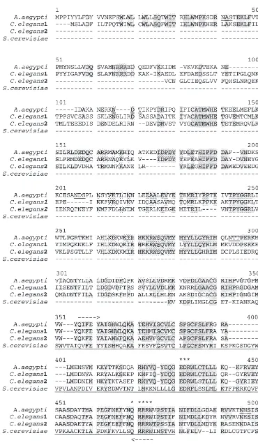
3.1. Analysis and comparison of Ae. aegypti C11A2b deduced amino acid sequence with known CS proteins from other organisms
The Ae. aegypti cDNA encodes a putative CS protein of 865 amino acids with a predicted molecular mass of 99.5 kDa. The Ae. aegypti CS deduced amino acid sequence was compared with CS proteins from nema-tode and yeast (Fig. 1). A conserved region of approxi-mately 130 amino acids between all CSs represents the catalytic site of the enzyme (Nagahashi et al., 1995) (Fig. 1). The Ae. aegypti potential coding region shares 90% similarity to two hypothetical CS proteins from
Caenor-habditis elegans (C. elegans1, Accession number
T32452, Bradshaw, H.; C. elegans2, Accession number T25284, McMurray, A.) and 50% similarity to CS of
Saccharomyces cerevisiae (S. cerevisiae, Accession
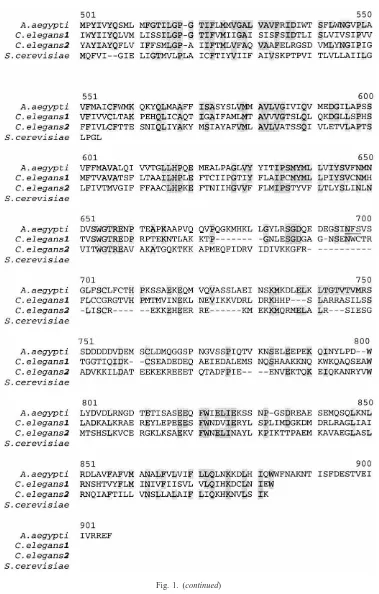
number AAA34844, CSD2, Bulawa, 1992) in the cata-lytic domain of CS enzymes. Additionally, sequence comparisons identified conserved residues in the cata-lytic domains. The N- and C-termini of Ae. aegypti CS-like protein and yeast CS are less conserved. Consistent with the fact that CS is membrane bound, the C-terminus of Aedes CS shows hydrophobic regions that are poten-tial membrane spanning domains (Fig. 2). Potenpoten-tial
Fig. 2. Hydrophobicity plot of Aedes aegypti CS obtained by a program developed by Han and Tashjian (1998). Alpha helicity index (Hα): Values above 1.6 are considered hydrophobic, and those below 0.8 are hydrophilic. Horizontal lines indicate stretches of more than 17 hydrophobic residues, denoting potential membrane-spanning domains.
trans-membrane positions include amino acids 473–500, 530–556 and 810–835 (Kihara et al., 1998).
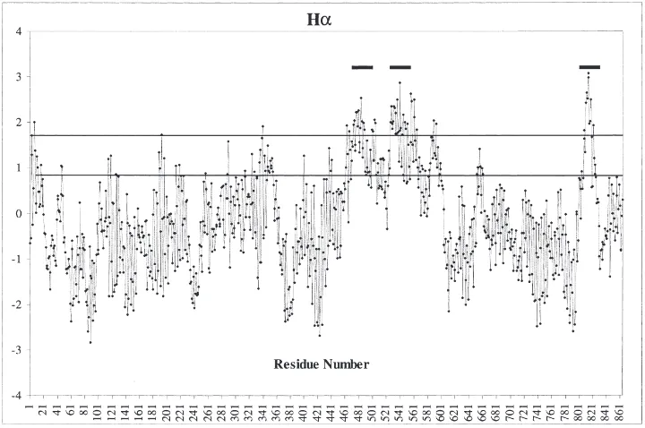
3.2. Southern blot analysis
Southern analyses of EcoRI restriction digests show that the CS PCR fragment hybridized to one or two gen-omic DNA fragments on an Ae. aegypti single animal blot (Fig. 3(A)). The hybridization pattern on the auto-radiograph of a Southern blot of pooled DNA samples from different strains of mosquitoes also shows three or fewer hybridizing bands. This suggests that CS is a sin-gle copy gene (Fig. 3(B)). The probe used on the South-ern blots was the 300 bp PCR fragment of CS cDNA. This DNA fragment contains an EcoRI site.
3.3. Analysis of CS mRNA by RT-PCR
Fig. 3. Autoradiographs of Southern blots of (A) Single animal DNA of Liverpool strain of Ae. aegypti and (B) pooled DNA samples from different strains of Aedes aegypti and Armigeres sabulbatus digested with EcoRI and hybridized with a chitin synthase PCR fragment. Strains=Bronze (A), Formosus (B), Gambiae (C), Hamburg (D), Liver-pool (E), Mini (F), Moyo-in-dry (G), Nagasaki (H), Red (I), Rocke-feller (J), and Silver (K) for Ae. aegypti and Malaysia (L) and Japan (M) for Ar. sabulbatus. Size (kb) as determined by32P-labeledλ-Hind
III DNA marker (Ma) is presented on the left.
3.4. In situ analysis
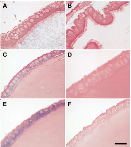
To localize CS mRNA, microscopic examination was carried out on midgut tissue sections hybridized with CS ribo-probes (Fig. 5). Midgut samples dissected from 3 h and 6 h post-blood-fed mosquitoes and hybridized with the anti-sense riboprobe showed localization to the per-iphery of the epithelial cells facing the midgut lumen (Fig. 5(C) and (E)). Staining in 6 h midguts (Fig. 5(E)) was more intense than in 3 h midguts (Fig. 5(C)). Non-blood-fed midguts showed faint staining (Fig. 5(A)). It is evident that blood feeding increased the amount of CS transcripts. Control samples hybridized with the sense ribo-probe showed no staining (Fig. 5(B), (D) and (F)).
Fig. 4. Transcription of chitin synthase RNA was analyzed using the cDNA samples generated from Ae. aegypti RNA by reverse transcrip-tion, and subject to RT-PCR using Ae. aegypti specific primers. CS expression profiles of: (A) Samples from dissected non-blood-fed midguts and from midguts dissected at different hours after blood feed-ing. The numbers at the top represent hours after blood-feedfeed-ing. (B) Samples from whole female mosquitoes collected at different hours after blood feeding as well as a sample of mosquito carcass (blood fed female minus midgut). Ma=123 bp DNA Marker, C=3 h carcass, Nbf=non-blood-fed females, Nbfm=non-blood-fed midguts, N=Neg-ative control (minus cDNA), P=Positive control (phage cDNA template).
4. Discussion
The PM represents a significant barrier to penetration and subsequent development of Plasmodium sp. (Billingsley and Rudin, 1992) and certain arboviruses (Whitfield et al., 1973). The effectiveness of this barrier depends on the time of its formation (Smartt et al., 1998). Due to the importance of chitin in the formation of the peritrophic matrix and its potential impact on vec-tor competence, chitin synthesis poses interesting and challenging questions. The multi-enzyme system that works to control the biosynthesis [chitin synthase] and degradation [chitinase] of chitin is likely complex. It is evident that both enzymes are zymogenic (Mayer et al., 1980; Shahabuddin et al., 1996) requiring proteolysis by trypsin for activation. Trypsin has been shown to con-tribute 75% of the proteolytic activity in midguts of Ae.
chi-Fig. 5. In situ hybridization of midgut sections showing the distribution of chitin synthase mRNA. (A): Non-blood-fed midgut sample probed with anti-sense (experimental), and (B): probed with sense riboprobe (negative control). Midgut dissected 3 h (C) and 6 h (E) after blood feeding and hybridized with an anti-sense riboprobe. Midgut dissected 3 h (D) and 6 h (F) after blood feeding and hybridized with sense riboprobe. Bar=25µm.
tinase acts to modulate PM thickness and permeability (Shen and Jacobs-Lorena, 1997). In support of this hypothesis, and in agreement with that of Shahabuddin et al. (1993), Shen and Jacobs-Lorena (1997) found that the PMs are stronger and persist longer in the guts of mosquitoes fed on a chitinase inhibitor. A thicker PM may be a more efficient barrier to parasite development in the mosquito, if induced in a temporally dependent fashion.
In the present study, we isolated the first mosquito
also are completely conserved in other proteins pos-sessing N-acetylglucosaminyltransferase activity, such as NodC proteins of Rhizobium bacteria, it has been sug-gested that they are located in the active pocket of the enzyme. Nagahashi et al. (1995) concluded, through mutant analysis, that these two catalytic domains are essential for catalytic activity and that residues corre-sponding in Ae. aegypti to Asp432, Gln470, Arg473 and Trp474 are the potential catalytic residues of the enzyme.
The presence of potential membrane-spanning domains in the carboxy terminal region of the Ae.
aegypti protein is consistent with the known membrane
localization of the enzyme. Analysis of the Ae. aegypti CS structural hydrophobicity plot (Han and Tashjian, 1998) reveals 3 potential transmembrane regions. Hydro-phobicity values calculated according to Kyte–Doolittle analysis in the GCG program (Wisconsin Package Ver-sion 10.0, Genetics Computer group (GCG), Madison, WI) also revealed these same three potential membrane-spanning regions (data not shown). The location of CS in the plasma membrane suggests that the enzyme could be a glycoprotein (Duran et al., 1975). The CS reported herein possesses 6 potential glycosylation sites in the coding sequence at amino acids 41 (NAS), 101 (NDT), 186 (NDS), 273 (NTT), 459 (NNS) AND 658 (NFS). Biochemical Con A-sepharose binding experiments in S.
cerevisiae negate the possibility that the enzyme is
gly-cosylated (Bulawa et al., 1986); however, whether Ae.
aegypti CS is glycosylated remains to be determined.
No structural similarities exist in the amino-termini of known S. cerevisiae CSs. Because this region is dispens-able for enzyme activity, it has been suggested (Silverman, 1989) that it may play a role in regulation or localization of respective enzymes. Three CS genes from S. cerevisiae lack a conventional amino terminal signal sequence and have been suggested to have a spe-cific transport system independent of the secretory path-way (Bulawa, 1992). Comparisons also show that the N-terminal region of Ae. aegypti CS is dissimilar to other CSs and seems to lack an N-terminal signal peptide. Membrane topology predictions indicate that the N-ter-minal region of the Aedes CS is oriented inside, facing the cytoplasm (PSORT prediction program- [Nakai and Horton, 1999]). We suggest that the Ae. aegypti cDNA clone encodes a chitin synthase.
Results of RT-PCR carried out on midguts showed that CS transcript was present in non-blood-fed samples and through 72 h post blood feeding, with a reduction of message seen at 96 h after a blood meal. Both non-blood-fed female, as well as non-non-blood-fed midgut samples, contained CS message; however female car-casses with the midgut removed showed little CS tran-script. CS RNA also could be detected in all develop-mental stages of the mosquito (data not shown). We propose that Ae. aegypti CS is midgut specific and
involved in midgut chitin formation. However, we believe there may be another gene for cuticular chitin synthase. We analyzed transcription of Ae. aegypti CS using RT-PCR because it is a more sensitive technique for detection of messages of low abundance.
In situ CS mRNA localization in midgut samples clearly showed low levels in non-blood-fed midguts and an increase in transcript following blood feeding. CS mRNA in midgut samples was distributed at the periph-ery of the epithelial cells facing the midgut lumen. Pre-liminary data suggest that Ae. aegypti chitin synthase is a single gene, however, as with yeast CS, the possibility remains that there may be a number of enzymes each encoded by a separate gene and each having a specific site of localization and chitin deposition (Robbins et al., 1993).
Acknowledgements
We thank J. Williams for providing the Aedes aegypti cDNA library and P. Robbins for a useful collaboration. We also thank L. Christensen for assistance with mos-quito rearing and J. Hillyer for image processing. This research was supported by grant AI44461-02 from the National Institutes of Health to B.M.C. The Ae. aegypti chitin synthase gene has been given the GenBank accession number AF223577.
References
Altschul, S.F., Gish, W., Miller, W., Myers, E., Lipman, D., 1990. Basic local alignment search tool. J. Mol. Biol. 215, 403–410. Billingsley, P.F., 1990. The midgut ultrastructure of hematophagus
insects. Annu. Rev. Entomol. 35, 219–248.
Billingsley, P.F., Rudin, W., 1992. The role of the mosquito peritrophic membrane in bloodmeal digestion and infectivity of Plasmodium species. J. Parasitol. 78, 430–440.
Briegel, H., Lea, A.O., 1975. Relationship between protein and proteo-lytic activity in the midgut of mosquitoes. J. Insect. Physiol. 9, 1597–1604.
Bulawa, C.E., 1992. CSD2, CSD3, and CSD4, genes required for chitin synthesis in Saccharomyces cerevisiae: the CSD2 gene product is related to chitin synthases and to the developmentally regulated proteins in Rhizobium species and Xenopus laevis. Mol. Cell. Biol. 12, 1764–1776.
Bulawa, C.E., 1993. Genetics and molecular biology of chitin synthesis in fungi. Annu. Rev. Microbiol. 47, 505–534.
Bulawa, C.E., Slater, M., Cabib, E., Au-Young, J., Sburlati, A., Lee Adair, W. Jr., Robbins, P.W., 1986. The S. cerevisiae structural gene for chitin synthase is not required for chitin synthesis in vivo. Cell 46, 213–225.
Candy, D.J., Kilby, B.A., 1962. Studies on chitin synthesis in the desert locust. J. Exp. Biol. 39, 129–140.
Chen, A.C., 1987. Chitin metabolism. Archives of Insect Biochemistry and Physiology 6, 267–277.
Christensen, B.M., Sutherland, D.R., 1984. Brugia pahangi: exsheath-ment and midgut penetration in Aedes aegypti. Trans. Am. Micros. Soc. 103, 423–433.
Cohen, E., 1991. Chitin biochemistry. In: Binnington, K., Retnakaran, A. (Eds.) Physiology of the Insect Epidermis. CSIRO Publishers, Melbourne, pp. 94–112.
Cohen, E., Casida, J.E., 1980. Properties of Tribolium gut chitin syn-thetase. Pestic. Biochem. Physiol. 13, 121–128.
De La Vega, H., Specht, C.A., Liu, Y., Robbins, P.W., 1998. Chitin-ases are a multi-gene family in Aedes, Anopheles and Drosophila. Insect Mol. Biol. 7, 233–239.
Duran, A., Bowers, B., Cabib, E., 1975. Chitin synthetase zymogen is attached to the yeast plasma membrane. Proc. Natl. Acad. Sci. 72, 3952–3955.
Ferdig, M.T., Li, J., Severson, D.W., Christensen, B.M., 1996. Mos-quito dopa decarboxylase cDNA characterization and blood-meal-induced ovarian expression. Insect Mol. Biol. 5, 119–126. Han, B., Tashjian, A.H., 1998. User-friendly and versatile software for
analysis of protein hydrophobicity. Biotechnique 25, 256–263. Hayes, R.O., 1953. Determination of a physiological saline for Aedes
aegypti (L). J. Eco. Entomol. 46, 624–627.
Kihara, D., Shimizu, T., Kanehisa, M., 1998. Prediction of membrane proteins based on classification of transmembrane segments. Pro-tein Engineering 11, 961–970.
Kogan, P.H., 1990. Substitute blood meal for investigating and main-taining Aedes aegypti (Diptera; Culicidae). J. Med. Entomol. 27, 709–712.
Lehane, M.J., 1997. Peritrophic matrix structure and function. Annu. Rev. Entomol. 42, 525–550.
Machida, S., Saito, M., 1993. Purification and characterization of mem-brane-bound chitin synthase. J. Biol. Chem. 268, 1702–1707. Maniatis, T., Frisch, E.F., Sambrook, J., 1982. Molecular Cloning: A
Laboratory Manual, 2nd ed. Cold Spring Harbor Laboratory, New York.
Mayer, R.T., Chen, A.C., DeLoach, J.R., 1980. Characterization of a chitin synthase from the stable fly, Stomoxys calcitrans (L.). Insect Biochem. 10, 549–556.
McCurrough, I., Flores-Carreon, A., Bartnicki-Garcia, S., 1971. Path-way of chitin synthesis and cellular localization of chitin synthetase in Mucor rouxii. J. Biol. Chem. 246, 3999–4007.
Nagahashi, S., Sudoh, M., Ono, N., Sawada, R., Yamaguchi, E., Uch-ida, Y., Mio, T., Takagi, M., Arisawa, M., Yamada-Okabe, H., 1995. Characterization of chitin synthase 2 of Saccharomyces
cere-visiae. Implication of two highly conserved domains as possible
catalytic sites. J. Biol. Chem. 270, 13961–13967.
Nakai, K., Horton, P., 1999. PSORT: a program for detecting the sort-ing signals of proteins and predictsort-ing their subcellular localization. Trends Biochem. Sci. 24, 34–35.
Peter, M.G., Schweikart, F., 1990. Chitin biosynthesis enhancement by the endochitinase inhibitor allosamidin. Biol. Chem. Hoppe Seyler 371, 471–473.
Ponnudurai, T., Billingsley, P.F., Rudin, W., 1988. Differential infec-tivity of Plasmodium for mosquitoes. Parasitol. Today 4, 319–321.
Ramasamy, M.S., Kulasekara, R., Wanniarachchi, I.C., Srikrishnaraj, A., Ramasamy, R., 1997. Interactions of human malaria parasites,
Plasmodium vivax and Plasmodium falciparum, with the midgut of Anopheles mosquitoes. Med. Vet. Entomol. 11, 290–296.
Richards, A.G., Richards, P.A., 1977. The peritrophic membrane of insects. Annu. Rev. Entomol. 22, 219–240.
Robbins, P.W., Bowen, A.R., Chen-Wu, J.L., Momany, M., Szaniszlo, P.J., Zwicker, J., 1993. The multiple chitin synthase genes of
Can-dida albicans and other pathogenic fungi — A review. In: Vanden
Bossche, H. et al. (Eds.), Dimorphic fungi in Biology and Medi-cine. Plenum Press, New York, pp. 51–59.
Rutledge, L.C., Ward, R.A., Gould, D.J., 1964. Studies on the feeding response of mosquitoes to nutritive solutions in a new membrane feeder. Mosquito News 24, 407–419.
Sambrook, J., Fritsh, E.F., Maniatis, T., 1989. Molecular Cloning: A Laboratory Manual, 2nd ed. Cold Spring Harbor Laboratory Press, New York.
Sentandreu, R., Martinez-Ramon, A., Ruiz-Herrera, J., 1984. Localiz-ation of chitin synthase in Mucor rouxii by an autoradiographic method. J. Gen. Microbiol. 130, 1193–1199.
Severson, D.W., Mori, A., Zhang, Y., Christensen, B.M., 1993. Link-age map for Aedes aegypti using restriction fragment length poly-morphisms. J. Hered. 84, 241–247.
Shahabuddin, M., Toyoshima, T., Aikawa, M., Kaslow, D.C., 1993. Transmission-blocking activity of a chitinase inhibitor and acti-vation of malarial parasite chitinase by mosquito protease. Proc. Natl. Acad. Sci. 90, 4266–4270.
Shahabuddin, M., Lemos, F.J., Kaslow, D.C., Jacobs-Lorena, M., 1996. Antibody mediated inhibition of Aedes aegypti midgut tryp-sins blocks sporogonic development of Plasmodium gallinaceum. Infect. Immunol. 64, 739–743.
Shen, Z., Jacobs-Lorena, M., 1997. Characterization of a novel gut-specific chitinase gene from the human malaria vector Anopheles
gambiae. J. Biol. Chem. 272, 28895–28900.
Silverman, S.J., 1989. Similar and different domains of chitin synth-ases 1 and 2 of S. cerevisiae: Two isozymes with distinct functions. Yeast 5, 459–467.
Smartt, C.T., Chiles, J., Lowenberger, C., Christensen, B.M., 1998. Biochemical analysis of a blood meal-induced Aedes aegypti gluta-mine synthetase gene. Insect Biochem. Mol. Biol. 28, 935–945. Sudah, P.M., Muthu, S.P., 1988. Damage to the mid-gut epithelium
caused by food in the absence of peritrophic membrane. Curr. Sci. 57, 624–625.
Sutherland, D.R., Christensen, B.M., Lasee, B.A., 1986. Midgut barrier as a possible factor in filarial worm vector competency in Aedes
trivittatus. J. Invertebr. Pathol. 47, 1–7.
Vardanis, A., 1979. Characteristics of the chitin-synthesizing system in insect tissue. Biochem. Biophys. Acta. 588, 142–147. Whitfield, S.G., Murphy, F.A., Sudia, W.D., 1973. St. Louis