UNILATERAL NEPHRECTOMY IN THE RAT
AS A TEACHING MODEL OF RENAL FUNCTION
Charles F. Zwemer
Depa rtm ent of Biology, Dick inson College, Ca rlisle, Pennsylva nia 17013
T
he white rat offers a valuable teaching model to demonstrate the in situ anatomy, homeostatic control, and function of the mammalian renal system. Students in the upper-level physiology class at Dickinson College are responsible for formulating a set of testable hypotheses as to the possible compensatory mechanisms that will result from a unilateral nephrectomy in the rat. To test their hypotheses, each group of students is made responsible for induction and maintenance of anesthesia, removal of the left kidney, closure, and recovery of the animal in their experiment. Before and 1 wk after nephrectomy, blood samples are collected via tail section for measurement of hematocrit and plasma creatinine concentrations. Pooling data across all student groups (n56) for pre- and postnephrectomy hematocrit and creatinine values allows students to run statistical comparisons testing their hypotheses. This laboratory experience in integra-tive physiology is a successful exercise in introducing fundamentals of experimental design and, most importantly, provides students with an appreciation for the process of humane use of animals in research and teaching.AM. J. PHYSIOL. 276 (ADV. PHYSIOL. EDUC. 21): S79–S85, 1999.
Key words:renal physiology experiments; kidney; creatinine; education
Animal research-based laboratory inquiry greatly en-hances the undergraduate physiology experience (1, 7, 10). At Dickinson College, we use a model of unilateral nephrectomy and recovery in the white rat to introduce students to fundamentals of experimen-tal design, hypothesis testing, and homeostatic func-tion of the mammalian renal system. The experiment is based, in part, on a research model popularized by Podrazik et al. (5, 6) and a model used in our research laboratory. The experiment covers two laboratory class periods in the upper-level undergraduate physiol-ogy class of 24 students. Introduced in the fall of 1995 as a nonrecovery observational study, the original experience has been expanded to a hypothesis-based recovery experiment. Although many schools of our size (2,000-student undergraduate liberal arts school) currently eschew the use of mammals in the teaching of physiology, I have found this exercise to be highly
successful at many didactic levels. This important point is argued eloquently by Woodhull-McNeal in a previous issue of this Journal (10), and, as I will demonstrate, the experience at Dickinson College fulfills major goals identified as common to the teach-ing of physiology in the laboratory settteach-ing (10).
homeostasis and renal function, learning the fundamen-tals of experimental design, inferring of scientific principles from experimental data, experiencing the nature of living tissue, and learning about the humane care and use of animals in physiology research. Additional goals include learning to critically analyze data through the use of statistics and gaining experi-ence in the effective communication of scientific results.
METHODS
Gr oup discussion.My students and I typically cover renal physiology in the latter third of the semester and spend approximately five lectures on the subject. In lecture on the Friday before the nephrectomy exer-cise (physiology lab is scheduled for Monday after-noons from 1:00 to 6:00 PM), I pose a question to the students about their impressions regarding renal func-tion and fluid and electrolyte homeostasis. Specifi-cally, I ask them whether normal renal function can be maintained when one kidney is removed. Because I ask this question on the first or second day of renal physiology, most students respond that the remaining kidney will1) not compensate or2) weakly compen-sate to maintain the body in an abnormal fluid and electrolyte balance at a new steady state. I allow a consensus to build and then ask them to articulate their hypothesis on the chalkboard.
With the hypothesis established, I engage the students in a discussion of experimental design. Our discussion first involves reliable assessment of renal function. This discussion provides a perfect opportunity to reinforce the concepts of renal clearance and glomeru-lar filtration rate and how to assess these functions using creatinine concentrations from the plasma and urine. We also discuss following plasma volume as a possible assessment of outcome. With outcome vari-ables and assessments determined and written on the board, we design the actual experiment by identifying the animals to be used and establishing a set of control or prenephrectomy assessments and a set of postne-phrectomy assessments. Additionally, I introduce the students to the need to establish a set of animals used as sham controls to identify any possible confounding influences from the operative preparation. The out-come variables that we measure are animal mass, plasma creatinine concentration (an indicator of renal
function), and hematocrit (an indicator of relative plasma volume). Although it would be even more effective, the determination of pre- and postnephrec-tomy creatinine clearance values was ruled out be-cause of the difficulty and unreliability of urine collec-tion procedures. Closing the lecture, we discuss issues of humane animal use and care in research and the details of the College’s Institutional Animal Care and Use Committee approval for our experiment (ap-proval no. B9706). As the students leave the class-room, they pick up a handout describing anesthetic choice, mechanism, dose, route, depth, and mainte-nance to be used during the procedure and a detailed description of the mechanics of the protocol. This handout is to be studied before the laboratory section on Monday afternoon.
The nephrectomy experiment is the last of three animal research experiments the students encounter over the semester (the other 9 laboratories rely on the students exploring principles on themselves) and is, by far, the most intensive because it involves recovery operative procedures. All students in my classes are well aware of their responsibility for the humane treatment of the rats, and they work in groups of four around one animal. I have reduced the number of animals used in this experiment from a total of 12 (students working in groups of 2) to 6 (students working in groups of 4) in an attempt to maximize the experience for students while using a minimum num-ber of animals. Some students have argued that the experience would be more meaningful if they worked in smaller groups but respond positively to the ratio-nale that the experience is essentially the same in the larger group and less animal intensive.
After this 30- to 40-min discussion, two students from each team prepare the operative area of their bench by cleaning it with bleach solution. They next place a cleaned heating pad, set on low, onto this clean area. Over the heating pad they place a clean absorbent drape, and on top of this they place a bleach-cleaned rodent operating board (Plas-Labs, Lansing, MI). The operative field is then illuminated using a dual goose-neck high-intensity fiber-optic light source (Dolan-Jenner Industries, Lawrence, MA).
While two students assemble the operative area, the other two students collect a male rat (Sprague-Dawley, Taconic Farms, Germantown, NY) from our colony, weigh it, and calculate an induction dose of injectable anesthesia for the animal (50 mg/kg ket-amine in combination with 5 mg/kg xylazine; Ref. 3). The rat is anesthetized in a covered induction jar (halothane) and is promptly removed when it loses its righting reflex. I have added this halothane step because it keeps the rat calm and reduces the chance of biting. It also reduces the chance that the rat will experience discomfort from the ketamine-xylazine injection. Once the rat is lightly anesthetized with halothane, the students inject it with ketamine-xylazine anesthesia using a dorsal subcutaneous injec-tion. The animal is placed into a cage, and the students wait until the rat is fully anesthetized. The students also draw a quarter-dose (25% of induction dose) of anesthesia for use intraoperatively, if needed, and a prophylactic dose of antibiotic (ampicillin, 10 mg/kg). An important note here is that the rats used in the experiment have had food but not water withheld (NPO) for an 18-h period before the procedure to equalize their nutritional status.
Once the animal enters a state of surgical anesthesia, determined by absence of response to clamping pressure applied to the proximal tail, it is gently restrained to the operative board and given antibiotic. The rat’s tongue is then pulled to the side of the mouth to maintain a clear airway. The rat’s tail is drawn onto the absorbent towel, and the distal end is coated with iodophore solution (Purdue Frederick, Norwalk, CT) and allowed to dry. Once the tail is dry, a 0.5- to 1-cm piece (measured from tip of tail) is quickly amputated using a pair of sharp operating scissors. Approximately 0.5 ml of blood is collected from the tail for determination of hematocrit
(micro-centrifuge method) and for plasma creatinine measure-ments (whole blood is spun for 5 min at 10,000 rpm, and plasma is then pipetted into labeled Eppendorf tubes and frozen at240°C). Hemostasis is achieved at the wound by applying direct pressure and sterile 2-in. 3 2-in. gauze, and the wound is dressed for recovery.
Next, the students concentrate on removing the left kidney of the rat. The procedure begins with the preparation of the surgical field. The ventral fur is clipped from the xiphoid process to the subumbilical region using clippers (Oster Professional Products, McMinnville, TN), and the clean abdomen is prepared by applying two coats of iodophore solution, which are allowed to dry. The field is draped off using a fenestrated drape (Sterile Suture Tray, Busse Hospital Disposables, Hauppauge, NY), and a midline incision is made using a sterile no. 10 scalpel blade. The abdominal muscles are cut along the linea alba using a pair of blunt/sharp operating scissors to expose the contents of the abdomen. The muscle walls are retracted laterally using sterile 4–0 silk suture on a tapered needle and are taped distally to the absorbent towel. Once the abdomen is opened, the students take a visual tour of the contents of the abdomen and find the stomach, small bowel, large bowel, spleen, pan-creas, and mesenteric arteries and veins. The kidneys are then exposed by pulling gently on the viscera and delivering them onto sterile gauze outside of the abdomen and pushing them to the right side of the animal (modified Kocher maneuver; Ref. 2). The left kidney, its blood supply, and the ureter are now exposed. At this point, I have the students stop and watch urine being evacuated from the renal pelvis into the ureter and down to the bladder by peristalsis. This observation captivates the students and provides a superb demonstration of peristalsis. The renal blood supply is then identified and isolated with two liga-tures (artery and vein are both tied off together at the hilus of the kidney and at the aorta/inferior vena cava using sterile 4–0 silk ligature). The ureter is ligated at the renal hilus and ,1 cm distal to the hilus. The
placed away from the field in a small beaker of saline (0.9 N NaCl) for further dissection. The student teams (n 5 2) working on sham animals manipulate the renal vessels and ureter but do not ligate the vessels or remove the left kidney.
After removal or manipulation of the left kidney in the nephrectomy groups, the abdominal contents in all rats are gently replaced, and the laparotomy wound is closed in layers using sterile 3–0 chromic gut suture with a cutting needle (Ethicon, Somerville, NJ) for the abdominal muscles and 3–0 Ethilon monofilament nylon suture with a cutting needle for the skin. The wound is then cleansed with iodophore solution, allowed to dry, and dressed using sterile 2-in.32-in. gauze and tape. The rat is placed on its side in a warmed recovery cage with ad libitum access to food and water. All animals are recovered completely by the next morning and are then moved to their normal cages in the vivarium until the following week.
With their animals now recovering, the students complete a thorough gross examination of the har-vested kidney. This examination includes a longitudi-nal dissection (in a frontal plane) and identification of the cortex, medulla, renal pyramid, renal pelvis, renal artery and vein, and ureter.
Operative pr ocedur e: week 2.One week later, the rats are retrieved from the colony, reanesthetized as previously described, and have another 0.5 ml of whole blood removed from their tail. From this sample, hematocrit is determined, and plasma is removed for creatinine analysis. After the blood sam-ple is removed, the rats are allowed to recover.
Each student group then determines plasma creati-nine concentration in their control and 1 wk postne-phrectomy samples using a spectrophotometric assay (Sigma Diagnostic Kit 555, Sigma, St. Louis, MO), standard micropipettes (Finnpipette, Labsystems, Hel-sinki, Finland), and a spectrophotometer (Spectronic 20D1series, Spectronic Instruments, Rochester, NY) at their laboratory benches. After the data collection, the class reassembles in the classroom and pools their data for mass, hematocrit, and plasma creatinine concentrations. Using commercially available statisti-cal software and a projection system (Statview, Aba-cus Concepts, Berkeley, CA), the students and I
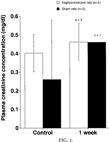
compare data between sham and experimental groups with pairwise comparisons and Student’s t-tests to compare mass hematocrit and plasma creatinine con-centrations (Fig. 1) at control versus 1 wk postnephrec-tomy time points. These data are then graphically presented using a commercially available software package (CA-Cricket Graph III, Computer Associates, Islandia, NY). A group discussion on the interpreta-tion of the data follows, concluding with an assign-ment for each student team in which they are asked to present the class data in the journal format of the Am erica n Journa l of Physiology.
DISCUSSION AND CONCLUSIONS
This laboratory experiment can be considered a cap-stone experience in the physiology class, and I con-sider the model to be highly successful on many fronts. As was mentioned in the introduction and as has been well articulated in previous articles in this Journal (1, 7, 10), successful whole animal physiology laboratories teach students at many different levels simultaneously (10). The experience described above
FIG. 1.
clearly meets this goal. From the outset, one of the primary purposes of the nephrectomy model is to provide hard evidence for the concept of homeosta-sis. In this 2-wk laboratory experience, homeostatic processes are demonstrated because mass, hemato-crit, and creatinine concentrations typically do not change significantly from prenephrectomy to 1 wk postnephrectomy or when compared with sham ani-mals. Figure 1 highlights the most recent creatinine data collected in the fall of 1998. These data show no significant changes in plasma creatinine between groups or within a group over time. The mortality rate for this class setting was higher than normal, and this outcome served to reduce the sample size in both groups. In looking at Fig. 1, it is possible that the more savvy students could ask whether they truly observed evidence of no change (homeostasis) or whether this was just the result of a cloaking effect of a small sample size. To address this possibility in the future, I plan to have students add their data to a larger running average of pre- and postnephrectomy values collected by students in previous years. This will serve to place their results in the scope of a larger sample population (different ages, masses, sources, and slightly different experimental design) and will reveal that with a larger sample size some differences may actually be seen. In comparing data from Fig. 1 with previous class creati-nine averages to date (n 5 14; average prenephrec-tomy creatinine 5 0.786 6 0.218 vs. postnephrec-tomy creatinine50.88660.317;P50.34), however, no significant difference is yet detected.
In our experience, the null hypothesis of no change between control and postnephrectomy values has been supported in all variables studied, and this provides a valuable lesson because it forces the students to reject their hypotheses on the basis of the data. In the class discussion after the statistical analy-sis, students are given a chance to reason as to the possible mechanisms of compensation. The discus-sion then moves beyond the classroom as students support their postulated mechanisms of increased renal blood flow, renal hypertrophy, and renal hyper-plasia of the remaining right kidney using the primary literature. Having each team submit the class results in the form of a research manuscript is critical to completing the experience because it forces the general discussion into a more succinct, clearly writ-ten form. The process also exposes the students to
writing as collaborators (the most common form of research writing), reinforces mechanics of scientific communication, and immerses them in the primary literature.
Beyond the demonstration of homeostasis and the inferring of scientific principles from the data, stu-dents also gain yet another exposure to the rudiments of statistical comparison and experimental design using live animals. Less tangible, but certainly no less important, students also gain experience in thinking and working as scientists and collaborators, build on skills of cooperation in intense settings, and strengthen a sense of accomplishment in learning. They also learn, or are exposed to, a great deal of technical information such as the mechanics of maintaining general surgical anesthesia and some of its idiosyncra-sies (no two animals require the same induction or maintenance dose), how to close a wound using suture and needles, and the rudiments of the Beer-Lambert law and the power of spectrophotometric technique.
missing a larger experience that many of us, who as professionals trained when animal-based experiments were commonplace, take for granted when we de-scribe the mechanics of how animals work. This experiment is successful from a rat survival standpoint and is associated with an operative mortality rate (usually caused by hemorrhage or overanesthetiza-tion) of 21% over the last 4 years. By students’ accounts, nothing I can say or write on a chalkboard or overhead projector can match this hands-on experi-ence.
In spite of its high popularity among students in my physiology classes, this laboratory experience has the potential to generate controversy among students who are opposed to animal research. Although I have yet to have a student reject participation in the nephrectomy exercise, I have planned an exercise to accommodate those who seek an equally rich but alternative experience. I plan to have the student (or students) evaluate the early primary literature on unilateral nephrectomy in both basic science and human clinical cases and to have them write a small review paper on these observations. I also plan to encourage them to share their findings with those in their research team who did operate, so that their review may be integrated into the nephrectomy manu-script being written by the entire team. I also intend to encourage the concerned student to watch as much of the procedure as possible so they get a feel for what the experience offers. This is an untested alternative and one that will seemingly maintain laboratory team cohesiveness while allowing the dissenting student to contribute to the exercise in a meaningful way.
Feedback.Aside from grading the team reports at the end of the exercise, I also rely on student course evaluations and anecdotal evidence from graduated students now in medical or graduate school for feedback. Student responses have been extremely positive. Students also have the opportunity to assess the quality of the laboratory by assigning a whole number of 1 through 6 to rate their satisfaction with the learning experience (15strongly disagree and 65 strongly agree). When asked to assign a score to the statements ‘‘The laboratory was a successful learning experience,’’ ‘‘As a result of this course, I have become more interested in the subject matter,’’ and ‘‘I would recommend this course to other students
looking for a worthwhile course,’’ students have responded with class average ratings of 5.8/6, 5.7/6, and 5.8/6, respectively, over the last 4 years. Finally, I have found that one of the best gauges of how students find a class is how they ‘‘vote with their feet.’’ Class enrollment and day-to-day attendance can be considered rough reflections of this notion. My physiology class has been overenrolled every semes-ter it has been taught, and attendance has not dropped below 80% in any lecture setting despite its 8:00 AM time slot. No student has missed the nephrectomy exercise. In evaluating all of the above indicators, I consider these strong evidence in support of animal research-based experiments in the undergraduate physiology curriculum.
In conclusion, the use of a unilateral nephrectomy and recovery in the rat has been a valuable addition to the teaching of physiology at Dickinson College. The project, which uses a minimal number of animals, comes at the middle or end of the semester and serves to tie together a large group of principles including experimental design, data analysis, inquiry-based learn-ing, and, above all, an appreciation for the use of animals in physiology research. Students not only rate this experiment as a positive experience, they consis-tently ask for further and more detailed animal re-search experiences. I interpret this as a clear indica-tion of growing interest in integrative physiology, and I look forward to and welcome comment from col-leagues who teach physiology at all academic levels on their impressions of this teaching model.
This laboratory exercise is supported, in part, by Dickinson College and by funding from the Merck Research Laboratories of Rahway, NJ, through the Merck Scholars Training Program.
Address for reprint requests and other correspondence: C. Zwemer, Dept. of Biology, Dickinson College, Carlisle, PA 17013 (E-mail: [email protected]).
Received 22 July 1998; accepted in final form 9 March 1999.
Refer ences
2. Gr eenfield, L. G., M. Mulholland, K. T. Oldham, G. B. Zelenock, and K. D. Lillemoe.Surgery: Scientific Principles a nd Pra ctice. Hagerstown, MD: Lippincott-Raven, 1997, p. 343. 3. Harkness, J. E., and J. E. Wagner.The Biology a nd Medicine of Ra bbits a nd Rodents. Baltimore, MD: Williams and Wilkins, 1995, p. 109.
4. Lenfant, C.Integrative physiology. Remember the big picture. Circula tion91: 1901, 1995.
5. Podrazik, R. M., J. E. Natale, G. B. Zelenock, and L. G. D’Alecy.Hyperglycemia exacerbates and insulin fails to pro-tect in acute renal ischemia in the rat.J. Surg. Res.46: 572–578, 1989.
6. Podrazik, R. M., R. S. Obedian, D. G. Remick, G. B. Zelenock, and L. G. D’Alecy. Attenuation of structural and functional damage from acute renal ischemia by the 21-amino steroid U74006F in rats.Curr. Surg.46: 287–292, 1989. 7. Randall, W. C., and T. Burkholder. Hands-on laboratory
experience in teaching-learning physiology.Am . J. Physiol.259 (Adv. Physiol. Educ.4): S4–S7, 1990.
8. Schafer, J. A. The state of the Society and its current chal-lenges.Physiologist39: 41, 46–55, 1996.
9. Smith, J. J., S. M. Koethe, and H. V. Forster.Pathophysiology links basic science, clinical medicine.Scientist12: 9, 1998. 10. Woodhull-McNeal, A. P. Project labs in physiology. Am . J.