Broad antiviral activity in tissues of crustaceans
Jingzhi Pan
a,*, Alexander Kurosky
b, Bo Xu
c, Ashok K. Chopra
a,
Dorian H. Coppenhaver
a, Indra P. Singh
a, Samuel Baron
aaDepartment of Microbiology and Immunology,Medical Branch,Uni6ersity of Texas,Gal6eston,TX77555-1019,USA bDepartment of Human Biochemistry and Genetics,Medical Branch,Uni6ersity of Texas,Gal6eston,TX77555-0645, USA
cProtein Expression and Purification Laboratory,Medical Branch,Uni6ersity of Texas,Gal6eston,TX77555-0645, USA
Received 24 April 2000; accepted 26 July 2000
Abstract
Innate antiviral substances occur in vertebrates and may function as host defenses. Virus infections are common among invertebrates, but little is known about the ability of invertebrates to control viral infections. Pre-existing antiviral substances may be particularly important, since invertebrates lack the antiviral defense conferred by specific immunity. In our study, we found that tissue extracts of blue crab (Callinectes sapidus), shrimp (Penaeus setiferus), and crayfish (Procambarus clarkii) contained antiviral activities that inhibit a variety of DNA and RNA viruses, i.e. Sindbis virus (SB), vaccinia virus (VAC), vesicular stomatitis virus (VS), mengo virus (MENGO), banzi virus (BANZI) and poliomyelitis (POLIO). The concentration of inhibitory activity was relatively high, ranging from 102 to 216 U/g tissue for Sindbis virus, using the various tissue extracts. The other viruses were somewhat less sensitive to the inhibitor. The main antiviral activity in the inhibitor preparation from blue crab resided in an approximately 440 kDa fraction. It was inactivated significantly by lipid extraction, but not by proteinase K or glycosidases. The antiviral mechanism of the inhibitor from the blue crab was inhibition of virus attachment to eukaryotic cells, as evidenced by inhibitory activity at 4°C. These studies are among the first to show the existence of broadly active antiviral activities in aquatic crustaceans. These antiviral substances may function as innate host defenses in these species that lack specific antibody immunity and, therefore, merit further study. © 2000 Published by Elsevier Science B.V.
Keywords:Antiviral; Broad spectrum; Crustaceans; Innate immunity
www.elsevier.com/locate/antiviral
1. Introduction
Non-specific viral inhibitors have been reported in various tissues and body fluids in vertebrates
(Krizanova and Rathova, 1969; Kitamura et al., 1973; Falkler et al., 1975; Yilma et al., 1985; Anders et al., 1990; Singh et al., 1992, 1993, 1995, 1999). They are active against a variety of DNA and RNA viruses, and contain protein, lipid and carbohydrates moieties as essentially functional groups in a complex of glycoprotein or glycolipid structures (Baron et al., 1989; Singh et al., 1992, * Corresponding author. Tel.: +1-409-7728497; fax: +
1-409-7725065.
E-mail address:[email protected] (J. Pan).
1995, 1999). These antiviral substances might be of significance as host defenses (Thormar et al., 1979; Anders et al., 1990; Singh et al., 1992; Baron et al., 1998). In comparison with verte-brates, invertebrates do not produce specific anti-bodies (Schapiro, 1975), and therefore, may rely on innate defense for host protection against viruses (Schnapp et al., 1996). Little is known about possible innate antiviral substances in invertebrates.
Viruses are the most common biological agents in the sea, they number 10 billion/l and probably infect many species (Fuhrman, 1999). Viral infec-tions are common in crustaceans. For instance, penaeid shrimps are infected by approximately 20 viruses (Lightner, 1996; Loh et al., 1997; Nadala et al., 1998; Johnson et al., 1999), while blue crabs are infected naturally or experimentally by ro-tavirus, enterovirus, Newcastle disease virus and poliovirus (Kingston and Dharsana, 1977; Mc-Cumber and Clem, 1977; Hejkal and Gerba, 1981; Seidel et al., 1983). Some viruses may infect multi-ple crustacean species. For instance, white spot syndrome virus is transferable experimentally from crabs to shrimp (Kanchanaphum et al., 1998). Some crustacean species are reported to contain antibacterial substances able to neutralize gram-positive and gram-negative bacteria in vitro (Chisholm and Smith, 1992, 1995). A few of these antibacterial substances were found to be active against specific viruses, i.e. vesicular stomatitis virus (VSV) and influenza virus (Murakami et al., 1991). Thus, crustaceans, such as crab, shrimp and crayfish, are appropriate invertebrate subjects to study innate immunity against viruses.
2. Materials and methods
2.1. Preparation of inhibitor
Fresh specimens of blue crab (Callinectes sapidus), shrimp (Penaeus setiferus), and crayfish (Procambarus clarkii) were purchased in Galve-ston, TX. Muscle tissues from different species were dissected, mixed with Hank’s balanced salt solution (1 g tissue per 3 ml HBSS) and homoge-nized and centrifuged at 15 000 rpm for 10 min.
The supernatant was passed through a 0.22 mm sterilizing filter, aliquoted and frozen at −70°C until used. The antiviral activity of U/ml of the supernatant was then converted into that of U/g tissue. These crustacean inhibitor preparations were extensively dialyzed against HBSS using a 12 – 14 kDa molecular weight cut off (MWCO) membrane tubing (Spectrum, Laguna Hills, CA) before antiviral studies.
2.2. Anti6iral assay
Six virus species were tested, a poxvirus, vac-cinia virus (VAC) strain IHDE; an alphavirus, Sindbis virus (SB) strain EgAr 339; a flavivirus, Banzi virus (BANZI) strain SAH 336; a rhab-dovirus, vesicular stomatitis virus (VS) strain In-diana and enteroviruses, poliomyelitis virus type I (POLIO) strain Mahoney and mengo virus (MENGO). Antiviral activity of the inhibitor preparations was titered by a standard plaque reduction assay (Baron and McKerlie, 1981; Singh et al., 1992), on Vero cells (African green monkey kidney cells, ATCC CRL 1587). Briefly, Vero cells (2×10⁴
per well) were placed in a 96-well microtiter plate and incubated until confluent cell monolayers formed.
2.3. Molecular size determination
The size of the inhibitor was determined by exclusion chromatography on Superdex G200 16/
60 FPLC column (Amersham Pharmacia Biotech, Inc., Piscataway, NJ) using sodium phosphate buffer (20 mM, pH 7.4) containing 200 mM of NaCl and 1 mM of EDTA. Samples were applied to the column and fractions were collected for antiviral assays. The column was calibrated with molecular weight markers as follows: blue dextran (2000 kDa), ferritin (440 kDa), aldolase (185 kDa), bovine serum albumin (BSA) (67 kDa) and chymotrypsinogen (25 kDa).
2.4. Proteolytic digestion, deglycosylation and lipid extraction
To determine the possible functional group (s) of the inhibitor, the effects of proteolytic diges-tion, deglycosylation and lipid extraction on the antiviral activity of the inhibitor preparation were studied as described previously (Singh et al., 1992, 1995; Baron et al., 1998). For proteolysis, 1 ml of the inhibitor preparation was incubated with in-soluble proteinase K (1.1 U, Sigma, St. Louis, MO) attached to 4% cross-linked beaded agarose. After incubation at 37°C overnight in a rotary shaker, the insoluble enzyme was removed by centrifugation and the supernatant was assayed for antiviral activity. For deglycosylation, 1 ml of the inhibitor preparation was incubated with 100 ml of a cocktail of glycosidases composed of a -galactosidase (2.6 U/ml, Boehringer Mannheim, Indianapolis, IN), b-galactosidase (1.7 U/ml, Boehringer Mannheim), b-glucosidase (3.7 U/ml, Boehringer Mannheim), b -N-acetylglucosamini-dase (4.9 U/ml, Sigma), endoglycosidase (1.7 U/
ml, Sigma), neuraminidase (1.7 U/ml, Sigma) and amannosidase (9.8 U/ml, Sigma). The reactions were stopped by adding dithiothreitol (DTT) to a final concentration of 100 mM DTT which was subsequently removed by dialysis prior to assay for residual antiviral activity. For lipid extraction, the inhibitor preparation was extracted three times with an equal volume of n-butanol. Antivi-ral activity in the aqueous phase was assayed after
evaporation of residual organic solvent in a speed vac (Savant Instruments, Hicksville, NY).
2.5. Stability
The denaturing effect of sulfhydyl and chaotropic reagents were determined by treating the inhibitor preparation with DDT (100 mM) and urea (4 M), respectively, for 4 h at room temperature. The treated samples were assayed after extensive dialysis to remove residual DTT or urea.
2.6. Mode of inhibition
Experiments to determine the possible mecha-nism of antiviral action were carried out as de-scribed previously (Hughes et al., 1981; Kumar et al., 1984; Singh et al., 1992, 1995; Baron et al., 1998). To test whether the inhibitor preparation was virucidal, 1 ml of the inhibitor preparation was mixed with 25 ml containing 103.3 PFU of virus (VAC, SB or VS) and incubated for 60 min at 37°C. Subsequently, the inhibitor-virus mixture was serially diluted in two-fold steps beyond the inhibitor titer and assayed for residual virus plaques.
To determine whether the inhibitor acts during attachment to cells or later in the viral replication cycle, the inhibitory effects of the inhibitor prepa-ration at 4 and 37°C were compared. Since virus replication at 4°C does not proceed beyond at-tachment because the cell membrane is quiescent, this test has traditionally been interpreted as indi-cating that an antiviral substance inhibits the attachment of virus to target cells. For this 4°C experiment, cold inhibitor preparation (or medium control) and viruses were added to Vero cells at 4°C for 1 h. The assay plates were main-tained at 4 for 2 h and then washed three times to remove the inhibitor preparation and unabsorbed virus. The plates were overlayed with medium containing methylcellulose and incubated at 37°C until viral plaques developed. For the 37°C exper-iment, the procedure was identical but carried out at 37°C.
inhibitor preparation was pre-incubated with Vero cells at 37°C overnight, then the cells were washed three times with media before challenging with 35 PFU of VAC, SB or VS, and incubated until plaques developed.
3. Results
3.1. Anti6iral spectrum
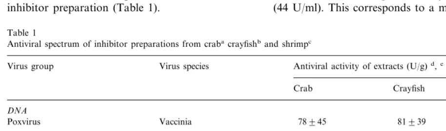
As shown in Table 1, the crab inhibitor prepa-ration was broadly inhibitory for the six viruses tested. These viruses represent a DNA virus, non-enveloped and non-enveloped RNA viruses. The titers of the inhibitor varied with the different viruses. The rank order of virus sensitivity was SB, VAC, MENGO, VS, BANZI and POLIO. We tested preliminarily the inhibitory effect of the inhibitor preparation on both cell free HIV and HIV in-fected CEM cells, and no antiviral activity was found. The inhibitory activity of tissue extracts from two other crustaceans, shrimp and crawfish, were found to be broadly active against the same viruses with titers comparable to that of crab inhibitor preparation (Table 1).
Confluent Vero cells were incubated with serial 2-fold dilutions of the samples or medium for 2 h, and then were decanted and incubated with new medium. The Vero cells were then stained with trypan blue at 2, 12 and 36 h, and trypan blue-stained cells were counted as dead cells. No differ-ence was found in the percent of dead cell counted (B10 dead cells per 2×10⁴cells) in presence and absence of the samples. In addition, Vero cells (2×10⁴ per well) were mixed with samples or medium controls in serial 2-fold dilutions and incubated overnight. The percent confluentcy was determined and there was no difference between the samples and medium control. The data indi-cated that cell toxicity was not the cause of virus inhibition.
3.2. Molecular size
The crab inhibitor preparation was fractionated by size-exclusion chromatography on a Superdex 200 26/60 FPLC column, and individual fractions were tested for the presence of antiviral activity. As showed in Fig. 1, the major peak of inhibitory activity against Sindbis virus was found in frac-tions 9 – 11 with the highest activity in fraction 10 (44 U/ml). This corresponds to a molecular mass
Table 1
Antiviral spectrum of inhibitor preparations from crabacrayfishband shrimpc
Virus species
Virus group Antiviral activity of extracts (U/g)d,e
Crab Crayfish Shrimp
DNA
Poxvirus Vaccinia 78945 81939 144969
RNA
Polio 18912 1899
Non-enveloped enterovirus 1298
Mengo 51927 39921 45921
132942
Enveloped alphavirus Sindbis 102927 216924
Flavivirus Banzi 24915 24912 –f
39924
Rhabdovirus Vesicular stomatitis 39924 1596
aC.sapidus. bP.clarkii. cP.setiferus.
dMean9S.D. of 3–5 experiments. Statistically differences of\2-fold are significant atPB0.05 by the Student’st-test. eNeuramide was used as positive control in all assays, and its titer of 64–128 (U/ml) served as indicator that the test system was
sensitive.
Fig. 1. Size exclusion chromatography of the inhibitor prepa-ration of blue crab. Crab (25 ml) inhibitor prepaprepa-ration (39 U/ml) was concentrated five times and was loaded on a column (2.5×90 cm) of Superdex 200 and eluted with sodium phosphate buffer (20 mM, pH 7.4) containing 200 mM of NaCl and 1 mM of EDTA. Fractions were collected and assayed for antiviral activity. The absorbance of eluted mate-rial at 280 nm is shown on top panel, and the arrows indicate the elution time of these molecular markers used to standard-ize the column. Antiviral titers (U/ml) against Sindbis virus are the average of three experiments (bottom panel).
addition, a secondary peak of relatively low an-tiviral activity (17 U/ml) was found in fractions 17 – 19, which corresponded to a molecular size of about 15 kDa.
3.3. Possible structure as determined by enzymatic and chemical treatments
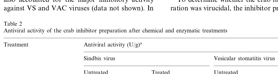
Lipid extraction with butanol significantly de-creased the antiviral activity in the residual aqueous phase of the inhibitor preparation (Table 2). Lipid extraction also resulted in the reduction of the size of the antiviral substance as evidenced by the filterability of the residual antiviral activity through a 10 K MWCO ultrafiltration membrane (data not shown). In contrast, neither insoluble proteinase K nor a cocktail of glycosidases signifi-cantly inactivated the antiviral activity of the in-hibitor preparation (Table 2).
In addition, a sulfhydryl reagent (DTT) and a chaotropic reagent (urea) were used to treat the inhibitor preparation, and no loss of antiviral activity was found (Table 2). Also, the inhibitor preparation remained active against SB and VS after incubation at 37°C for 12 h.
3.4. Mode of action
To determine whether the crab inhibitor prepa-ration was virucidal, the inhibitor prepaprepa-ration was of approximately 440 kDa. The same fractions
also accounted for the major inhibitory activity against VS and VAC viruses (data not shown). In
Table 2
Antiviral activity of the crab inhibitor preparation after chemical and enzymatic treatments
Treatment Antiviral activity (U/g)a
Sindbis virus Vesicular stomatitis virus
Treated Untreated Treated
Untreated
Proteinase K 60921 57930 78933 48933
60932
48921 39918
Glycosidases 39915
Butanolb 99933 1596 51927 15913
69930 96948
100 mM DTTc 2796 39915
99933 123966
90924 132939
4 M Urea
aMean9S.D. of 4–10 experiments, titer differences of\2-fold are significantly different (PB0.05).
bAntiviral activity in the aqueous phase of the treated sample were determined and were significantly lower than the untreated
sample.
cSamples were incubated for 2 h with a final concentration of DTT or urea at room temperature. DTT and urea were removed
mixed with a high dose of the viruses, incubated for 2 h and then serial two fold dilutions were made beyond the effective concentration of the inhibitor to assay for residual virus PFU. If the inhibitor preparation is virucidal, the virus plaque numbers in the higher dilutions of the mixture which are beyond the inhibitor concentration, would be much lower than that in the medium control. In our experiments, no significant differ-ence (P\0.05) of virus plaque numbers was found between the mixture with and without the inhibitor preparation, for the three viruses tested, SB (log10 5.5 with and log10 5.1 without sample), VAC (log105.1; log10 5.2) and VS (log105.7; log10 5.6). Thus, the inhibitor preparation appeared not to be virucidal with the viruses tested.
To determine whether the inhibitor preparation acts during virus attachment or thereafter, antivi-ral activity at 4 and 37°C was compared. Virus replication at 4°C does not proceed beyond at-tachment because the cell membrane and cell metabolism are quiescent at this temperature (Singh et al., 1995). Therefore, a comparable an-tiviral activity at 4 and 37°C has traditionally been interpreted as indicating that an antiviral substance inhibits the attachment of virus to target cells. Our experiments showed that the antiviral titers of the crab inhibitor preparation were at the same level (P\0.05) at 4 and 37°C with the viruses tested, i.e. SB (192912 at 4°C; 327954 at 37°C), VAC (231987 at 4°C; 2799 72 at 37°C) and VS (144960 at 4°C; 144933 at 37°C).
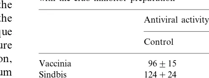
To determine whether the inhibitor preparation induced durable antiviral activity in host cells, the inhibitor preparation was preincubated with Vero cells overnight and then washed before virus chal-lenge. As shown in Table 3, the inhibitor prepara-tion pretreated Vero cells were fully resistant to subsequent infections of SB and VAC compared with control group. This finding indicates that the crab inhibitor preparation induces a durable an-tiviral state in Vero cells against subsequent infec-tion of SB and VAC. In contrast, VS was not inhibited by the same pretreated cells, indicating a reversible and probably different mechanism of inhibition.
Table 3
Cell associated antiviral activity after preincubation of cells with the crab inhibitor preparation
Antiviral activity (U/g)a
Preincubatedb
Vesicular stomatitis 2496 390
aMean9S.D. of 3–4 experiments. Differences between the
control and the preincubated sample are not significant statis-tically for VAC and SB (P\0.05).
bVero cells were incubated overnight with serial dilutions of
the inhibitor preparation, and washed three times with medium (preincubation) or left unwashed (control) before virus challenge.
4. Discussion
activity only on enveloped viruses (Hiraki et al., 1997). Also, blue crabs were reported to clear radio-labeled viruses (bacteriophages and po-liovirus) within a period of time that was too short for significant cell division or protein syn-thesis to occur. Therefore, the authors concluded that naturally occurring defenses exist in this spe-cies (McCumber and Clem, 1977).
Our studies focused on the large molecular size inhibitor, because the highest antiviral titers against viruses were found in the 440 kDa frac-tion of the crab inhibitor preparafrac-tion. Therefore, the 440 kDa molecule accounts for the most of the virus inhibition of the crab inhibitor prepara-tion. Consequently in our studies, small sub-stances (B12 kDa) were removed by extensive dialysis using 12 – 14 kDa MWCO dialysis tubing before antiviral assay.
The antiviral activity of the inhibitor prepara-tion was reduced by treatment with butanol lipid extraction, but was unaffected by treatments with DTT (a sulfhydryl reagent), urea (a protein solu-bilizing agent), proteinase K and carbohydrases. These data provided evidence that lipid compo-nents are important for a large portion of the antiviral activity in the inhibitor preparation. In addition, the lipid extraction appeared to reduce the size of the residual antiviral substance in the inhibitor preparation. A similar finding has been reported earlier. Lipid extraction of a large molec-ular size viral inhibitor from mouse and lamb brains released a low molecular weight moiety possessing broad antiviral activity (Singh et al., 1995; Baron et al., 1998). Lipids or lipid-contain-ing compounds have been reported to be antiviral with diverse mechanisms of inhibition. For in-stance, lipids isolated from human milk were ef-fective against enveloped viruses such as HIV, Herpes simplex, Vesicular stomatitis and Visna viruses, by affecting the viral envelope and caus-ing leakage (Thormar et al., 1987). In contrast, lipoproteins in human serum were broadly active against both enveloped and non-enveloped viruses, by preventing virus attachment or pene-tration of target host cells (Singh et al., 1995, 1999).
The inhibitor preparation does not directly neu-tralize virus infectivity, as evidenced by the
recov-ery of infectious viruses when the virus-inhibitor mixture was diluted beyond the inhibitory level. The inhibitor appears to act during the virus attachment stage, as evidenced by the equal an-tiviral titer of the inhibitor preparation at 4 and 37°C. If the inhibitor acts at both attachment and post-attachment stages, its titer should be signifi-cantly higher at 37 than at 4°C but it was not. Our data, therefore, suggest that the inhibitor preparation inhibits virus infection at an early stage of virus replication, probably by preventing virus attachment to Vero cells.
It is of interest to note that Vero cells pre-treated with the inhibitor preparation, were resis-tant to subsequent infection with SB and VAC, but were sensitive to infection with VS (Table 3). This finding indicates that the crab inhibitor preparation induces a durable antiviral state to Vero cells against subsequently infection of SB and VAC. This cellular resistance could not be due to interferon, since Vero cells do not produce it (Hughes et al., 1981). It remains to be deter-mined whether this cellular antiviral state may be due to inhibition of virus attachment, since inhibi-tion also occurred at 4°C (see above). Among naturally occurring non-specific antiviral sub-stances in vertebrates, only interferon induces a durable antiviral state in host cells (Kumar et al., 1984; Baron and Dianzani, 1994; Baron et al., 1989; Anders et al., 1990; Harmsen et al., 1995; Singh et al., 1999). However, a viral inhibitor from mosquitoes was reported to have cell associ-ated antiviral activity (Luo and Brown, 1993, 1994). The mechanisms by which Vero cells gain cell-associated resistance to two of the three viruses is an interesting subject for future studies. Two actions by one inhibitor preparation has been reported for an antiviral substance, isolated from horseshoe crabs, which exerted an inhibitory effect on HIV in MT-4 cells by prevention of virus adsorption, while it also directly inactivated VS by destroying its envelope subunits (Morimoto et al., 1991; Murakami et al., 1991). However, fur-ther studies are important to define whefur-ther the extracts contain multiple inhibitors or a single inhibitor.
crus-taceans. They may be important host defense substances and thereby may contribute to survival despite continuing exposure to extremely high dose of viruses (Fuhrman, 1999). Therefore, fur-ther study of the broadly antiviral inhibitors in invertebrates seems warranted.
Acknowledgements
The authors acknowledge the excellent techni-cal assistance from J. Poast, D. Nguyen and X. Liang.
References
Anders, E.M., Hartley, C.A, Jackson, D.C., 1990. Bovine and mouse serum beta inhibitors of influenza A viruses are mannose-binding lectins. Proc. Natl. Acad. Sci. USA 87, 4485 – 4489.
Baron, S., McKerlie, L., 1981. Broadly active inhibitor of viruses spontaneously produced by many cell types in culture. Infect. Immun. 32, 449 – 453.
Baron, S., Niesel, D., Singh, I.P., McKerlie, L., Poast, J., Chopra, A., Antonelli, G., Dianzani, F., Coppenhaver, D.H, 1989. Recently described innate broad spectrum virus inhibitors. Microb. Pathog. 7, 237 – 247.
Baron, S., Dianzani, F., 1994. The interferons: a biological system with therapeutic potential in viral infections. An-tiviral Res. 24, 97 – 110.
Baron, S., Chopra, A.K., Coppenhaver, D.H., Gelman, B.B., Poast, J., Singh, I.P., 1998. A host defense role for a natural antiviral substance in the nervous system. J. Neu-roimmunol. 15, 168 – 173.
Chisholm, J.R., Smith, V.J., 1992. Antibacterial activity in the haemocytes of the shore crab Carcinus maenas. J. Mar. Biol. Assoc. UK 72, 529 – 542.
Chisholm, J.R., Smith, V.J., 1995. Comparison of antibacterial activity in the hemocytes of different crustacean species. Comp. Biochem. Physiol. A. Physiol. 110, 39 – 45. Falkler, W.A., Jr, Diwan, A.R., Halstead, S.B., 1975. A lipid
inhibitor of dengue virus in human colostrum and milk; with a note on the absence of dengue secretory anti-body. Arch. Virol. 47, 3 – 10.
Fuhrman, J.A., 1999. Marine viruses and their biogeochemical and ecological effects. Nature 399, 541 – 548.
Johnson, M.C., Maxwell, J.M., Loh, P.C., Leong, J.A., 1999. Molecular characterization of the glycoproteins from two warm water rhabdoviruses: snakehead rhabdovirus (SHRV) and rhabdovirus of penaeid shrimp (RPS)/spring viremia of carp virus (SVCV). Virus Res. 64, 95 – 106. Ginsberg, H.S., 1960. Serum and tissue inhibitors of virus.
Bacteriol. Rev. 24, 141 – 150.
Grossberg, S.E., Jameson, P., Sedmak, J.J., 1974. Interferon bioassay methods and the development of standard proce-dures: a critique and analysis of current observations. The Tissue Culture Monogr. No. 3, The Tissue Culture Assoc., Rockville, M.D.
Harmsen, M.C., Swart, P.J., de Bethune, M.P., Pauwels, R., De Clercq, E., The, T.H., Meijer, D.K., 1995. Antiviral effects of plasma and milk proteins: lactoferrin shows potent activity against both human immunodeficiency virus and human cytomegalovirus replication in vitro. In: The Tissue Culture Monogr. No. 3. The Tissue Culture, Rockville, MD.
Hejkal, T.W., Gerba, C.P., 1981. Uptake and survival of enteric viruses in the blue crab,Callinectes sapidus. Appl. Environ. Microbiol. 41, 207 – 211.
Hiraki, A., Yukawa, M., Kim, J., Ueda, S., 1997. Antiviral substance from silkworm faeces: purification and its chem-ical characterization. Biol. Pharm. Bull. 20, 547 – 555. Hughes, T.K., Blalock, J.E., McKerlie, M.L., Baron, S., 1981.
Cell-produced viral inhibitor: possible mechanism of action and chemical composition. Infect. Immun. 32, 454 – 457. Kanchanaphum, P., Wongteerasupaya, C., Sitidilokratana, N.,
Boonsaeng, V., Panyim, S., Tassanakajon, A., Withy-achumnarnkul, B., Flegel, T.W., 1998. Experimental trans-mission of white spot syndrome virus (WSSV) from crabs to shrimp (Penaeus monodon). Dis. Aquat. Organ. 11, 1 – 7. Kingston, D.J., Dharsana, R., 1977. Mortality of crabs in the presence of Newcastle disease virus. Vet. Rec. 100, 433 – 434.
Kitamura, T., Tanaka, Y., Sugane, M., 1973. Studies on a heat-labile variola virus inhibitor in normal sera. II. Fur-ther characterization of the inhibitor and its activity. Inter-virology 1, 288 – 296.
Krizanova, O., Rathova, V., 1969. Serum inhibitors of myx-oviruses. Curr. Top. Microbiol. Immunol. 47, 125 – 151. Kumar, S., McKerlie, M.L., Albrecht, T.B., Goldman, A.S.,
Baron, S., 1984. A broadly active viral inhibitor in human and animal organ extracts and body fluids. Proc. Soc. Exp. Biol. Med. 177, 104 – 111.
Laille, M., Gerald, F, Debitus, C., 1998. In vitro antiviral activity on dengue virus of marine natural products. Cell. Mol. Life Sci. 54, 167 – 170.
Lightner, D.V., 1996. Epizootiology, distribution and the im-pact on international trade of two penaeid shrimp viruses in the Americas. Rev. Sci. Tech. 15, 579 – 601.
Loh, P.C., Tapay, L.M., Lu, Y., Nadala, E.C., 1997. Viral pathogens of the penaeid shrimp. Adv. Virus. Res. 48, 263 – 312.
Luo, T., Brown, D.T., 1993. Purification and characterization of a Sindbis virus-induced peptide which stimulates its own production and blocks virus RNA synthesis. Virology 194, 44 – 49.
Luo, T., Brown, D.T., 1994. A 55-kDa protein induced in Aedes albopictus(mosquito) cells by antiviral protein. Vi-rology 200, 200 – 206.
(Tyr5,12,Lys7)-polyphemusin II. Biochem. Biophys. Res. Commun. 15, 845 – 850.
McCumber, L.J., Clem, L.W., 1977. Recognition of viruses and xenogeneic proteins by the blue crab, Callinectes sapidus. I. Clearance and organ concentration. Dev. Comp. Immunol. 1, 5 – 14.
Miller, D.G., Miller, A.D., 1993. Inhibitors of retrovirus infec-tion are secreted by several hamster cell lines and are also present in hamster sera. J. Virol. 67, 5346 – 5352. Morimoto, M., Mori, H., Otake, T., Ueba, N., Kunita, N.,
Niwa, M., Murakami, T., Iwanaga, S., 1991. Inhibitory effect of tachyplesin I on the proliferation of human im-munodeficiency virus in vitro. Chemotherapy 37, 206 – 211. Murakami, T., Niwa, M., Tokunaga, F., Miyata, T., Iwanaga, S., 1991. Direct virus inactivation of tachyplesin I and its isopeptides from horseshoe crab hemocytes. Chemotherapy 37, 327 – 334.
Nadala, E.C., Jr, Tapay, L.M., Loh, P., 1998. Characteriza-tion of a non-occluded baculovirus-like agent pathogenic to penaeid shrimp. Dis. Aquat. Organ. 33, 221 – 229. Riedel, B., Brown, D.T., 1979. Novel antiviral activity found
in the media of Sindbis virus-persistently infected mosquito (Aedes albopictus) cell cultures. J. Virol. 29, 51 – 60. Schapiro, H.C., 1975. Immunity in decapod crustaceans. Am.
Zool. 15, 13 – 19.
Schnapp, D., Kemp, G.D., Smith, V.J., 1996. Purification and characterization of a proline-rich antibacterial peptide, with sequence similarity to bactenecin-7, from the haemo-cytes of the shore crab,Carcinus maenas. Eur. J. Biochem. 240, 532 – 539.
Seidel, K.M., Goyal, S.M., Rao, V.C., Melnick, J.L., 1983. Concentration of rotavirus and enteroviruses from blue crabs (Callinectes sapidus). Appl. Environ. Microbiol. 46, 1293 – 1296.
Singh, I.P., Coppenhaver, D.H., Chopra, A.K., Baron, S., 1992. Further characterization of a broad-spectrum antivi-ral substance in human serum. Viantivi-ral. Immunol. 5, 293 – 303.
Singh, I.P., Coppenhaver, D.H., Chopra, A.K., Baron, S., 1993. Generalized occurrence of the broadly antiviral sub-stance UTI in mammalian sera. J. Biol. Regul. Homeost. Agents. 7, 7 – 14.
Singh, I.P., Chopra, A.K., Coppenhaver, D.H., Smith, E., Poast, J., Baron, S., 1995. Vertebrate brains contain a broadly active antiviral substance. Antiviral. Res. 27, 375 – 388.
Singh, I.P., Chopra, A.K., Coppenhaver, D.H., Ananathara-maiah, G.M., Baron, S., 1999. Lipoproteins account for part of the broad non-specific antiviral activity of human serum. Antiviral. Res. 42, 211 – 218.
Thormar, H., Wisniewski, H.M., Lin, F.H., 1979. Sera and cerebrospinal fluids from normal uninfected sheep contain a visna virus inhibiting factor. Nature 279, 245 – 246. Thormar, H., Isaacs, C.E., Brown, H.R., Barshatzky, M.R.,
Pessolano, T., 1987. Inactivation of enveloped viruses and killing of cells by fatty acids and monoglycerides. Antimi-crob. Agents. Chemother. 31, 27 – 31.
Yilma, T., Owens, S., Adams, D.S., 1985. Preliminary charac-terization of a serum viral inhibitor in goats. Am. J. Vet. Res. 46, 2360 – 2362.