P
ATHOPHYSIOLOGY OF
M
OOD
D
ISORDERS
Postmortem Studies in Mood Disorders Indicate Altered
Numbers of Neurons and Glial Cells
Grazyna Rajkowska
The influence of stress and glucocorticoids on neuronal pathology has been demonstrated in animal and clinical studies. It has been proposed that stress-induced changes in the hippocampus may be central to the development of depression in genetically vulnerable individuals. New evi-dence implicates the prefrontal cortex (PFC) in addition to the hippocampus as a site of neuropathology in depression. The PFC may be involved in stress-mediated neurotoxicity because stress alters PFC functions and glucocorticoid receptors, the PFC is directly interconnected with the hip-pocampus, and metabolic alterations are present in the PFC in depressed patients. Postmortem studies in major depres-sion and bipolar disorder provide the first evidence for specific neuronal and glial histopathology in mood disorders. Three patterns of morphometric cellular changes are noted: cell loss (subgenual PFC), cell atrophy (dorsolateral PFC and orbitofrontal cortex), and increased numbers of cells (hypothalamus, dorsal raphe nucleus). The relevance of cellular changes in mood disorders to stress and prolonged PFC development and a role of neurotrophic/neuroprotec-tive factors are suggested, and a link between cellular changes and the action of therapeutic drugs is discussed. The precise anatomic localization of dysfunctional neurons and glia in mood disorders may reveal cortical targets for novel antidepressants and mood stabilizers. Biol Psychiatry
2000;48:766 –777 © 2000 Society of Biological Psychiatry
Key Words: Prefrontal cortex, neuropathology, mor-phometry, neurotrophic factors, stress
Stress and Morphopathology of Depression
C
ortical regions such as the hippocampus (limbic archicortex) and prefrontal cortex (PFC; association neocortex) have been implicated in the neuropathology of depression and the response to stress. Reductions in the volume of the hippocampus are reported in subjects with a history of depression (Bremner et al 2000; Krishnan et al 1991; Shah et al 1998; Sheline et al 1996; 1999).Inter-estingly, the loss of hippocampal volume is correlated with the total lifetime duration of depression but not with the age of the patients (Sheline et al 1999). It has been proposed that repeated stress during recurrent depressive episodes may inflict cumulative hippocampal injury as reflected in the loss of structural volume (Duman 1999).
The volume reduction in the hippocampus in depression may be a result of a loss in the number of neurons due to a neurotoxic effect of glucocorticoids (McEwen 1997; Sapolsky et al 1990, 1991). Glucocorticoids are adrenal steroids that are secreted in increased amounts during stress and are essential for maintaining homeostasis through the hypothalamic–pituitary–adrenal (HPA) axis. The biological actions of glucocorticoids are mediated via mineralocorticoid receptors (MRs) and glucocorticoid re-ceptors (GRs) located in the hippocampus, frontal cortex, and other regions (Lopez et al 1998; McEwen 1991). In a recent study, significant reductions in MR messenger RNA (mRNA) in the hippocampus and GR mRNA in the PFC were detected in suicide victims with a history of mood disorders (Lopez et al 1998; 42). Thus, stress-induced alterations in glucocorticoids and subsequent decreases in MR or GR populations in the hippocampus and PFC may be a mechanism whereby stress triggers or exacerbates depression.
Chronic stress or the repeated administration of glu-cocorticoids to animals results in the degeneration of specific types of hippocampal neurons (Sapolsky et al 1990, 1991; Watanabe et al 1992). Stress- or glucocorti-coid-related cellular changes observed in the primate and rodent hippocampus include dendritic atrophy, shrinkage of the neuronal cell body, and nuclear pyknosis (Sapolsky et al 1990; Watanabe et al 1992). Stressful experience also suppresses the normal production of granule cells in the hippocampal dentate gyrus during postnatal development and in adulthood (Gould and Tanapat 1999). Such post-natal reductions in the production of new cells are likely to alter the structure and function of the adult hippocampal formation and related structures. Thus, it has been pro-posed that changes in the hippocampus, secondary to stress, may be central to the development of depression in genetically vulnerable individuals (Duman 1999).
The influence of stress and glucocorticoids on neuronal
From Laboratory of Quantitative Neuroanatomy, Department of Psychiatry and Human Behavior, University of Mississippi Medical Center, Jackson. Address reprint requests to Grazyna Rajkowska, Ph.D., University of Mississippi
Medical Center, Department of Psychiatry & Human Behavior, Box 127, 2500 North State Street, Jackson MS 39216.
Received February 17, 2000; revised June 5, 2000; accepted June 12, 2000.
pathology in depression is likely to involve several other brain regions and circuits in addition to those in the hippocampus. The PFC may be another site in which stress-mediated neurotoxicity contributes to depression. The PFC has strong reciprocal connections with the hippocampus and other stress-related structures. More-over, animal and clinical studies indicate that stress alters prefrontal cognitive functions, and depressed suicide vic-tims have a decrease in GR mRNA in the PFC. Finally, glucocorticoids may play a role in metabolic disturbances detected by imaging techniques in the PFC in living patients with mood disorders and produce the changes in cell morphometry that are reported in postmortem studies of mood disorders.
Evidence for Involvement of the Prefrontal
Cortex in the Neuropathology of Depression
Several lines of evidence suggest that the PFC is involved in the neuropathology of major depressive disorder (MDD) and bipolar disorder (BPD, manic– depressive). In both MDD and BPD abnormal symptoms such as distur-bances of social behavior, depressed moods, and deficits in working memory suggest pathophysiologic involve-ment of the PFC. Direct evidence obtained from neuroim-aging studies further suggests that the PFC may be a common site of neuropathology in mood disorders. In BPD and MDD the PFC displays reductions in gray and/or white matter volume and sulcal widening as well as alterations in glucose metabolism and blood flow (Cohen et al 1989; Drevets et al 1997; Elkis et al 1996; Swayze et al 1990). Volumetric changes in the PFC in depression, similar to those described in the human hippocampus, are also reported in familial mood disorders (Drevets et al 1997). Recent examinations of the PFC in MDD and BPD in postmortem tissue provide the first morphometric evi-dence that specific changes in cell number packing density and size are associated with mood disorders (Ongur et al 1998; Rajkowska et al 1999; G. Rajkowska et al, unpub-lished data). These cellular changes in MDD and BPD may be the basis for morphometric alterations detected by the neuroimaging studies in gray and white matter in these disorders.
Stress is implicated in the volumetric and cellular alterations in the PFC because of the reciprocal cortical connections with the hippocampus and with other key structures involved in the stress response (e.g., paraven-tricular nucleus of the hypothalamus, amygdala, monoam-inergic nuclei of the brainstem; for a review, see Lopez et al 1999). Moreover, stress alters the level of GRs in the PFC. The exposure to chronic unpredictable stress results in downregulation of GR mRNA in the rat PFC (Herman et al 1995; J.F. Lopez, unpublished observations, 2000).
Decreases in GR mRNA are also detected in the PFCs of depressed suicidal individuals (Lopez et al 2000). It is suggested that downregulation of GRs in the PFC may be a compensatory response to elevated levels of circulating glucocorticoids. Interestingly, GR mRNA downregulation is not observed in the hippocampus from these same individuals (Lopez et al 2000). Instead, marked decreases in MR mRNA are found in the hippocampus of depressed suicide victims.
The involvement of different types of GRs in the PFC and hippocampus pathology suggests different sensitivity to circulating glucocorticoids of cells in these two regions. Another possibility is that other mechanisms may also contribute to PFC dysfunction during stress. For example, increases in catecholamine release during stress are re-ported in the PFC by behavioral and biochemical animal studies. Exposure to mild to moderate uncontrollable stress impairs working memory performance as well as other prefrontal functions in monkeys and rats (Arnsten and Goldman-Rakic 1998; Arnsten et al 1999; Mizoguchi et al 2000; Seamans et al 1998). Neurocognitive prefrontal deficits are observed in unmedicated patients with major depression, and these deficits are correlated with the severity of depression (Merriam et al 1999). It has been proposed that stress may impair cognitive functions through altering catecholamine actions on the dendritic stem of PFC neurons and that this mechanism involves G protein–linked intracellular pathways (for a review, see Arnsten and Goldman-Rakic 1998). It has been further suggested that increases in catecholamine release in the PFC during stress may occur due to blockage of catechol-amine transporters by circulating steroids. This mecha-nism may have a survival value. The PFC may be taken offline during stress exposure, allowing faster, more ha-bitual mechanisms regulated by subcortical and posterior cortical structures to adjust behavioral responses (Arnsten and Goldman-Rakic 1998). Further neuroanatomic, bio-chemical, and genetic studies will elucidate the unique role of the PFC in the stress response and the neurobiology of depression.
and after puberty. Thus, the development of the catechol-amine and other prefrontal systems is influenced by gonadal hormones as well as glucocorticoids, providing further support for the relationship between stress, hor-mones, mood, and the PFC dysfunctions.
Cell Pathology in Depression
Recent postmortem studies demonstrate that mood disor-ders are characterized by specific histopathologic changes in both neurons and glial cells. These alterations at the microscopic level may give rise to the volume reductions and metabolic abnormalities reported in mood disorders in neuroimaging studies and contribute to identifying dys-functional neuronal circuits and their cellular components in these disorders.
Two independent postmortem studies morphometrically estimated cell number and density in the subgenual pre-frontal region (Ongur et al 1998), dlPFC, and orbitopre-frontal region (ORB; Rajkowska et al 1999). Significant reduc-tions in glial cells were noted in these brain regions in subjects with mood disorders as compared with psychiat-rically normal control subjects. In addition, neuronal cell packing density and the size of neuronal cell bodies are decreased in the lateral orbitofrontal cortex (areas 10 and 47) and in the dlPFC (Brodmann’s area 9) and in MDD and BPD (Rajkowska et al 1999; G. Rajkowska et al, unpublished data). The reduction in cell number and density is accompanied by a 38 – 40% decrease in the volume of gray matter in the subgenual region (Drevets et al 1997; Ongur et al 1998) and a 12–15% reduction of cortical thickness in the lateral orbitofrontal cortex (Rajkowska et al 1999).
Glial Pathology
The unexpected prominent reductions in glial cell number and packing density are reported in independent laborato-ries in postmortem brains from subjects with mood disor-ders in different regions of the PFC and in the anterior cingulate cortex (Cotter et al 2000; Ongur et al 1998; Rajkowska et al 1999). Such a striking cellular deficit in mood disorders suggests that glia may be unique targets for novel strategies in the treatment of major depression. Stereological estimates of the number of glial cells in MDD and BPD patients reveal significant reductions in glial number in the subgenual medial prefrontal region in both mood disorders (Ongur et al 1998). The most prominent glial reductions are found in subgroups of subjects with familial MDD (24%) or BPD (41%).
Parallel morphometric studies of glial cells in the dlPFC and ORB cortex also reveal marked decreases in overall (11–15%) and laminar (20 –30%; layers III–VI) cell
pack-ing densities in subjects with MDD, as compared with control subjects (Rajkowska et al 1999). Comparable reductions in glial densities are detected in the dlPFC from subjects with BPD (G. Rajkowska et al, unpublished data). These reductions in glial density in MDD and BPD are accompanied by a significant enlargement in the size of glial nuclei in specific cortical layers.
Increases in glial nuclear sizes are also reported in the same prefrontal region in subjects with a neurodegenera-tive disorder—Huntington’s disease (Rajkowska et al 1998). In Huntington’s disease, however, the enlargement of glial cells is accompanied by marked gliosis manifested by increases in glial cell densities (Selemon et al 1995). Therefore, the cortex from subjects with mood disorders does not exhibit the classic morphometric signature of gliosis (i.e., glial hypertrophy in conjunction with glial proliferation). Rather, fewer but larger glial cells are present, and these cells may have more elaborate cytoplas-mic processes, although these studies are yet to be com-pleted. The lack of glial proliferation in mood disorders suggests that the glial pathology in mood disorders is not a response to ongoing neurodegenerative changes in the cortex; however, if fewer neurons are present in the cortices of depressed patients, the reductions may have occurred before the disease onset or may represent a more prolonged or moderate process of degeneration such that full-scale gliosis has not been triggered.
Taken together, the data suggest that there is disease-specific glial pathology in mood disorders. A link between reductions in glial cells revealed by postmortem analyses and altered glucose metabolism reported in neuroimaging studies can be suggested, since glial cells are involved in the active processing of glucose metabolism and are reported as the primary sites of glucose uptake and phosphorylation during neuronal activity (Tsacopoulos and Magistretti 1996).
It is also interesting to speculate that glial pathology might be related to the dysfunction of serotoninergic and/or noradrenergic systems reported extensively in de-pression (Heninger and Charney 1987; Hollister and Clag-horn 1993), since glia may play a role in serotonin and norepinephrine neurotransmission via postsynaptic recep-tors distributed on their cell bodies and processes (Griffith and Sutin 1996; Milner et al 1998; Shimizu et al 1996; Whitaker-Azmitia et al 1993; Wilkin et al 1991). Recog-nition and precise anatomic localization of dysfunctional glial types and their receptors may offer a new cortical model for development of antidepressant and mood-stabi-lizing medications.
Neuronal Pathology
BPD reveals significant reductions, in comparison to control subjects (Rajkowska et al 1999; G. Rajkowska et al, unpublished data). These neuronal reductions are more subtle than the corresponding glial alterations, and they are detected only when specific morphological size-types of neurons are analyzed in individual cortical layers. For example, marked reductions in the density of large neu-rons (corresponding to pyramidal glutamatergic excitatory neurons) are found in layers III and V of the dlPFC in BPD and MDD. In other prefrontal regions such as the rostral ORB, the most prominent neuronal reductions in MDD are confined to layer II cells (mostly corresponding to nonpy-ramidal inhibitory local circuit neurons; Rajkowska et al 1999). Interestingly, reductions in the density of specific populations of layer II nonpyramidal neurons containing the calcium-binding protein calretinin are reported in the anterior cingulate cortex in subjects with a history of mood disorders (S. Diekmann et al, unpublished observations, 1998). Thus, the layer-specific cellular changes in the PFC reported in mood disorders imply the involvement of several neural circuits and neurotransmitter systems in the neuropathology of depression (for more details, see Rajkowska 2000).
The reductions in neuronal densities are paralleled by smaller sizes of neuronal somatas and significant 12–15% decreases in cortical thickness observed in the rostral and middle parts of the ORB region in MDD (Rajkowska et al 1999, submitted). Neuronal loss (in contrast to glial loss) is not reported in the subgenual region in familial mood disorders (Ongur et al 1998); however, in this study specific types of neurons were not analyzed in individual cortical layers.
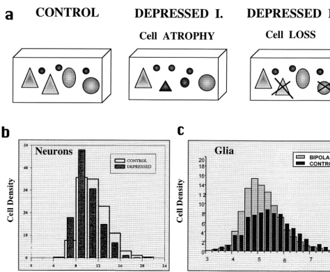
Cell Loss versus Cell Atrophy
Recent postmortem studies reveal several patterns of morphometric cellular changes in mood disorders: cell loss (subgenual prefrontal cortex), cell atrophy (and pos-sibly cell loss, dlPFC and ORB), or increased numbers of cells (hypothalamus, dorsal raphe nucleus) are reported.
Loss of glial but not neuronal cells is observed in mood disorders in the subgenual prefrontal region, whereas lamina-specific reductions in the density of both neurons and glia are reported in the dlPFC and ORB regions in MDD and BPD (Figure 1a). Whether these prominent reductions in cell density represent cell loss or only atrophy of cell bodies and/or their processes has not been established. It is not entirely clear whether cell loss accounts for the reductions in cell packing density because density measurements are de-pendent not only on the total number of cells present but also on the total volume in which cells are counted. For the estimation of total number of neurons or glia in a particular brain region, it is essential that the borders of this region be
established so that sampling is confined to the region within these borders (Gundersen et al 1988; West 1993). Since the entire extent and borders of the previously studied cortical areas were not available for examination, estimates of total cell number were not possible. On the other hand, indirect evidence from morphometric analyses of cell sizes and cortical and laminar thickness suggests that some cell loss in addition to cell atrophy takes place in the PFC in mood disorders.
In MDD, the densities of the largest neurons are significantly reduced by 22–37% in the rostral part of the lateral ORB and in the dlPFC region (Rajkowska et al 1999). In contrast, the densities of small neurons are
increased by 6 –27% in those regions. The latter
observa-tion suggests that either neuronal shrinkage or a develop-mental deficiency rather than neuronal loss accounts for the overall smaller sizes of neuronal soma in those cortical layers (Figure 1b). If neuronal loss had occurred, it is likely that the density of large neurons would have been decreased without associated increases in the density of small neurons, as was demonstrated in Huntington’s dis-ease (Rajkowska et al 1998).
In contrast to MDD, in BPD the decreased densities of large- and medium-sized neurons are not accompanied by increases in small neurons’ density (G. Rajkowska et al, unpublished data). Therefore, in BPD these decreases in the densities of large and medium types of neurons suggest neuronal loss rather than a diminution in neuronal size in this disorder (Figure 1c). Another indication of cell loss in BPD comes from the observation that the width of layer IIIc in the dlPFC is increased as compared with control subjects. In this layer, marked increases in the size of glial nuclei are also observed in BPD. In light of the increased width of layer IIIc, an increase in interneuronal neuropil, perhaps including the processes of hypertrophied glial cells and/or enlarged dendritic trees, might account for the reduction in neuronal and glial densities or cell loss in this layer; however, definitive answers regarding cell loss in the PFC in mood disorders await stereological studies in which the total numbers of specific types of neurons and glia will be estimated.
con-centrations in cerebrospinal fluid (Banki et al 1987). These findings of increases in neurons are consistent with the evidence of activation of the HPA axis in some subsets of depressed patients (Holsboer et al 1992). Increased num-bers of CRH, AVP, or OXT cells suggest an increase in related cell function, which may in turn have a modulatory effect on cortical or brain stem neurons. The PFC may be influenced by hypothalamic overactivation because this region is directly connected with the PVN and indirectly via monoaminergic brain stem nuclei. Moreover, the PFC can modulate the activity of the HPA axis (Herman and
Cullinan 1997; Herman et al 1996), and messenger RNA for CRH receptors has been detected in the PFC (Steckler and Holsboer 1999).
Increased AVP and OXT cell numbers are related to the increased production of these neuropeptides in the HPA axis (Frolkis et al 1982; Hoogendijk et al 1985; Lucassen et al 1994) and in turn may be associated with the increased activity of signaling systems reported in mood disorders (Hughes and Dragunow 1995; Hyman and Nestler 1996; Manji and Lenox 1999; Pandey et al 1999).
Neuron–Glia Interactions
The cellular changes described here indicate that both types of brain cells, neurons and glia, are abnormal in mood disorders. The question remains whether depressed patients are genetically predisposed for the cellular changes detected postmortem and had smaller neurons and/or less glia from birth, or whether the cellular changes are a consequence of MDD. Alternatively, those geneti-cally predisposed to the greatest histopathologic alter-ations may exhibit a greater vulnerability to depression. Cell loss or atrophy may also be related to developmental factors such as diminished amounts of neurotrophic fac-tors and/or malfunctions in programmed cell death (apoptosis).
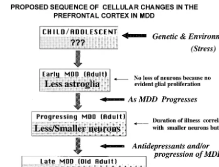
It is unknown at this stage whether cellular changes in the brains of depressed adults can also be found in depressed adolescents. There are no reports on morphom-etry in postmortem tissues from adolescent patients. Re-ductions in the density and size of neurons and glia found in adults could be a contributing factor to the pathophys-iology of depression or could worsen as the illness progresses. It is interesting to speculate that the PFC or hippocampus from adolescents with depression will ex-hibit primarily glial pathology due to genetic and/or early adverse environmental events that over time (and with recurrence of depressive episodes) leads to neuronal pa-thology. Lack of sufficient glial support (e.g., less struc-tural support due to a reduced number of glial cells or a shortage in glucose, the energy substrate provided to neurons by glia) may lead to neuronal pathology. Thus, it can be hypothesized that glial pathology in adolescents precedes neuronal changes (Figure 2). Alternatively, if the pathology in depressed adolescents is found in both glia and neurons, it may suggest that depression-induced re-ductions in activity of neurons with altered morphology will require less glial support, which may be reflected in a reduced number or density of glial cells. Pathology of both neurons and glia may worsen with increasing duration of illness and recurrent depressive episodes, and that pathol-ogy may eventually lead to cell loss.
The hypothesis that glial changes precede neuronal changes is supported by our recent observations on the distribution of glial fibrillary acid protein (GFAP), a marker for immunoreactive astroglial cells, in the dlPFC in MDD (Miguel-Hidalgo et al, in press). In MDD, there is a significant positive correlation between age and GFAP immunoreactivity (i.e., the GFAP areal fraction and pack-ing density of GFAP-immunoreactive glia). The correla-tion between age and GFAP immunoreactivity suggested that glial pathology in younger subjects might be different from that in older subjects. In younger adults (23– 45 years) with MDD, the GFAP areal fraction is smaller than
the smallest value of the control subjects, whereas there is a tendency for a larger GFAP areal fraction in older (46 – 86) MDD subjects, as compared with age-matched control subjects. Thus, GFAP-immunoreactive astroglia may be differentially involved in the pathology of MDD at the early and late stages of this disorder.
Cellular Changes and Prolonged PFC
Development
The most pronounced reductions in neuronal density in MDD and BPD are observed in superficial prefrontal layers II and III in both mood disorders. Neurons of these layers show greater plasticity than neurons of deep layers V and VI due to their late neurogenesis and extremely prolonged postnatal development. The prolonged postnatal development may render these neurons more susceptible to environmental factors related to the appearance of depression. The maturation and stabilization of neural elements and synapses on cells in layers II and III continue until adulthood (Koenderink et al 1994; Kostovic et al
1988; Mrzljak et al 1990, 1992). For example, during the developmental progression to adolescence there is an increase in the number of myelinated axons and an outgrowth of dendritic trees on layer III pyramidal neu-rons. These changes in neuronal and synaptic density may play critical roles in the remodeling of the basic cyto- and chemoarchitecture of the PFC.
The late structural and chemical maturation of prefron-tal neurons and associated glia, especially those located in upper cortical layers, makes them more vulnerable to postnatal environmental and experience-dependent insults. The final formation of the prefrontal framework is stimu-lated during postnatal development by environmental factors such as personal experience and neuroendocrine factors. Exposure to chronic stress during the maturation of cellular elements in the PFC may lead to overactivation of the HPA axis and to hypercortisolemia. Stress may alter functioning of the developing PFC in a manner similar to that in the hippocampus. In the developing hippocampus, stress inhibits the proliferation of granule cell precursors (Tanapat et al 1998). Adult neurogenesis in the dentate gyrus is also regulated by adrenal steroids (Cameron and Gould 1994). Most recently, postnatal generation of new prefrontal neurons in the monkey PFC (Gould et al 1999) suggests prolonged plasticity in this region. Thus, circu-lating glucocorticoids or adrenal steroids may alter pre-frontal cell morphology or even suppress adult cell pro-duction, as revealed in animal studies (Kritzer et al 1999; Kritzer and Kohama 1999; Lopez et al 1999).
Neurotrophic/Neuroprotective Factors and
Cell Pathology
Experimental data with in situ hybridization histochemis-try indicate that the development of cortical neuronal circuits may be related to the expression of specific target-derived neurotrophic factors such as brain-derived neurotrophic factor (BDNF; Huntley et al 1992). Expres-sion of BDNF mRNA increases during later stages of prefrontal cortical development and continues into adult-hood (Friedman et al 1991; Maisonpierre et al 1990), and the deprivation of neurotrophic factors activates cell death in neurons. Thus, any reduction in the supply of the neurotrophic factor could lead to a greater degree of neuronal death. Neurotrophic factors act by suppressing the latent biochemical pathway (a suicide program) present in all cells (for a review, see Jessel and Sanes 2000). Once the program is activated, cells die by apopto-sis, and one of the early features of the apoptotic process is cell shrinkage. Therefore, it can be speculated that the neuronal shrinkage observed in the PFC in mood disorders represents an early stage of apoptosis. Currently, however,
there is no postmortem evidence for apoptotic markers in depression.
There is a temporal correlation between the ingrowth of afferent axons into the PFC and detectable expression of BDNF mRNA (Murer et al 1999; Siuciak et al 1998). Therefore, it is likely that BDNF production by prefrontal neurons is required for a normal growth of afferent systems targeting those or neighboring neurons. The PFC is a target of overlapping cortical afferents coming from multimodal sensory areas, motor and limbic cortices and subcortical afferents such as thalamocortical axons, cho-linergic projections from the basal ganglia, dopamine axons of the ventral tegmental area, serotonin axons of the dorsal raphe, and norepinephrine axons of the locus coeruleus (for a review, see Fuster 1997, 6 – 42). The last three mentioned systems constitute monoaminergic pro-jection systems implicated in the pathophysiology of depression (Heninger and Charney 1987; Hollister and Claghorn 1993). Parallel studies of the locus coeruleus (Klimek et al 1997) or the dorsal raphe nucleus (Stock-meier et al 1999) in MDD, however, do not reveal severe morphological changes or the loss of norephinephrine or serotonin cell bodies. Rather, the number of serotonin neurons in the dorsal raphe nucleus may actually be increased in MDD (Underwood et al 1999). Thus, altered input from brain stem monoaminergic systems as well as other cortical regions may contribute to the pathology of prefrontal neurons in depression.
The survival of appropriate populations of synaptically connected neurons and supporting glial cells depends on neurotrophic factors such as BDNF (Ghosh et al 1994; Ohgoh et al 1998). For example, separation-induced cell death can be suppressed by BDNF. Separation of astroglial cells from cortical neurons in culture was shown to lead to neuronal death (Ohgoh et al 1998). Inasmuch as separa-tion-induced cell death is suppressed by neurotrophic factors such as BDNF, glial cell production of neurotro-phic factors such as glial-derived neurotroneurotro-phic factor appears crucial to cortical neuron survival (Ohgoh et al 1998).
brains of depressed subjects will exhibit reduced levels of BDNF, as compared with matched control subjects.
Other neurotrophic factors such as astroglial neuropro-tective protein S-100-bor fibroblast growth factor (FGF) may also be involved in neuron– glial interactions associ-ated with the pathophysiology of depression. For example, loss of astroglial protein S-100-b was evoked in the hippocampi of rats that underwent serotonin depletion during early postnatal development (Mazer et al 1997). The glial FGF factor was shown in other animal studies to regulate the size of the cerebral cortex during embriogen-esis (Vaccarino et al 1999). The cortical localization of glia– derived trophic factors has not yet been examined in the human brain.
A Link between Cellular Changes and the
Action of Therapeutic Drugs
The neurotrophins and monoamine neurotransmitters ap-pear to play related roles in stress, depression, and thera-pies for treating depression. From animal studies reporting that stress and antidepressant treatments regulate specific neurotrophin-related target genes within the central ner-vous system, it has been proposed that, in individuals genetically predisposed to clinical depression, cellular changes may be related to stress-induced changes in neurotrophin-related intracellular mechanisms (Duman 1999). These researchers further proposed that in depres-sion precipitated by stress, hypoxia–ischemia, neurotox-ins, or viral infections vulnerable neurons and glia may undergo atrophy or damage caused by increased levels of glucocorticoids and decreased levels of BDNF (Duman 1999). It is further hypothesized that upregulation of the BDNF gene could reverse the atrophy or damage of vulnerable neurons or protect these neurons from further damage. A role for BDNF in treatments for depression is also revealed in studies where the repeated treatment of rats with chronic electroconvulsive seizure or antidepres-sant drugs is shown to block the stress-induced decrease in BDNF in the hippocampus (Nibuya et al 1995).
Upregulation of BDNF occurs via increased serotonin and norephinephrine neurotransmission and upregulation of the cyclic adenosine monophosphate (cAMP)– cAMP response element binding protein cascade. A large body of evidence supports a role for BDNF in regulating the physiology and morphology of the serotonin system (Celada et al 1996; Mamounas et al 1995; Siuciak et al 1998; Vaidya et al 1997). For example, the infusion of BDNF into the neocortex substantially increases the sur-rounding density of serotonin axons (i.e., induces the sprouting of mature, uninjured serotonin axons; Mamou-nas et al 1995). The administration of 2,5-dimethoxy-4-iodoamphetamine (DOI), a serotonin2A receptor agonist,
dramatically increases the expression of BDNF mRNA in the rat frontal and parietal cortices in layers II/III and V/VI (Vaidya et al 1997). These are precisely the layers in which prominent reductions in neuronal and glial cell density and enlargements of glial nuclei are observed in postmortem brains of depressed subjects (Rajkowska et al 1999). Thus, serotonin receptor–mediated regulation of BDNF suggests that serotonin may play a role in the neuronal and glial changes reported in the neocortex in depressed patients.
Therapeutic medications are likely to exert their thera-peutic actions via networks of interconnected neurotrans-mitter pathways and their signal transduction systems (in particular, guanine nucleotide– binding proteins, adenylyl cyclases, and protein kinase C isozymes). Recent molec-ular studies in human cell culture and the animal brain suggest that antidepressant and mood-stabilizing medica-tions alter the genomic level of various neurotrophins, receptors, and enzymes involved in neurotransmitter bio-synthesis (Hughes and Dragunow 1995; Hyman and Nes-tler 1996; Manji and Lenox 1999; Manji et al, in press). For example, genes of several endogenous proteins, in-cluding neurotrophin receptors, are known to be regulated by the activator protein 1 family of transcription factors. Activator protein 1 DNA binding activity was markedly increased in the frontal cortex and hippocampus of rats treated chronically with mood stabilizers (Manji and Lenox 1999). Moreover, recent experiments with mice chronically treated with lithium, the major therapeutic mood stabilizer, reveal enhanced production of new cells in the dentate gyrus (Chen et al, in press). Thus, lithium has significant effects on the regulation of gene expression in the central nervous system.
Evidence has emerged suggesting that lithium may also have neuroprotective or neurotrophic actions in BPD (Manji et al 1999). Robust increases are noted in the level of bcl2, a major neuroprotective protein, in layers II/III of the rat PFC after chronic lithium treatment (Chen et al 1999). These increases in bcl2, suggesting a neuroprotec-tive effect for lithium, are found in the same cortical layer III in which pathologic changes are detected postmortem in BPD.
related to increases in the width of specific cortical layer(s) observed postmortem in BPD subjects. Most BPD subjects used for this study took lithium months or years before their deaths (Rajkowska et al, submitted). Of relevance to this hypothesis is the observation that the thickness of sublayer IIIc is greater and pyramidal cell density tends to be lower in subjects with a long exposure to lithium. Thus, a compensatory increase in dendritic (and/or glial) neuropil and consequent decrease in neuro-nal density may be a response to medication. Other therapeutic drugs may have a similar neuroprotective effect on cortical cells. Treatment with deprenyl, a neuro-protectant and antidepressant drug, enhances performance in cognitive tasks and is linked to increased dendritic tree aborization in the primate PFC (Shankaranarayana Rao et al 1999).
In summary, alterations in cell number or density revealed postmortem in mood disorders may be relevant to stress-induced changes in gene expression related to signal transduction pathways and cell survival (neurotrophic/ neuroprotective) factors. These changes may be reversed by antidepressant and mood-stabilizing medications by altering genomic level of various neurotrophins, receptors, and enzymes involved in neurotransmitter biosynthesis. The precise anatomic localization of dysfunctional neu-rons and glia, in conjunction with molecular genetic approaches, may facilitate our understanding of the neu-robiology of mood disorders and may reveal new cortical targets for action of novel antidepressant and mood-stabilizing medications.
The work reviewed here was supported by a National Alliance for Research on Schizophrenia and Depression (NARSAD) Young Investi-gator Award and a NARSAD Independent InvestiInvesti-gator Award, National Institute of Mental Health Grant No. 55872, and the American Founda-tion for Suicide PrevenFounda-tion.
The author thanks Craig Stockmeier, Ph.D., for helpful comments on the manuscript and editorial assistance.
Aspects of this work were presented at the conference “Depression in the Twenty-First Century: New Insights into Drug Development and Neurobiology,” February 21–22, 2000, Dana Point, California. The conference was sponsored by the Society of Biological Psychiatry through an unrestricted educational grant provided jointly by Pharmacia & Upjohn and Janssen Pharmaceutica.
References
Arnsten AF, Goldman-Rakic PS (1998): Noise stress impairs prefrontal cortical cognitive function in monkeys: Evidence for a hyperdopaminergic mechanism. Arch Gen Psychiatry 55:362–368.
Arnsten AF, Mathew R, Ubriani R, Taylor JR, Li BM (1999): alpha-1 noradrenergic receptor stimulation impairs prefrontal cortical cognitive function. Biol Psychiatry 45:26 –31. Banki CM, Bissette G, Arato M, O’Connor L, Nemeroff CB
(1987): CSF corticotropin-releasing factor-like immunoreac-tivity in depression and schizophrenia. Am J Psychiatry 144:873– 877.
Bremner JD, Narayan M, Anderson ER, Staib LH, Miller HL, Charney DS (2000): Hippocampal volume reduction in major depression. Am J Psychiatry 157:115–118.
Cameron HA, Gould E (1994): Adult neurogenesis is regulated by adrenal steroids in the dentate gyrus. Neuroscience 61: 203–209.
Celada P, Siuciak JA, Tran TM, Altar CA, Tepper JM (1996): Local infusion of brain-derived neurotrophic factor modifies the firing pattern of dorsal raphe serotonergic neurons. Brain
Res 712:293–298.
Chen G, Rajkowska G, Seraji-Bozorgzad N, Du F, Manji HK (in press): Enhancement of hippocampal neurogenesis by lith-ium. J Neurochem.
Chen G, Zeng WZ, Yuan PX, Huang LD, Jiang YM, Zhao ZH, Manji HK (1999): The mood-stabilizing agents lithium and valproate robustly increase the levels of the neuroprotective protein bcl-2 in the CNS. J Neurochem 72:879 – 882. Cohen RM, Semple WE, Gross M, Nordahl TE, King AC, Pickar
D, Post RM (1989): Evidence for common alterations in cerebral glucose metabolism in major affective disorders and schizophrenia. Neuropsychopharmacology 2:241–254. Cotter D, Mackay D, Beasley C, Kerwin I, Everall I (2000):
Reduced glial density and neuronal volume in major depres-sive disorder and schizophrenia in the anterior cingulate cortex. Schizophr Res 41:105.
Drevets WC, Price JL, Simpson JR Jr, Todd RD, Reich T, Vannier M, Raichle ME (1997): Subgenual prefrontal cortex abnormalities in mood disorders. Nature 386:824 – 827. Duman RS (1999): The neurochemistry of mood disorders:
Preclinical studies. In: Charney DS, Nestler EJ, Bunney BS, editors. Neurobiology of Mental Illness. New York: Oxford University Press, 333–347.
Elkis H, Friedman L, Buckley PF, Lee HS, Lys C, Kaufman B, Meltzer HY (1996): Increased prefrontal sulcal prominence in relatively young patients with unipolar major depression.
Psychiatry Res 67:123–134.
Friedman WJ, Olson L, Persson H (1991): Cells that express brain-derived neurotrophic factor mRNA in the developing postnatal brain. Eur J Neurosci 3:688 – 697.
Frolkis VV, Golovchenko SF, Medved VI, Frolkis RA (1982): Vasopressin and cardiovascular system in aging. Gerontology 28:290 –302.
Fuster JM (1997): The Prefrontal Cortex: Anatomy Physiology,
and Neuropsychology of the Frontal Lobe, 3rd ed.
Philadel-phia: Lippincott-Raven.
Ghosh A, Carnahan J, Greenberg ME (1994): Requirement for BDNF in activity-dependent survival of cortical neurons.
Science 263:1618 –1623.
Gould E, Reeves AJ, Graziano MS, Gross CG (1999): Neuro-genesis in the neocortex of adult primates. Science 286:548 – 552.
Gould E, Tanapat P (1999): Stress and hippocampal neurogen-esis. Biol Psychiatry 46:1472–1479.
Gundersen HJ, Bagger P, Bendtsen TF, Evans SM, Korbo L, Marcussen N, et al (1988): The new stereological tools: Disector, fractionator, nucleator and point sampled intercepts and their use in pathological research and diagnosis. APMIS 96:857– 881.
Heninger GR, Charney DS (1987): Mechanisms of action of antidepressant treatments: Implications for the etiology and treatment of depressive disorders. In: Meltzer HY, editor.
Psychopharmacology: The Third Generation of Progress.
New York: Raven, 535–544.
Herman JP, Adams D, Prewitt C (1995): Regulatory changes in neuroendocrine stress-integrative circuitry produced by a variable stress paradigm. Neuroendocrinology 61:180 –190. Herman JP, Cullinan WE (1997): Neurocircuitry of stress:
central control of the hypothalamo-pituitary-adrenocortical axis. Trends Neurosci 20:78 – 84.
Herman JP, Prewitt CM, Cullinan WE (1996): Neuronal circuit regulation of the hypothalamo-pituitary-adrenocortical stress axis. Crit Rev Neurobiol 10:371–394.
Hollister LE, Claghorn JL (1993): New antidepressants. Annu
Rev Pharmacol Toxicol 33:165–177.
Holsboer F, Spengler D, Heuser I (1992): The role of cortico-tropin-releasing hormone in the pathogenesis of Cushing’s disease, anorexia nervosa, alcoholism, affective disorders and dementia. Prog Brain Res 93:385– 417.
Hoogendijk JE, Fliers E, Swaab DF, Verwer RW (1985): Activation of vasopressin neurons in the human supraoptic and paraventricular nucleus in senescence and senile demen-tia. J Neurol Sci 69:291–299.
Hughes P, Dragunow M (1995): trkC may be an inducible transcription factor target gene. Neuroreport 6:465– 468. Huntley GW, Benson DL, Jones EG, Isackson PJ (1992):
Developmental expression of brain derived neurotrophic factor mRNA by neurons of fetal and adult monkey prefrontal cortex. Brain Res Dev Brain Res 70:53– 63.
Hyman SE, Nestler EJ (1996): Initiation and adaptation: A paradigm for understanding psychotropic drug action. Am J
Psychiatry 153:151–162.
Jessel T, Sanes J (2000): The generation and survival of nerve cells. In: Kandel E, Schwartz J, Jessel T, editors. Principles of
Neural Science. New York: McGraw-Hill, 1041–1062.
Kathol RG (1985): Etiologic implications of corticosteroid changes in affective disorder. Psychiatr Med 3:135–162. Kawamoto Y, Nakamura S, Kawamata T, Akiguchi I, Kimura J
(1999): Cellular localization of brain-derived neurotrophic factor-like immunoreactivity in adult monkey brain. Brain
Res 821:341–349.
Klimek V, Stockmeier C, Overholser J, Meltzer HY, Kalka S, Dilley G, Ordway GA (1997): Reduced levels of norepineph-rine transporters in the locus coeruleus in major depression.
J Neurosci 17:8451– 8458.
Koenderink MJ, Uylings HB, Mrzljak L (1994): Postnatal maturation of the layer III pyramidal neurons in the human prefrontal cortex: A quantitative Golgi analysis. Brain Res 653:173–182.
Kostovic I, Skavic J, Strinovic D (1988): Acetylcholinesterase in the human frontal associative cortex during the period of cognitive development: Early laminar shifts and late innerva-tion of pyramidal neurons. Neurosci Lett 90:107–112.
Krishnan KR, Doraiswamy PM, Figiel GS, Husain MM, Shah SA, Na C, et al (1991): Hippocampal abnormalities in depression. J Neuropsychiatry Clin Neurosci 3:387–391. Kritzer MF, Adler A, Marotta J, Smirlis T (1999): Regionally
selective effects of gonadectomy on cortical catecholamine innervation in adult male rats are most disruptive to afferents in prefrontal cortex. Cereb Cortex 9:507–518.
Kritzer MF, Kohama SG (1999): Ovarian hormones differen-tially influence immunoreactivity for dopamine beta-hydrox-ylase, choline acetyltransferase, and serotonin in the dorso-lateral prefrontal cortex of adult rhesus monkeys. J Comp
Neurol 409:438 – 451.
Lopez JF, Akil H, Watson SJ (1999): Neural circuits mediating stress. Biol Psychiatry 46:1461–1471.
Lopez JF, Chalmers DT, Little KY, Watson SJ (1998): A.E. Bennett Research Award. Regulation of serotonin1A, glu-cocorticoid, and mineralocorticoid receptor in rat and human hippocampus: implications for the neurobiology of depres-sion. Biol Psychiatry 43:547–573.
Lopez JF, Little KY, Watson SJ (2000, May): Corticosteroid receptor regulation in the brain of suicide victims. Abstract presented at the 55th annual convention of the Society of Biological Psychiatry, Chicago.
Lucassen PJ, Salehi A, Pool CW, Gonatas NK, Swaab DF (1994): Activation of vasopressin neurons in aging and Alzheimer’s disease. J Neuroendocrinol 6:673– 679. Maisonpierre PC, Belluscio L, Friedman B, Alderson RF,
Wie-gand SJ, Furth ME, et al (1990): NT-3, BDNF, and NGF in the developing rat nervous system: Parallel as well as recip-rocal patterns of expression. Neuron 5:501–509.
Mamounas LA, Blue ME, Siuciak JA, Altar CA (1995): Brain-derived neurotrophic factor promotes the survival and sprout-ing of serotonergic axons in rat brain. J Neurosci 15:7929 – 7939.
Manji HK, Lenox RH (1999): Ziskind-Somerfeld Research Award. Protein kinase C signaling in the brain: Molecular transduction of mood stabilization in the treatment of manic-depressive illness. Biol Psychiatry 46:1328 –1351.
Manji HK, Moore GJ, Chen G (1999): Lithium at 50: Have the neuroprotective effects of this unique cation been over-looked? Biol Psychiatry 46:929 –940.
Manji HK, Moore GJ, Rajkowska G, Chen G (in press): Neuroplasticity and cellular resilience in mood disorders. Mol
Psychiatry.
Mazer C, Muneyyirci J, Taheny K, Raio N, Borella A, Whitaker-Azmitia P (1997): Serotonin depletion during synaptogenesis leads to decreased synaptic density and learning deficits in the adult rat: A possible model of neurodevelopmental disorders with cognitive deficits. Brain Res 760:68 –73.
McEwen BS (1991): Stress and hippocampus. An update on current knowledge. Presse Med 20:1801–1806.
McEwen BS (1997): Possible mechanisms for atrophy of the human hippocampus. Mol Psychiatry 2:255–262.
Merriam EP, Thase ME, Haas GL, Keshavan MS, Sweeney JA (1999): Prefrontal cortical dysfunction in depression deter-mined by Wisconsin Card Sorting Test performance. Am J
Psychiatry 156:780 –782.
sepa-rates young from old adults with major depressive disorder.
Biol Psychiatry.
Miguel-Hidalgo JJ, Rajkowska G (1999): Immunohistochemistry of neural markers for the study of the laminar cytoarchitecture in celloidin sections from the human cerebral cortex. J
Neu-rosci Methods 93:69 –79.
Milner TA, Lee A, Aicher SA, Rosin DL (1998): Hippocampal alpha2a-adrenergic receptors are located predominantly pre-synaptically but are also found postpre-synaptically and in selec-tive astrocytes. J Comp Neurol 395:310 –327.
Mizoguchi K, Yuzurihara M, Ishige A, Sasaki H, Chui DH, Tabira T (2000): Chronic stress induces impairment of spatial working memory because of prefrontal dopaminergic dys-function. J Neurosci 20:1568 –1574.
Moore GJ, Bebchuk JM, Hasanat K, Chen G, Seraji-Bozorgzad N, Wilds IB, et al (2000): Lithium increases N-acetyl-aspartate in the human brain: In vivo evidence in support of bcl-2’s neurotrophic effects? Biol Psychiatry 48:1– 8. Moore GJ, Bebchuk JM, Wilds IB, Chen G, Manji HK (in press):
Pharmacologic increase in human gray matter. Lancet. Mrzljak L, Uylings HBM, Kostovic I, van Eden CG (1992):
Prenatal development of neurons in the human prefrontal cortex. II. A quantitative golgi study. J Comp Neurol 316: 485– 496.
Mrzljak L, Uylings HBM, Van Eden CG, Judas M (1990): Neuronal development in human prefrontal cortex in prenatal and postnatal stages. Prog Brain Res 85:185–222.
Murer MG, Boissiere F, Yan Q, Hurot S, Villares J, Faucheux B, et al (1999): An immunohistochemical study of the distribu-tion of brain-derived neurotrophic factor in the adult human brain, with particular reference to Alzheimer’s disease.
Neu-roscience 88:1015–1032.
Murphy BEP (1991): Steroids and depression. J Steroid Biochem 38:537–559.
Nibuya M, Morinobu S, Duman RS (1995): Regulation of BDNF and trkB mRNA in rat brain by chronic electroconvulsive seizure and antidepressant drug treatments. J Neurosci 15: 7539 –7547.
Ohgoh M, Kimura M, Ogura H, Katayama K, Nishizawa Y (1998): Apoptotic cell death of cultured cerebral cortical neurons induced by withdrawal of astroglial trophic support.
Exp Neurol 149:51– 63.
Ongur D, Drevets WC, Price JL (1998): Glial reduction in the subgenual prefrontal cortex in mood disorders. Proc Natl
Acad Sci U S A 95:13290 –13295.
Pandey GN, Dwivedi Y, Pandey SC, Teas SS, Conley RR, Roberts RC, Tamminga CA (1999): Low phosphoinositide-specific phospholipase C activity and expression of phospho-lipase C beta 1 protein in the prefrontal cortex of teenage suicide subjects. Am J Psychiatry 156:1895–1901.
Purba JS, Hoogendijk WJ, Hofman MA, Swaab DF (1996): Increased number of vasopressin- and oxytocin-expressing neurons in the paraventricular nucleus of the hypothalamus in depression. Arch Gen Psychiatry 53:137–143.
Raadsheer FC, Hoogendijk WJ, Stam FC, Tilders FJ, Swaab DF (1994): Increased numbers of corticotropin-releasing hor-mone expressing neurons in the hypothalamic paraventricular nucleus of depressed patients. Neuroendocrinology 60:436 – 444.
Raadsheer FC, van Heerikhuize JJ, Lucassen PJ, Hoogendijk WJ, Tilders FJ, Swaab DF (1995): Corticotropin-releasing hor-mone mRNA levels in the paraventricular nucleus of patients with Alzheimer’s disease and depression. Am J Psychiatry 152:1372–1376.
Rajkowska G (2000): Histopathology of the prefrontal cortex in major depression: What does it tell us about dysfunc-tional monoaminergic circuits? Prog Brain Res 126:397– 412.
Rajkowska G, Miguel-Hidalgo JJ, Wei J, Dilley G, Pittman SD, Meltzer HY, et al (1999): Morphometric evidence for neuro-nal and glial prefrontal cell pathology in major depression.
Biol Psychiatry 45:1085–1098.
Rajkowska G, Selemon L, Goldman-Rakic P (1998): Neuronal and glial somal size in the prefrontal cortex: A postmortem morphometric study of schizophrenia and Huntington’s dis-ease. Arch Gen Psychiatry 55:215–224.
Rubinow DR, Schmidt PJ (1995): The neuroendocrinology of menstrual cycle mood disorders. Ann N Y Acad Sci 771:648 – 659.
Sapolsky RM, Stein-Behrens BA, Armanini MP (1991): Long-term adrenalectomy causes loss of dentate gyrus and pyramidal neurons in the adult hippocampus. Exp Neurol 114:246 –249. Sapolsky RM, Uno H, Rebert CS, Finch CE (1990):
Hippocam-pal damage associated with prolonged glucocorticoid expo-sure in primates. J Neurosci 10:2897–2902.
Seamans JK, Floresco SB, Phillips AG (1998): D1 receptor modulation of hippocampal-prefrontal cortical circuits inte-grating spatial memory with executive functions in the rat.
J Neurosci 18:1613–1621.
Selemon LD, Rajkowska G, Goldman-Rakic PS (1995): Abnor-mally high neuronal density in the schizophrenic cortex: A morphometric analysis of prefrontal area 9 and occipital area 17. Arch Gen Psychiatry 52:805– 818.
Shah PJ, Ebmeier KP, Glabus MF, Goodwin GM (1998): Cortical grey matter reductions associated with treatment-resistant chronic unipolar depression. Controlled magnetic resonance imaging study. Br J Psychiatry 172:527–532. Shankaranarayana Rao BS, Lakshmana MK, Meti BL, Raju
TR (1999): Chronic (2) deprenyl administration alters dendritic morphology of layer III pyramidal neurons in the prefrontal cortex of adult Bonnett monkeys. Brain Res 821:218 –223.
Sheline Y, Wang P, Gado M, Csernansky J, Vannier M (1996): Hippocampal atrophy in recurrent major depression. Proc
Natl Acad Sci U S A 93:3908 –3913.
Sheline YI, Sanghavi M, Mintun MA, Gado MH (1999): De-pression duration but not age predicts hippocampal volume loss in medically healthy women with recurrent major depres-sion. J Neurosci 19:5034 –5043.
Shimizu M, Nishida A, Zensho H, Yamawaki S (1996): Chronic antidepressant exposure enhances 5-hydroxytryptamine7 re-ceptor-mediated cyclic adenosine monophosphate accumula-tion in rat frontocortical astrocytes. J Pharmacol Exp Ther 279:1551–1558.
Siuciak JA, Clark MS, Rind HB, Whittemore SR, Russo AF (1998): BDNF induction of tryptophan hydroxylase mRNA levels in the rat brain. J Neurosci Res 52:149 –158. Steckler T, Holsboer F (1999): Corticotropin-releasing hormone
Stockmeier CA, Dilley GE, Kulnane LS, Miguel-Hidalgo JJ, Rajkowska-Markow G (1999): Morphometric evaluation of the midbrain dorsal raphe nucleus (DR) in suicide victims with major depression (MD). Abstr Soc Neurosci 25:2098.
Swaab DF, Hofman MA, Lucassen PJ, Purba JS, Raadsheer FC, Van de Nes JA (1993): Functional neuroanatomy and neuro-pathology of the human hypothalamus. Anat Embryol (Berl) 187:317–330.
Swayze VW II, Andreasen NC, Alliger RJ, Ehrhardt JC, Yuh WT (1990): Structural brain abnormalities in bipolar affective disorder. Ventricular enlargement and focal signal hyperin-tensities. Arch Gen Psychiatry 47:1054 –1059.
Tanapat P, Galea LA, Gould E (1998): Stress inhibits the proliferation of granule cell precursors in the developing dentate gyrus. Int J Dev Neurosci 16:235–239.
Tsacopoulos M, Magistretti PJ (1996): Metabolic coupling be-tween glia and neurons. J Neurosci 16:877– 885.
Underwood MD, Khaibulina AA, Ellis SP, Moran A, Rice PM, Mann JJ, Arango V (1999): Morphometry of the dorsal raphe nucleus serotonergic neurons in suicide victims. Biol
Psychi-atry 46:473– 483.
Vaccarino FM, Schwartz ML, Raballo R, Nilsen J, Rhee J, Zhou M, et al (1999): Changes in cerebral cortex size are governed by fibroblast growth factor during embryogenesis. Nat
Neu-rosci 2:246 –253.
Vaidya V, Marek G, Aghajanian G, Duman R (1997): 5-HT2A receptor-mediated regulation of brain-derived neurotrophic factor mRNA in the hippocampus and the neocortex. J
Neu-rosci 17:2785–2795.
Watanabe Y, Gould E, Cameron HA, Daniels DC, McEwen BS (1992): Phenytoin prevents stress- and corticosterone-induced atrophy of CA3 pyramidal neurons. Hippocampus 2:431– 435.
West MJ (1993): New stereological methods for counting neu-rons. Neurobiol Aging 14:275–285.
Whitaker-Azmitia P, Clarke C, Azmitia E (1993): Localization of 5-HT1A receptors to astroglial cells in adult rats: Implica-tions for neuronal-glial interacImplica-tions and psychoactive drug mechanism of action. Synapse 14:201–205.
Wilkin GP, Marriott DR, Cholewinski AJ, Wood JN, Taylor GW, Stephens GJ, Djamgoz MB (1991): Receptor activation and its biochemical consequences in astrocytes. Ann N Y Acad