APOPTOSIS IN SEPSIS
Rizka Humardewayanti-Nugroho
Tropical and Infectious Division of Internal Medicine Gadjah Mada University/Sardjito General Hospital Yogyakarta
INTRODUCTION
The word sepsis is derived from the Greek term for rotten or “to make putrid”. Sepsis, defined as the systemic host response to microorganisms in previously sterile tissues, is a syndrome related to severe infections and is characterized by end-organ dysfunction away from the primary site of infection. To meet the definition of sepsis, patients need to satisfy at least two of the Systemic Inflammatory Response Syndrome (SIRS) criteria in association with having a suspected or confirmed infection. The severity and mortality increase when this condition is complicated by predefined organ dysfunction (severe sepsis) and cardiovascular collapse (septic shock). The normal host response to infection is complex, aiming to both identify and control pathogen invasion and start immediate tissue repair. Both the cellular and humoral immune systems are activated, giving rise to anti-inflammatory and proinflammatory responses. Exacerbating these mechanisms can cause a chain of events that leads to sepsis, promoting massive liberation of mediators
1
and the progression of multiple organ dysfunction . Sepsis remains a critical problem with significant morbidity and mortality even in the modern era of critical care management. Despite intense efforts, sepsis remains a serious clinical problem and still associated with a high mortality rate. Septic shock and sequential multiple organ failure/dysfunction syndrome (MOF/MODS) correlate with poor outcome, and septic shock is the most common cause of death in intensive care units. A recent review by Angus et al estimated the 1995 incidence of sepsis in the United States to be 751,000 cases, resulting in 215,000 deaths. The average cost per case of sepsis was $22,100 with total costs of $16.7 billion nationally. A more recent analysis of hospital records indicates that the total number of patients who are dying is actually increasing. This study also confirmed the work of Angus et al that the
incidence of sepsis is increasing and projected to continue to grow as the population ages. These studies concluded that “severe sepsis is a common, expensive, and frequently fatal condition, with as many deaths annually as those from acute
2,3
myocardial infarction .
The immunological cascade resulting in the sepsis response can be initiated by tissue injury, ischemia reperfusion injury, gram-positive organisms, and fungi as well as gram-negative organisms and their constituent endotoxin. The sepsis response may begin with an infectious nidus, which may either invade the bloodstream, leading to dissemination and positive blood cultures, or proliferate locally and release various microbial
3
products into the bloodstream .
Multiple derangements exist in sepsis involving several different organs and systems, although controversies exist over their individual contribution to the disease process. Septic patients have substantial, life-threatening alterations in their coagulation system. Previously, it was believed that sepsis merely represented an exaggerated, hyperinflammatory response with patients dying from inflammation-induced organ injury. More recent data indicate that substantial heterogeneity exists in septic patients' inflammatory response, with some appearing immuno-stimulated, whereas others appear suppressed. Cellular changes continue the theme of heterogeneity. Some cells work too well such as neutrophils that remain activated for an extended time. Other cellular changes become accelerated in a detrimental fashion including
2
lymphocyte apoptosis .
Acta Interna - The Journal of Internal Medicine
syndromes. In particular, increased apoptosis, particularly in lymphoid tissues and potentially in some parenchymal tissues from solid organs, may
3
contribute to the sepsis-associated MODS
APOPTOSIS: Physiological cell death
All tissues must be able to tightly control cell numbers and tissue size and to protect themselves from rogue cells that threaten
4
homeostasis . Cells can die in two ways: by damaging their cell membrane and then undergoing necrosis or by shrinking and blebbing the intact cell membrane, which leads to apoptosis. Physical or chemical injury, such as the deprivation of oxygen that occurs in the heart muscle during myocardial infarction or in sepsis when the microvasculature is disrupted by fibrin deposition and intravascular coagulopathies, often leads to cell disintegration or necrosis. The necrotic tissue is phagocytosed and degraded by phagocytic cells, often leading to
3
inflammation .
An alternative form of cell death is known as programmed cell death or apoptosis. Apoptosis is a normal cellular process that is crucial for tissue remodeling and/or development. For example, most thymocytes undergo an apoptotic cell death when they fail positive selection or are negatively selected as a result of recognizing self-antigens. During apoptosis, cells undergo a non-necrotic cellular suicide that, in contrast to necrosis, generally does
3
not produce inflammation and injury in the tissue . The term apoptosis, from Greek origins (apo5for, ptosis5falling), was chosen to describe the cellular process of programmed cell death. Apoptosis is a tightly regulated intracellular program in which cells destined to die activate enzymes that degrade the cell's DNA and nuclear and cytoplasmic proteins. Programmed cell death eliminates unwanted cells or potentially reactive cell lines either before or after maturation. This process is vital to fetal and embryonic development and to
4
tissue remodeling . Cell populations that normally have a high rate of proliferation, such as the intestinal epithelium, depend upon apoptosis to maintain the necessary number of cells. The number of activated immune cells must be controlled to contain the inflammatory response. Hormone-dependent apoptosis occurs during estrus and causes
4
prostatic atrophy after castration .
Apoptosis must be distinguished from necrosis, which is also a form of cellular death. In contrast to apoptosis, necrosis is not a genetically programmed function, it affects groups of neighboring cells, and produces an inflammatory response. The death of a cell by necrosis leads to the release of alarm signal molecules that stimulate 1 or m o r e p a t t e r n - r e c o g n i t i o n r e c e p t o r s o n macrophages, dendritic cells, and natural killer cells. The presence of necrotic cells in a tissue is frequently interpreted by the immune system as dangerous and therefore acts as a signal to initiate an immune response. Unlike apoptosis, with necrosis there is cellular swelling with loss of cell membrane integrity, organelle swelling, and lysosomal leakage. The degradation of DNA is random and lysed cells are ingested by macrophages. Whether a cell survives or dies by apoptosis is determined by the balance between pro-apoptotic (stress ordeath) signals and anti-apoptotic (mitogenic or survival)
4
signals within and around the cell .
Apoptosis can be genetically encoded or can occur in response to cellular or external stimuli. There are 3 features that characterize apoptosis: protein cleavage or hydrolysis, breakdown of nuclear DNA, and recognition of the apoptotic cell
4
by phagocytic cells .
MECHANISMS OF APOPTOSIS
Initially, apoptosis was considered to be a mechanism by which selected cell populations could be actively eliminated from specific tissues during morphogenesis and tissue remodeling; however, it is also thought to have a role in the resolution of the
5
immune response .
Apoptotic cell death occurs primarily through three different pathways: the extrinsic death receptor (type I cells), the intrinsic (mitochondrial) pathway (type II cells), and the endoplasmic
5
reticulum (ER) or stress-induced pathway .
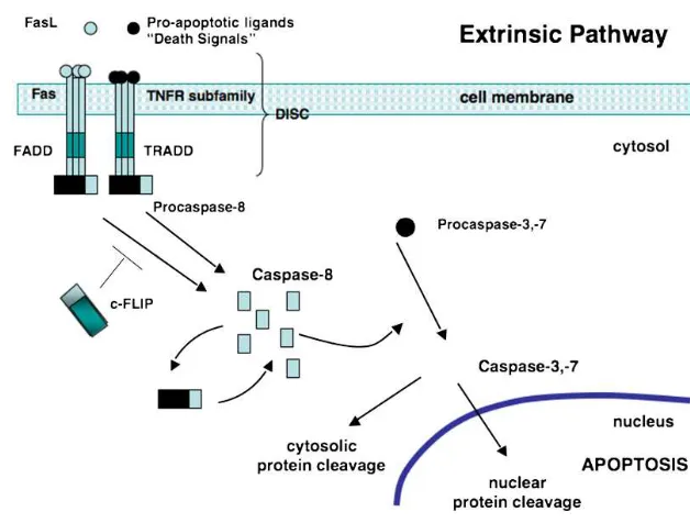
The extrinsic pathway (see Figure 1) begins with pro-apoptotic receptors on the cell's surface activated by a pro-apoptotic molecule or ligands specific for that receptor. These cell DRs belong to the tumor necrosis factor (TNF) receptor superfamily, with the Fas receptor and TNFR1 as the most intensely studied members. Fas is present on a Rizka Humardewayanti-Nugroho
variety of cell types including activated B cells and T cells. Ligands that activate pro-apoptotic receptors include the Fas ligand (FasL) and TNF-a37–42 (Table 1). FasL is expressed by a variety of cell types, including activated T cells and natural killer cells. TNF-a is produced predominantly by activated monocyte/macrophages and lymphocytes. The intracellular portion of the DR is known as the death domain (DD). Once 3 or more DR-ligand complexes bunch, their DDs are brought into close proximity and a binding site for an adaptor protein is formed.
The adaptor protein is specific for that receptor (eg, Fas-associated DD [FADD] or TNF receptor-associated DD [TRADD]). This complex of ligand-receptor-adaptor protein is called the death-inducing signaling complex (DISC), leading to the recruitment and assembly of initiator caspases 8 and 10. These caspases can now undergo self-processing and release active caspase enzyme molecules into the cytosol. Here, they activate the effector caspases 3, 6, and 7. Figure 1 illustrates the sequence of events
4
that trigger the extrinsic pathway .
Fig. 1. The extrinsic or death receptor (DR) pathway.
Pro-apoptotic ligands, death signals, and Fas bind to Fas or TNFRs. The intracellular portion of the DR is known as the death domain (DD). Bunching of the receptor-ligand complexes groups their DDs and a binding site for an adaptor protein is formed. This ligand-receptor-adaptor protein complex is called the death-inducing signaling complex (DISC). It recruits and assembles initiator caspase-8 that releases active caspase enzyme molecules into the cytosol. Here, they activate the effector caspases-3 and -7, resulting in nuclear protein cleavage and the initiation of apoptosis. FasL, Fas ligand; TNFR tumor necrosis factor receptor; FADD, Fas-associated death domain; TRADD, TNF-Fas-associated death domain; c-FLIP, FLICE-like inhibitory protein; DISC, death-inducing signaling complex. Source: O'Brien, M.A., et al.Apoptosis: A review of pro-apoptotic and anti-apoptotic pathways and dysregulation in disease. Journal of Veterinary Emergency and Critical Care 18(6). 2008; 572–585.
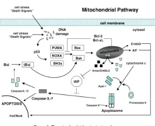
Intrinsic mitochondrial pathway: The
intrinsic mitochondrial pathway (see Figure 2)
is initiated from within the cell in response to
cellular stresses such as DNA damage, radical
oxygen species, radiation, hormone or
growth-factor deprivation, chemotherapeutic agents,
cytokines, and glucocorticoids. Initiation of this
pathway eventually results in the release of
pro-apoptotic proteins from the mitochondria that
will activate caspase enzymes and trigger
apoptosis. The success of the pathway in
inducing apoptosis depends on the balance of
activity between pro-apoptotic and
anti-apoptotic members of the B-cell lymphoma-2
4
(Bcl-2) superfamily of proteins .
apoptosis activating factor-1 (Apaf-1). Cytochrome c and Apaf-1 combine with procaspase-9 for activation of this caspase (Figure 2). The binding of these 3 substances forms an apoptosome, which
4
then activates procaspase-34 .
The third and least understood pathway is referred to as the endoplasmic reticulum or ER pathway. It involves the activation of caspase 12 by
+
Ca2 and oxidant stress and is said to be able to function independently of the mitochondria. Cellular stresses such as hypoxia, glucose starvation, disturbances in calcium homeostasis, and exposure to free radicals injure the ER, resulting in the unfolding of proteins and reduction in protein
4,5
synthesis .
In normal cells, an adaptor protein, TNF receptor associated factor 2 (TRAF2), is bound to procaspase-12, rendering it inactive. Stress of the ER leads to the dissociation of TRAF2, activation of caspase-12. Once activated, caspase-12 cleaves procaspase-9, which then cleaves procaspase-3. This mechanism is independent of the mitochondria although there is evidence that caspase 12 can cause the release of cytochrome c from the mitochondria
4
and thus stimulate the intrinsic pathway .
SYSTEMIC INFLAMMATORY RESPONSE SYNDROME (SIRS) AND MULTIPLE ORGAN DYSFUNCTION SYNDROME (MODS)
The theory that apoptosis contributes to the multiple organ dysfunction of sepsis was formulated originally by Bone in 1996. The argument put forth postulated that certain i m m u n o m o d u l a t o r y f a c t o r s p r e s e n t i n overwhelming amounts in SIRS could contribute to a generalized systemic increase in cellular apoptosis, thereby accounting for the organ failure
6
of MODS . Bone proposed that the anergic or hypoimmune aspect of sepsis, when apoptosis becomes most critical, must also be addressed, Apoptotic loss of B cells, T cells, and dendritic cells in sepsis decreases antibody reduction, macrophage activation, and antigen presentation,
4
respectively . As SIRS is essentially an imbalance between pro- and anti-inflammatory immune activity, it follows that the induction (or even inhibition) of apoptosis occurring in SIRS is
inappropriate and ultimately more autotoxic than
6
beneficial
A number of studies suggest that dysregulated apoptotic immune cell death may play a role in contributing to the immune dysfunction and MOF observed during sepsis. These include increased proinflammatory [TNF, interleukin 1β, interferon-γ (IFN- γ, interleukin 8, etc.] and anti-inflammatory (interleukin 6, interleukin 10, etc.) cytokines, elevated glucocorticoid levels secondary to adrenal cortex stimulation, increased production of ROS associated with ischemia/reperfusion, and the presence of bacterial wall products in the systemic circulation, heat shock, oxygen free radicals, nitric oxide (NO), Fas ligand (FasL), and cytotoxic T lymphocytes, which express FasL on their surface as a method of killing a Fas
receptor-5
expressing cell .
Apoptosis in Sepsis
Key cells involved in the inflammatory process (neutrophils, macrophages, dendritic cells, and lymphocytes) are also cells targeted for apoptosis. Apoptosis of immune cells is normally not a pathologic process because inflammatory cells must be eliminated so that inflammation does not continue unabated. However, in sepsis or other overwhelming inflammatory processes (like trauma and severe burns) there is extensive cell death of lymphocytes and dendritic cells and delayed cell death of neutrophils. This leads to a blunted immune response coinciding with increased cellular
4
damage .
L e u k o c y t e s a r e r e s p o n s i b l e f o r opsonization and phagocytosis of infected cells and antigens at the site of inflammation. Neutrophils are one of the main mediators of tissue injury and they play an important role in the inflammatory response. Neutrophils are one of the first cells to migrate to the site of inflammation with an average half life of 6–12 hours when unstimulated. The primary role of the neutrophil is to serve as an innate defense against infection by eliminating pathogens. As they kill pathogens using ROIs and a mixture of lytic enzymes, they can potentially contribute to bystander cell/organ injury. Their release of proteolytic enzymes, toxic oxygen metabolites, and anti-microbial peptides is associated with extensive tissue damage. Under steady state conditions and in the absence of cytokines or other proinflammatory agents, aged neutrophils typically undergo spontaneous apoptosis. Apoptotic neutrophils are Pro-apoptotic proteins of the Bcl-2 family
initiate apoptosis by blocking the anti-apoptotic activity of Bcl-2 and Bcl-xL by binding to their mitochondrial binding sites or by triggering the activation of pro-apoptotic Bax/Bak. A third type of pro-apoptotic activity is through the cytoplasmic protein, Bid. This molecule is found in the cytoplasm in an inactive form. When cleaved by activated caspase-8 from the extrinsic pathway, Bid (once activated, referred to as t-Bid) causes a structural change to Bax making it similar to the structure of the anti-apoptotic molecule, Bcl-2, allowing Bax to translocate to the mitochondria. This is but one method of cross-talk that occurs
4
between the intrinsic and extrinsic pathways . There are at least 3 current theories describing the exact mechanism by which the Bcl-2 pro-apoptotic proteins lead to increased
Figure 2: The mitochondrial or intrinsic pathway.
Activation of the pro-apoptotic proteins Bax and Bak occurs through conversion of Bid to tBid by caspase-8 or-10 and through activation of PUMA, Noxa, or other BH3 initiator proteins when p53 is induced by DNA damage. Activated Bax and Bak oligomerize at the mitochondrial membrane and cause the release of several mitochondrial factors. Cytochrome c combines with Apaf-1 and procaspase-9 forming an apoptosome. Also released from the mitochondria are Smac/DIABLO, proteins that inactivate IAPs. Activated caspase-9 then is able to activate caspase-3 or -7 allowing apoptosis to proceed. Also released from the mitochondria are EndoG and AIF that stimulate apoptosis independent of caspases. Bcl-2 and Bcl-xL block the activation of Bax and Bak. Bcl-2, B-cell lymphoma-2; IAP, inhibitor of apoptosis protein; Apaf-1, apoptosis-activating factor-1; Smac, second mitochondrial-derived activator of caspases; DIABLO, director inhibitor of apoptosis-binding protein with LOw pI; BH3, Bclhomology- 3; tBid, truncated Bid; EndoG, endonuclease G; AIF, apoptosis-inducing factor; Bax, Bcl-2-associated protein x; Bak, Bcl-2- associated protein k. Source: O'Brien, M.A., et al.Apoptosis: A review of pro-apoptotic and anti-apoptotic pathways and dysregulation in disease. Journal of Veterinary Emergency and Critical Care 18(6). 2008; 572–585.
Acta Interna - The Journal of Internal Medicine
then phagocytosed by macrophages that prevent them from releasing their cytotoxic contents into the extracellular milieu, which would occur if the cells died by necrosis. This safe removal of apoptotic neutrophils helps to limit tissue damage during the resolution of inflammation. Thus, the normal, constitutive apoptotic death of neutrophils and their clearance is thought to be an important limit on their
4-6.
potential to cause damage .
Leukocyte-endothelial interactions, which may also contribute to inflammation-mediated injury, involve two sets of adhesion molecules, selectins, and integrins. P-selectin is expressed on platelets; E- and P-selectins are expressed by endothelial cells, whereas L-selectin is expressed on leukocytes. Selectins mediate neutrophil rolling along activated endothelial surfaces as circulating neutrophils decelerate to engage endothelial receptors. The beta-2 intergrins (CD11/CD18 complexes) mediate tight adhesion to endothelial membranes, allowing subsequent egress of neutrophils to extravascular sites of inflammation. Activated neutrophils stimulate transendothelial albumin transport through intracellular adhesion molecule-1 mediated, Scr-dependent caveolin
7
phosphorylation .
N e u t r o p h i l s c o n t r i b u t e t o b l o o d coagulation in localized inflammation and in generalized sepsis. During systemic inflammation, homeostatic mechanisms are compromised in the m i c r o c i r c u l a t i o n i n c l u d i n g e n d o t h e l i a l hyperactivity, fibrin deposition, microvascular occlusion, and cellular exudates that further impede adequate tissue oxygenation. Neutrophils participate in these rheologic changes through their augmented binding to blood vessel walls and through the formation of platelet-leukocyte aggregates. Neutrophil elastase, other proteases, glycases and inflammatory cytokines degrade endogenous anticoagulant activity, and impair fibrinolysis on endothelial surfaces favoring a
7
procoagulant state .
Neutrophil apoptosis in sepsis
Once a neutrophil is released into circulation, its apoptotic program has been
4
activated . Although neutrophils are committed to apoptosis, their death can be delayed in sites of
inflammation by external factors including pro-nflammatory cytokines, bacterial membrane components such as lipopolysaccharide (LPS; endotoxin), and pro-granulocyte differentiation factors such as granulocyte and monocyte colony stimulating factor (GM-CSF). A hallmark of sepsis is the loss of normal apoptosis of neutrophils. Studies have shown that sepsis can shorten as well as prolong the life span of the activated neutrophils (early or delayed apoptosis, respectively). Early apoptosis of neutrophils dampens respiratory burst activity and may lessen secondary tissue injury. Delayed apoptosis of neutrophils is associated with the persistence of inflammation and localised tissue injury, contributes to increased cellular damage, especially in the lung, liver, kidneys, and
6,7
gastrointestinal tract .This prolonged life produces neighboring cell damage and contributes to activation of pro-inflammatory cytokines. Normally, pro-inflammatory mediators (TNF-α, IL-1β, IL-6, IL-8, and IFN-γ) released from macrophages and neutrophils have overlapping effects and function to limit damage, combat pathogenic organisms, eliminate foreign antigens, and promote repair. Anti-inflammatory cytokines (IL-4, IL-10, TGF-β, soluble receptors and receptor antagonists) are also quickly released to try to reduce and locally contain the inflammatory response. Many of these inflammatory components are the key factors responsible for the dysregulated
4
apoptosis of immune cells in sepsis .
Acute respiratory distress syndrome (ARDS) is marked by significant pulmonary accumulation of neutrophils, and is considered to be a direct effect of neutrophil-induced injury. Cells retrieved from the lungs of septic patients show reduced rates of neutrophil apoptosis with the degree of inhibition paralleling the severity of sepsis. Additional studies have shown that increased apoptosis (via Fas/FasL-dependent mechanism) of pulmonary epithelial cells will lead to permeability
4
changes characteristic of ARDS . The cytokines produced by activated neutrophils summon macrophages to the area of inflammation; cytokines are also produced by tissue macrophages in
4
response to foreign invasion .
Macrophages are antigen presenting cells (APC) capable of engulfing foreign material, Rizka Humardewayanti-Nugroho
infected cells, and apoptotic cells through
4
recognition of specific cell surface molecules . In as many as 30% of bacteremic patients who die from systemic inflammatory response syndrome or multiple organ dysfunction syndrome, no focus of infection is identified. Premature B cell and intestinal epithelial cell death through apoptosis in the intestines is one theory to explain intestinal bacterial translocation and loss of the first line of
4
intestinal defense.
Lymphocyte apoptosis in sepsis
Lymphocytes (B and T cells) are central to the adaptive immune response and rapidly expand in response to cytokines and antigen-specific
5
stimulation . Lymphocytes need 2 signals to stimulate differentiation and initiation of the immune response. The first signal is the presentation of antigen thus accounting for the specificity of the response. The second signal involves costimulatory molecules on APCs or APC secretion of cytokines. The costimulatory molecules interact with specific T cell sites producing a pro-apoptotic or anti-apoptotic state. Failure of the appropriate second signal after markedly reduced capacity of recombination activating gene (RAG)_/_. (lymphocyte-deficient) mice to survive cecal ligation and perforation
5
(CLP) . Normally, apoptosis of lymphocytes is a process to delete auto reactive lymphocytes or to
5
contain/resolve immune cell activation .
During normal health, the immediate fate of each lymphocyte is determined through continuous summation of a stream of proapoptotic and antiapoptotic signals that arrive from its external environment and from its internal cytoplasmic milieu. A shift toward initiation of apoptosis should therefore be expected during the early phase of sepsis, when bacteria or their by products stimulate macrophages to release proapoptotic substances such as TNF-, nitric oxide, and glucocorticoids. As the disease develops, accumulating products of
lymphocyte apoptosis can act as antiinflammatory s t i m u l i , w h i c h c o n t r i b u t e t o t h e immunosuppression commonly observed as sepsis progresses to septic shock, and which can lead to a
6
state of immune paralysis before death .
Overwhelming infection should lead to massive clonal expansion of B and T lymphocytes but instead there is significant loss of these cell lines in sepsis. Lack of stimulation by APC cells experiencing apoptosis leads to poor B cell and T cell stimulation. These unstimulated T cells are immune suppression, leaving the patient vulnerable to subsequent infections or unable to fight existing sepsis, resulting in MOF. In this respect, it was initially proposed that an increase in lymphocyte apoptosis as well as the loss of functionally responsive cells might serve to decrease the ability of the septic host to regulate the development of an immune response to opportunistic pathogens, thereby impairing the development of adaptive and innate immune system cross-talk needed to clear the
5
infection .
Hotchkiss and co-workers have shown an increase in splenic lymphocyte apoptosis in septic mice, which is associated with an increase in mortality. Lymphocyte apoptosis may be associated with immune dysfunction as a result of decreased proliferation and interferon-γ (IFN-γ) release capability. IFN-γ is a potent macrophage activator and induces a Th1 response. As seen in apoptotic and necrotic splenocyte-adoptive transfer experiments, necrotic and apoptotic cells exercise their effects through variation of IFN-γ levels. Transfer of apoptotic splenocytes retro-orbitally in CLP mice decreased their survival, whereas adoptive transfer of necrotic splenocytes increased splenocyte IFN-γ and in doing so, improved survival. This survival benefit was blocked in IFN-γ-deficient mice or in mice treated with an anti-IFN-γ antibody. These results are interesting, as this adoptive transfer study illustrates the potential impact of apoptotic cells in vivo in sepsis and thus, points at another mechanism (besides loss/ death of functional immune cells; Fig. 3A) by which
immune suppression might be promulgated in the septic animal (Fig. 3B). Alternatively, the inability to clear these dying lymphocytes/cells appropriately (as a result of dysfunction in these cell phagocytic capacities; often seen following sepsis and shock; refs.) may allow them to progress to a state of secondary necrosis, producing localized
5
bystander injury in the tissue (Fig. 3C) .
Numerous studies have demonstrated a massive apoptotic loss of lymphocytes during sepsis. A prospective investigation in adult patients compared spleens obtained either intraoperatively or within 6 hours after death from sepsis or trauma
and found that those from sepsis patients showed a marked decrease in B cells and CD4 T cells (Figure 4). The degree of splenic B-cell depletion corresponded with the duration of sepsis. Active caspase-9 was present in splenic lymphocytes with apoptotic features, suggesting a mitochondrial-mediated pathway of cell death, although evidence indicates that apoptotic cell death in patients with sepsis can also proceed by the death receptor pathway. In most patients, loss of cells from the spleen corresponded with a premortem decrease in
6,8
circulating lymphocytes .
Fig 3.A depiction of several possible mechanisms of immune suppression.
A depiction of several possible mechanisms of immune suppression. (A) The simple hypothesis (mechanism) that the immune dysfunction observed is a result of advertant/inadvertent apoptotic (Ao) loss of immune cell potential/capacity resultant from extrinsic and/or intrinsic Ao pathway activation. Here, no consideration is made for Ao cell clearence. (B) Alternatively, the effect that clearance of necrotic and/or apoptotic cell materials has on the developing macrophage phenotype (proinflammatory vs. anti-inflammatory/immune-suppressive) is considered when phagocytic function is normal. HSP, Heat shock protein(s); TLR, Toll-like receptor; CD1d/MICA, nonvariant MHC I-like antigen family; FcR, immunoglobulin constant region receptor(s); CR, complement receptor(s); ScavR, scavenger receptor(s), which bind; PS, phosphatidyl serine; TGF-β, transforming growth factor-β. (C) Finally, a scheme in which phagocytic function is compromised, so as to block apoptotic cell clearance, subsequently allowing apoptotic cells to move into secondary necrosis, which in turn, produces bystander tissue injury. TSP: Thrombospondin; M, macrophage(s). Source: Wesche, D.E., et al. Leukocyte apoptosis and its significance in sepsis and shock. Journal of Leukocyte Biology Volume 78. August 2005; 325-37.
Although there is a consensus that apoptosis is increased in lymphoid cell populations during
sepsis syndrome, the humoral or endocrine factors that stimulate this increased apoptosis are still not
3
fully known .
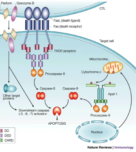
Fig4. Apoptotic pathways activated by cytolytic T cells.
parenchymal cell apoptosis. This increased hepatocellular apoptosis was an important signal for the transmigration of primed neutrophils sequestered in sinusoids. TNF-α induced neutrophil sequestration in hepatic sinusoids during sepsis and endotoxemia, which was also associated with
3
hepatocyte necrosis .
Endothelial Cell Failure and Apoptosis in Other Cells
Endothelial cells reside at the critical interface between the blood and tissue. Intact endothelial cells exhibit anticoagulant properties through elaboration of anticoagulant molecules such as protein C. These cells also serve as a barrier between blood products and procoagulant molecules, such as heparin, residing in the extracellular matrix. Endothelial disruption comes about because of increased expression of adhesion molecules on the endothelial cells, resulting in attachment of white blood cells. It has also become increasingly clear that abundant cross talk exists between the coagulation system and the inflammation system in sepsis. Endothelial cells will undergo apoptosis in response to several mediators
in vitro, including some infectious agents. However, endothelial cells are relatively resistant to the effects of endotoxin, and several investigators have failed to demonstrate convincing evidence of endothelial cell apoptosis during sepsis. Although it is strongly suspected that endothelial cells are dysfunctional in septic patients, clear-cut documentation during in
2 vivo settings has been difficult to obtain .
CONCLUSION
Apoptosis is a normal biologic process necessary to maintain cellular homeostasis. Apoptosis, or programmed cell death, is c h a r a c t e r i z e d b y n u c l e a r d e g e n e r a t i o n , condensation, and nuclear DNA degradation; phagocytosis of cell residua. There are characteristic pathways that lead to this form of cell death continually influenced by local cellular events, growth factors, and neighboring stresses. This complicated system has numerous built-in avenues
and failsafe mechanisms, including pro-apoptotic and anti-apoptotic factors. During sepsis, just 2 of the many diseases causing dysregulation of the apoptotic process, cells are either killed too quickly or survive too long. MODS or even MOF are often associated with an increased rate of apoptosis in lymphoid cells and, to a lesser extent, in organ parenchyma.
REFERENCES
1. Silva, E., Passos, R. D. H., Ferri, M. B., de Figueiredo L.F. P., Sepsis: From Bench to Bedside. Clinics 2008;63(1):109-20
2. Remick, D.G., Pathophysiology of Sepsis. The American Journal of Pathology, Vol. 170, No. 5, May 2007: 1435-44.
3. Oberholzer, C., Oberholzer, A., Clare-Salzler, M., Moldawer, L. L., Apoptosis in Sepsis: A New Target for Therapeutic Exploration. The FASEB Journal Vol. 15 April 2001: 879-92.
4. O'Brien, M.A. and Kirby, R.Apoptosis: A review of pro-apoptotic and anti-apoptotic pathways and dysregulation in disease. Journal of Veterinary Emergency and Critical Care 18(6) 2008, pp 572–585.
5. Wesche, D.E., Lomas-Neira, J.L., Perl, M., Chung, C., and Ayala, A. Leukocyte apoptosis and its significance in sepsis and shock. Journal of Leukocyte Biology Volume 78, August 2005, pp 325-37.
6. Thome, M & Tschoop, J. 2001. (cited march 2011) Apoptotic pathways activated by cytolytic T cell (1 s c r e e n ) . Av a i l a b l e f r o m ; h t t p : / / w w w . nature.com/…/fig_tab/nri1001-050a/_F1.html 7. Power, C., Fanning, N., Redmond, H. P., Cellular
Apoptosis and Organ Injury in Sepsis : A Review. Shock, Vol. 18, No. 3. 2002, pp 197-211.
8. Cinel, I., Steven M. Opal, M. S.Molecular biology of inflammation and sepsis: A primer. Crit Care Med 2009 Vol. 37, No. 1; pp 291-304
9. Parrino, J., Hotchkiss, R.S., and Bray, M. Prevention of Immune Cell Apoptosis as Potential Therapeutic Strategy for Severe Infections. Emerging Infectious Diseases www.cdc.gov/eid Vol. 13, No. 2, February 2007 pp 191-8.
Volume 2, Number 1, June 2012 Apoptosis in Sepsis
Fig 5. A summary of the general changes in the levels of apoptosis (Ao) and/or activation-induced cell death (AICD) reported in experimental sepsis (filled arrows) or shock/trauma (open arrows) in mice and septic-critically ill patients, as well as the mediators reported to affect the onset and frequency of apoptosis in various immune as well as nonimmune cell types. GALT, Gut-associated lymphoid tissue; PMN, polymorphonuclear neutrophil; PAF, platelet-activating factor. Source: Wesche, D.E., et al. Leukocyte apoptosis and its significance in sepsis and shock. Journal of Leukocyte Biology Volume 78. August 2005; 325-37.
Dendritic cell apoptosis in sepsis
Dendritic cells, another type of APC, are viewed as the sentinels of the immune system. The dendritic cell is a critical link between the innate and adaptive immunity. Dendritic cells not only migrate to lymphoid organs and stimulate T cells after maturation, but they also play an integral part in
8
lymphocyte apoptosis and immune suppression . Like macrophages, mature dendritic cells are able to activate lymphocytes through the presentation of antigen.
Immature dendritic cells are capable of ingesting apoptotic cells but this will render them incapable of maturing and stimulating T cells. Macrophages and dendritic cells will secrete IL-10 after engulfing apoptotic cells. IL-10, considered an anti-inflammatory cytokine selectively blocks the maturation of dendritic cells. This has been shown to suppress the phagocytic activity as well as pro-inflammatory cytokine production of alveolar macrophages. IL-6 is also secreted by dendritic cells after ingesting apoptotic cells, leading to autocrine blockage of maturation. This lack of maturation leads to a tolerogenic state with no further
4
stimulation of the immune system . After ingesting apoptotic cells, dendritic cells can mature only when there are danger signals expressed by the apoptotic cell or when dendritic cells are engulfing an
excessive number of apoptotic cells. This leads to secretion of pro-inflammatory cytokines IL-1β and TNF-α by these signaled dendritic cells. Inadequate clearance of surplus apoptotic cells results in these cells becoming necrotic and inducing a
pro-4
inflammatory response . Studies have demonstrated apoptosis of intestinal and splenic B cells, CD4 T cells and dendritic cells in sepsis. Dendritic cells, particularly those that are CD8+ lymphoid-derived, appear to be lost in the spleens of septic patients and mice]. This loss of dendritic cells by apoptosis has been seen to occur after CD3+ CD4+ T cell activation. However, the significance and the nature of this apoptotic dendritic cell response in septic
5
animals and patients remain to be fully explored .
Apoptosis In Parenchymal Cells