Pulmonary artery vegetation in a pediatric
patient with ventricular septal defect: a
case report
Haryo Aribowo1*
1Thoracic and Cardiovascular Surgery Division, Department of Surgery, Faculty of Medicine, Univrsitas Gadjah Mada/Dr. Sardjito General Hospital, Yogyakarta, Indonesia
ABSTRACT
Infective endocarditis (IE) is one of the congenital heart disease complications which is frequently seen in ventricular septal defects (VSD). The Duke criteria are the diagnostic criteria for IE. One of the major criteria is evidence of vegetation. In VSD complicated with IE, vegetation is frequently found on the opening of the defect, on the right ventricular side of the opening, on the tricuspid valve, and less frequently it is found on the pulmonary valve. Vegetation found in the lumen of pulmonary artery is rarely reported. In this article, we reported a rare case of pulmonary artery vegetation in a boy with moderate VSD and treated with combination of parenteral antibiotic followed by successful surgical vegetation evacuation and VSD closure. A 6 years old boy was consulted with congenital
heart disease. His chief complaint was shortness of breath. He came with unspecific signs
and symptoms with a history of frequent hospitalization due to pneumonia and paleness. Chest X-ray showed enlargement of heart chambers. Transthoracic echocardiography
(TTE) revealed moderate size VSD and multiple vegetation on right ventricle outflow
tract, pulmonary artery valve, and inside the lumen of main pulmonary artery and right pulmonary artery. The blood culture showed a positive result for Streptococcus viridans.
He was treated with parenteral antibiotic and operated on later. We successfully performed evacuation of the vegetation and VSD closure.
ABSTRAK
Endokarditis infektif merupakan salah satu komplikasi penyakit jantung bawaan yang sering menyertai defek septum ventrikel (VSD). Kriteria Duke merupakan kriteria diagnosis untuk endokarditis infektif. Salah satu kriteria mayor nya adalah bukti adanya vegetasi. Pada VSD dengan komplikasi endokarditis infektif, vegetasi sering ditemukan pada pembukaan dari defek, pada ventrikel kanan, pada katup trikuspid, dan yang paling jarang pada katup pulmonalis. Vegetasi pada arteri pulmonalis jarang dilaporkan. Di artikel ini, kami melaporkan kasus vegetasi arteri pulmonal pada anak laki-laki dengan VSD sedang dan diterapi dengan kombinasi antibiotik parenteral diikuti dengan pembedahan evakuasi vegetasi dan penutupan VSD. Anak laki-laki berusia 6 tahun dikonsultasikan dengan penyakit jantung bawaan. Keluhan utamanya adalah sesak nafas. Pasien datang dengan tanda dan gejala yang tidak khas dengan riwayat rawat inap berulang karena pneumonia dan pucat. Hasil X-ray dada menunjukkan pembesaran ruangan-ruangan jantung. Transthoracic echocardiography menunjukkan VSD berukuran sedang dan vegetasi multiple pada right
dan arteri pulmonalis kanan. Kultur darah positif untuk Streptococcus viridans. Pasien diberi antibiotik parenteral selanjutnya dilakukan pembedahan. Kami berhasil melakukan evakuasi vegetasi dan penutupan VSD.
Keywords:pulmonary artery – vegetation - ventricular septal defect - infective endocarditis - surgery
INTRODUCTION
Infective endocarditis (IE) is one of the congenital heart disease complications. Incidence of IE is approximately 0.05-0.12 case/1000 patients/year.1 IE is found in 60% of congenital heart disease cases, and 14% of them are ventricular septal defects.2 The Duke criteria are used to established IE diagnosis. Vegetation is one of the major diagnostic criteria for infective endocarditis.3,4 Vegetation
is defined as an intra-cardiac mass composed
of microorganisms (bacterial or fungal) and
surrounded by layers of platelet/fibrin which
are attached to the endothelium.5 In cases
of VSD complicated with IE, vegetation is
frequently found on the opening of the defect, on the right ventricular side of the opening, on the tricuspid valve and the less frequently it is on the pulmonary valve. Vegetation found in the lumen of pulmonary artery is rarely reported.6 In this article, we reported a rare case of pulmonary artery vegetation in
a pediatric patient with a ventricular septal
defect.
CASE REPORT
A 6 years old boy was consulted to
thoracic and cardiovascular surgery division of Dr. Sardjito General Hospital, Yogyakarta
with congenital heart disease. The patient’s chief complaint was shortness of breath. There
was a history of paleness, frequent cough and cold, weight loss, and fatigue/intolerance to exercise. Those symptoms were reported since he was 2 years old and worsened in the past week. There were also several histories
of prior hospitalization due to pneumonia and anemia. On physical examination, blood
pressure was 83/46 mmHg, heart rate was 116 bpm, respiratory rate was 32x/minutes, and there was no febrile. On auscultation, there was inconstant S2 split and grade 4/6 pan
systolic murmur in the left parasternal line.
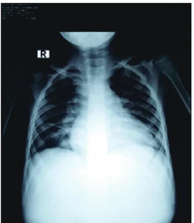
Other physical exams showed no abnormality. Chest x-rays showed cardiomegaly with
enlargement of the left atrium and ventricle (FIGURE 1). Transthoracic echocardiography
revealed a perimembranous VSD with the size of 1.4 cm with multiple vegetation
and moderate tricuspid regurgitation. The
vegetation was located on the right ventricle outflow tract (RVOT), pulmonary artery valve,
and inside the lumen of the main pulmonary artery (MPA) and the right pulmonary artery (RPA) (FIGURE 2 and 3). Vegetation on the
pulmonary artery valve was moving into the
main pulmonary artery at the systolic phase.
Blood culture was done following the TTE finding which suggested the diagnosis of IE. The blood culture result showed a growth of S. viridans and was tested sensitive to ampicillin
FIGURE 1. Chest x-ray showed enlargement of the heart with cardiac-thoracic ratio >0.5
FIGURE 2. Transthoracic echocardiography showed moderate size of VSD. RV: right ventricle; LV: left ventricle; RA: right atrium; LA: left atrium; VSD: ventricular septal defect.
The diagnosis of IE was established regarding the Duke criteria. We found two
major criteria of IE: positive blood culture
and evidence of vegetation. A final diagnosis
of VSD, IE of RVOT and the pulmonary
artery valve with the involvement of MPA and RPA, and moderate tricuspid insufficiency was considered. Due to the IE, intravenous antibiotic treatment was given. He was treated with intravenous ampicillin (50 mg/kg/day) and gentamycin (3mg/kg/day) for 4 weeks.
We evaluated the patient after the antibiotic
therapy, and dyspnea was less than before, blood culture was negative for any pathogens, however, some remaining vegetation was
still to be found on pulmonary artery valve
and main pulmonary artery. We switched the
antibiotics to ceftriaxone (50 mg/kg/day) for
2 weeks and gentamycin (3 mg/kg/day) for 3
days. On the TTE evaluation, the vegetation
was not significantly reduced and tended to be permanently attached. Later on, we decided to do surgical evacuation due to no significant
reduction in size and the mobility of the vegetation.
The VSD was closed through the median
sternotomy approach. Superior vena cava,
inferior vena cava, and aorta were cannulated
and connected to a cardiopulmonary bypass
machine. Cardioplegic agent was administered
and the heart became asystole. First incision
was made on the pulmonary artery, where we found multiple vegetation with size varied between 2-5 mm. The remaining vegetation
in the pulmonary valve and pulmonary
artery was manually evacuated. We found perimembranous VSD with diameter of 14
mm. We performed VSD closure using a
GoreTex patch sewn to the rightward aspect
of the defect. We closed the heart and placed a pericardium drain.
DISCUSSION
We found IE with pulmonary artery involvement, which is rarely to be found. Once it exists, it is most likely to be associated with
pulmonary artery valve endocarditis. Isolated pulmonary valve endocarditis and pulmonary artery endarteritis is a very rare condition.6 IE pathogenesis involves pathogen access to the
systemic blood flow, endothelial damage, and formation of vegetation which may become an emboli and flow to other site.7 In this study, the patient has VSD as a risk factor for developing
IE. Children with congenital heart disease
(CHD) have a greater risk for developing IE,
with 42% of pediatric IE cases reported with
CHD as an underlying disease.8 VSD is the third most common defect among patients
with IE, after cyanotic CHD and Atrium septal
defect.9 The abnormal hemodynamic from the heart defect shunting may cause injury to the endothelial layer and provide suitable medium for bacterial colonization resulting in IE.
We found nonspecific complaints related to IE in this patient. One study shows various clinical presentations of pediatric IE, where it is classified into two categories, subacute and acute. Subacute IE shows low grade fever and nonspecific symptoms such as fatigue, arthralgia, myalgia, weight loss, fatigue/ exercise intolerance, and diaphoresis, while Acute IE shows rapidly progressive disease with high fever and severely ill appearance.10
However, low-grade fever is only present in
3-15% of patients.11
The murmurs on specific locations that we found in this case were suggestive to be
caused by the VSD and tricuspid regurgitation.
Murmurs were also found in 80% to 85% of IE
patients as a sign of valvular regurgitation.12
Other signs of IE such as Osler’s nodes, Janeway lesions, and Roth spots are rarely to
vegetation in the heart and pulmonary artery. TTE is a diagnostic modality used in detecting the presence of vegetation in pediatric patients
with IE. TTE had a mean sensitivity of 97%
for the detection of vegetation in pediatric patients.14 Vegetation is further defined as
an echogenic mass adhering to the wall or valve leaflet with different characteristics
from the remaining original heart tissue.15 In
this case, we found the vegetation extended from the right ventricular outflow tract, to
the pulmonary artery. The characteristic of the vegetation is best described as tending to extend from the heart defect into the upstream chamber after the defect.16
We found no complications related to the presence of vegetation in the pulmonary
artery. However, prior research shows
it may cause pulmonary hypertension, lung embolization, and pulmonary artery
dissection which increases the patient’s
morbidity and mortality rate.10,17 In this case,
we found positive blood culture result for S. viridans. Blood culture is one of the major Duke criteria for IE. The causative pathogen
is found in 86% of IE cases, and the most
frequently found pathogens are gram-positive bacteria. The remaining are fungi and gram-negative bacteria.2 In community-acquired IE, Streptococcus viridans is the most
isolated bacteria, followed by S. aureus.18
Other causative pathogens are classified into the HACEK group which consists of Haemophillus spp, Aggregatibacter spp, C. hominis, E. corrodens, and Kingella spp.19 Our
finding is concordant with a prior study which
states that S. viridans infection manifests as subacute IE.10
We administered a combination of
antibiotics followed by surgical therapy in
for at least 4 weeks and may be continued to 6-8 weeks. Continuing the antimicrobial
treatment despite the negative blood culture is reasonable as a prophylactic against reinfection.19 Surgical interventions are a secondary therapy in IE. The right timing to perform surgical intervention in IE is still
controversial, however, earlier intervention
should be considered in order to reduce the risk of severe complications and improves patient prognosis.17 Surgical interventions in IE include: removal of vegetation, repair of damaged heart tissue, and correction of the abnormal structure.20
CONCLUSION
We reported a rare case of pulmonary
artery vegetation in a boy with moderate VSD that we treated with a combination of parenteral antibiotics followed by successful
surgical vegetation evacuation and VSD closure.
ACKNOWLEDGEMENT
We would like to thank the patient who
has participated in this study.
REFERENCE
1. Rosenthal LB, Feja KN, Levasseur SM, Alba LR, Gersony W, Saiman L. The changing epidemiology of pediatric endocarditis at a children’s hospital over seven decades. Pediatr Cardiol 2010; 31(6):813-20. http:// dx.doi.org/10.1007/s00246-010-9709-6
3. Durack DT, Lukes AS, Bright DK. New criteria for diagnosis of infective endocarditis:
utilization of specific echocardiographic
findings: Duke Endocarditis Service. Am J Med 1994; 96(3):200-9 http://dx.doi. org/10.1016/0002-9343(94)90143-0
4. Li JS, Sexton DJ, Mick N, Nettles R, Fowler VG Jr., Ryan T, et al. Proposed modifications to the Duke criteria for the diagnosis of infective endocarditis. Clin Infect Dis 2000; 30(4):633-8. http://dx.doi.org/10.1086/313753
5. McCormick JK, Tripp TJ, Dunny GM, Schlievert PM. Formation of vegetations during infective endocarditis excludes binding of bacterial-specific host antibodies to enterococcus faecalis. J Infect Dis 2002; 185(7):994-7. http://dx.doi.org/ 10.1086/ 339604
6. Farsak B, Yilmaz M, Oc M, Ozkutlu S, Demircin M. Non-valvular isolated pulmonary artery vegetations. Med Sci Monit 2002; 8(4):CS39-41.
7. Werdan K, Dietz S, Loffler B, Niemann S, Bushnaq H, Silber RE, et al. Mechanisms of infective endocarditis: pathogen-host interaction and risk states. Nat Rev Cardiol 2013; 11(1):35-50. http://dx.doi.org/10.1038/ nrcardio.2013.174
8. Day MD, Gauvreau K, Shulman S, Newburger JW. Characteristics of children hospitalized with infective endocarditis. Circulation 2009;
119(6): 865-70. http://dx.doi.org/10.1161/
circulationaha.108.798751
9. Rushani D, Kaufman JS, Ionescu-Ittu R, Mackle AS, Pilote L, Therrien J,et al. Infective endocarditis in children with congenital heart disease. Circulation 2013;
128(13):1412-9. http://dx.doi.org/10.1161/
circulationaha.113.001827
10. O’Brien SE, Fulton DR, Edwards MS, Armsby C. Infective endocarditis in children. July 2016. from: URL: http://www.uptodate. com/contents/infective-endocarditis-in-childr en?source=search&search=infective+endoca
11. Brusch JL, Bronze MS. Infective
Endocarditis: Practice Essentials, Background, Pathophysiology. 2015. Cite from: URL: http://emedicine.medscape. com/ article/216650
12. Karchmer A: Infective endocarditis. In: Zipes DP, Libby P, and Bonow RO (eds); Braunwald’s heart disease: a textbook of cardiovascular medicine. Philadelphia: Elsevier Saunders 2005; 1633-56.
13. Cahill TJ, Prendergast BD. Infective Endocarditis. Lancet 2016; 387(10021):882-93 http://dx.doi.org/10.1016/s0140-6736(15) 00067-7
14. Penk JS, Webb CL, Shulman ST, Anderson EJ. Echocardiography in pediatric infective endocarditis. Pediatr Infect Dis J 2011; 30(12):1109-11.
h t t p : / / d x . d o i . o r g / 1 0 . 1 0 9 7 / inf.0b013e31822d320b
15. Sanfilippo AJ, Picard MH, Newell JB,
Rosas E, Davidoff R, Thomas JD, et al. Echocardiographic assessment of patients with infectious endocarditis: prediction of risk for complications. J Am Coll Cardiol 1991; 18(5):1191-9. http://dx.doi. org/10.1016/0735-1097(91)90535-h
16. Sampedro MF, Patel R. Infections associated with long-term prosthetic devices. Infect Dis
Clin North Am 2007; 21(3):785-819. http://
dx.doi.org/10.1016/j.idc.2007.07.001
17. Shi X, Wang X, Wang C, Zhou K, Li Y, Hua Y. A rare case of pulmonary artery
dissection associated with infective
endocarditis. Medicine (Baltimore) 2016;
95(19):e3358. http://dx.doi.org/10.1097/
md.0000000000003358
18. Baddour LM, Freeman WK, Suri RM, Wilson WR. Cardiovascular Infections. In: Mann DL: Braunwald’s Heart Disease: A textbook of Cardiovascular Medicine 10th edition. Elsevier Saunders. 2015; pp1524-50.
et al. Infective endocarditis in childhood: 2015 update, a scientific statement from the American heart association. Circulation2015; 132(15):1487-515.
h t t p : / / d x . d o i . o r g / 1 0 . 1 1 6 1 / cir.0000000000000298
20. Kang DH. Timing of surgery in infective endocarditis. Heart 2015;
101(22):1786-91. http://dx.doi.org/10.1136/
