INTERSTITIAL LUNG DISEASE IN SYSTEMIC SCLEROSIS
Puji Astuti Tri K1, Anak Agung Arie1, Cleopas Martin Rumende21)Internal Medicine Department, Cipto Mangunkusumo National General Hospital-Faculty of Medicine, Universitas Indonesia
2)Division of Respirology and Critical Care, Internal Medicine Department, Cipto Mangunkusumo National General Hospital-Faculty of Medicine, Universitas Indonesia
A CASE REPORT
ABSTRACT
Systemic Sclerosis (SSc) is achronic tissue disorder characterized by immune dysfunction, microvascular injury, and
fibrosis. Organ involvement in patients with SSc is variable; however, pulmonary involvement occurs in up to 90% of patients with SSc. Interstitial lung disease (ILD) is a majorcomplication in SSc and has ahigh mortality rate. The SSc-ILD therapy is basically consistent with the progress of scleroderma pathophysiology. In this case, we examine a case of 59-years-old female patientwith a blackened ulcer on her left hand ring finger with disappearing of her distal finger segment, and also a chronic white phlegm cough followed by dysnea in exertion. Clinical examination and evaluation explored that she had a scleroderm, accompanied with ILD. Her complaint did not improve, so she got an immunosup-presant and supportive therapy to control the worsening of her disease.
Keywords: systemic sclerosis, interstitial lung disease
Introduction
Systemic Sclerosis (SSc) is a chronic connective
tissue disorder involving multisystem, with a variety
of manifestations and often leads to disability and mortality.Systemic Sclerosis is also characterized by
an inflammatory process, followed with functional
and structural impairment of the blood vessels to the
visceral organ, then continous with fibrosis.1 Lung disease is the most common complication of SSc. About 80% of patients with SSc have lung disease, especially fibrotic lung disease, and is the most common cause of death in SSc patients (33%). Fibrotic lung diseasesthat manifested in SSC are In-terstitial Lung Disease (ILD) and pulmonary hyper-tension.Both of these diseases cause high mortality rates in SSC, 33% and 28% of death.2,3
Prevalence of SSc in the US is 50-300 cases per 1 million inhabitants, with 90% of cases diagnosed with ILD. The 5 years survival rate was 84,1% and 74,9% in 10 years.4The incidence of SSc-associated ILD (SSc-ILD) mostly concerns women aged 30 to
55 years.5
The clinical manifestations of SSc-ILD are difficult to distinguish from idiopathic pulmonary fibrosis or other lung diseases.5The most sensitive investiga-tion to detect ILD is by means ofHigh Resoluinvestiga-tion CT (HRCT) scan, with a ground glass appearance as
its primary feature. The SSc-ILD therapy is basi-cally consistent with the progress of scleroderma pathophysiology.
Address for Correspondance : Puji Astuti Tri K, Anak Agung Arie, Cleopas Martin Rumende
Email : [email protected]
HOWTOCITETHISARTICLE :
fibrosis. Organ involvement in patients with SSc is variable; however, pulmonary involvement occurs in up to 90% of patients with SSc. Interstitial lung disease (ILD) is a majorcomplication in SSc and has ahigh mortality rate. The SSc-ILD therapy is basically consistent with the progress of scleroderma pathophysiology. In this case, we examine a case of 59-years-old female patientwith a blackened ulcer on her left hand ring finger with disappearing of her distal finger segment, and also a chronic white phlegm cough followed by dysnea in exertion. Clinical examination and evaluation explored that she had a scleroderm, accompanied with ILD. Her complaint did not improve, so she got an immunosup-presant and supportive therapy to control the worsening of her disease.
systemic sclerosis, interstitial lung disease
tissue disorder involving multisystem, with a variety
an inflammatory process, followed with functional
visceral organ, then continous with fibrosis.1 Lung disease is the most common complication of SSc. About 80% of patients with SSc have lung disease, especially fibrotic lung disease, and is the most common cause of death in SSc patients (33%). Fibrotic lung diseasesthat manifested in SSC are In-terstitial Lung Disease (ILD) and pulmonary hyper-tension.Both of these diseases cause high mortality rates in SSC, 33% and 28% of death.2,3
Prevalence of SSc in the US is 50-300 cases per 1 million inhabitants, with 90% of cases diagnosed with ILD. The 5 years survival rate was 84,1% and 74,9% in 10 years.4The incidence of SSc-associated ILD (SSc-ILD) mostly concerns women aged 30 to
The clinical manifestations of SSc-ILD are difficult to distinguish from idiopathic pulmonary fibrosis or other lung diseases.5The most sensitive investiga-tion to detect ILD is by means ofHigh Resoluinvestiga-tion
its primary feature. The SSc-ILD therapy is basi-cally consistent with the progress of scleroderma pathophysiology.
Email : [email protected]
Introduction
Systemic Sclerosis (SSc) is a chronic connective tissue disorder involving multisystem, with a variety of manifestations and often leads to disability and mortality.Systemic Sclerosis is also characterized by an inflammatory process, followed with functional and structural impairment of the blood vessels to the visceral organ, then continous with fibrosis.1 Lung disease is the most common complication of SSc. About 80% of patients with SSc have lung disease, especially fibrotic lung disease, and is the most common cause of death in SSc patients (33%). Fibrotic lung diseasesthat manifested in SSC are Interstitial Lung Disease (ILD) and pulmonary hypertension.Both of these diseases cause high mortality rates in SSC, 33% and 28% of death.2,3
Prevalence of SSc in the US is 50-300 cases per 1 million inhabitants, with 90% of cases diagnosed with ILD. The 5 years survival rate was 84,1% and 74,9% in 10 years.4The incidence of SSc-associated ILD (SSc-ILD) mostly concerns women aged 30 to 55 years.5
The clinical manifestations of SSc-ILD are difficult to distinguish from idiopathic pulmonary fibrosis or other lung diseases.5The most sensitive investigation to detect ILD is by means ofHigh Resolution CT (HRCT) scan, with a ground glass appearance as its primary feature. The SSc-ILD therapy is basically consistent with the progress of scleroderma pathophysiology.
Case Illustration
A 59-years-old female patient admitted to the hospital with a blackened ulcer on her left hand ring finger since 3 weeks before arrival to hospital, accompanied with swelling, redness, pain (visual analog scale: 6), and disappearing of her distal finger segment. Patient also complained a chronic white phlegm cough followed by shortness of breath which was getting more cumbersome. The patient felt nauseated and bloated. There was no fever complained. Such complaints often reoccured for 3 years, accompanied by stiff on the forehead and around the mouth. There was no other diseases histories. She was hemodinamically stable with peripheral oxygen saturation 98% on room air.
Physical examination showed fibrosis in the forehead and around the mouth. From chest auscultation, a basilar crackle was obtained. There was also an atrophy, gangrene, and autoamputation the tip of the thumb both hands.
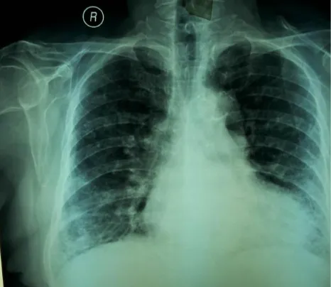
Figure 1. Chest radiography showing with aortic calcification and reticular infiltrates in
both lungs
Figure 2. High resolution computed tomography (HRCT) scan of the chest showed graound glass appearance in a).
Axial view, b). Coronal view
During treatment, the complaints of cough and dyspnea worsened. She then treated with targeted dose of unfractionated heparin, antiplatelet, nifedipin, and beraprost for her digital ulcer. For the SSc-ILD, she got mycophenolate mofetil, and morphine sulphate for her pain treatment.
Discussion
SSc is characterized by three mechanisms, such as a vasculopathy of small blood vessels, autoantibody formation, and fibroblast dysfunction that increases the deposit of extracellular matrix.6 The initial stages was inflammation, then it involves the blood vessel tissue’s structure and function, and progressively lead to visceral organ dysfunction due to fibrosis.1
Table 1. The American College of Rheumatology / European League Against Rheumatism Collaborative Initiative 2013 Criteria for the classification of systemic sclerosis6
Item Sub-item(s) Weight/score
Skin thickening of the fingers of both hands extending proximal to the metacarpophalangeal joints (sufficient criterion)
- 9
Skin thickeninng of the fingers (only count the higher score)
Puffy finger
Sclerodactyly of the fingers (distal to the metacarpophalangeal joints but proximal to the proximal interphalangeal joints)
2
’s structure and
Fingertip lesions (only count the higher score) Digital tip ulcers
Fingertip pitting scras
2
3
Telangiectasia - 2
Abnormal nailfold capillaries - 2
Pulmonary arterial hypertension and/or interstitial lung disease (max score: 2)
Pulmonary arterial hypertension
Interstitial lung disease
2
2
Raynaud’s phenomenon - 3
SSc-related autoantibodies (anticentromere, anti-topoisomerase I (anti Scl-70), anti RNA polymerase III) (maximum score:3)
Anticentomere
Anti-topoisimerase I
Anti-RNA polymerase III
3
In this case, there was a thickening of the skin of the fingers of both hands up to the metacarpophalangeal joint (9 points), finger swelling (4 points), fingertip ulcers (2 points), nail abnormalities (2 points), interstitial lung disease (2 points), and Raynaud's phenomenon (3 points). With a total of 22 points, the diagnosis of SSc in this patient is upright.
ILD is very common (90% of cases in SSC) and is the leading cause of death in SSc. It occursin the interstitial tissue abnormalities of the lung parenchyma. Early ILD enforcement helps to improve the prognosis of SSc.1,3 The pathogenesis of SSc-ILD is not yet fully understood.There are three mechanisms of SSc-ILD: 1. Repeated and persistent endothelial cell damage; 2. Activation of the natural and adaptive immune system; 3. Activation of fibroblasts, resulting from injury and accumulation of extracellular matrix.7
The epithelial or endothelial cell death is known to be a major cause of SSc-ILD. It causes necrosis, apoptosis, pyroptosis, activation of the coagulation pathway, and
inducing the expression of 6epithelial integrins, then activating TGF, which ultimately activates the response immune to fibrosis. Another mechanism is immune system dysregulation, imbalance and dysfunction of T cell in the circulation and bronchoalveolar fluid. Increased T cells (Th17 and Th22) in the circulation cause pulmonary fibrosis. Activation of myofibroblasts also plays a role in pulmonary fibrosis.8,9,10
The SSc-ILD manifestations are characterized by dyspnea on exertion and may be accompanied with a non-productive cough and inspiratory fine crackles at the bilateral basilar area on auscultation, supported by typical SSc symptoms. However, crackles usually can not be detected at the beginning of symptoms.SSc-ILD patients have positive antinuclear antibody (ANA) results, Scl 70, anti-topoisomerase I (anti-Topo I) and anti-Th / To, and anti-centromere antibodies.11
titial
case, with non-specific interstitial pneumonia (NSIP) appearance. At the beginning of the disease, there is a 'ground glass opacities' in the peripheral region of the lung, then progress into a reticular and honeycomb feature on the pulmonary basal. HRCT also could determine ILD prognosis. Pulmonary involvement increases more than 20% of mortality rate.12
Pulmonary function tests are also very important in determining the severity SSc-ILD. On spirometry test, it showed restrictive pattern with decreased forced expiratory volume in 1 sec (FEV1) and forced vital capacity (FVC), as well as a normal or slightly increased FEV1/FVC ratio, due to parenchymal damage.13
Inflammatory cells and other inflammation mediators (neutrophil and eusonophil, and may be lymphocytes and mast cells) were present in the brochoalveolar lavage (BAL) fluid of patients with SSc-ILD, reflecting ongoing inflammation in the lungs. In histopathology, a nonspecific interstitial pneumonitis (NSIP) is the most common histological finding. Fewer than 20% of patients with SSc-ILD showed inflammation-dominant lesions at lung biopsy.2,12
Inflammation and fibrosis are the main mechanisms of ILD in SSC. Inflammation is a reversible phase, so the treatment could be given in this phase. But, in the event of pulmonary fibrosis, which most patients had, there is no specific treatment to overcome. Therefore, the prevention of disease progression becomes the main goal of therapy.14
Cyclophosphamide is used as an induction regimen. Oral administration of 2 mg/kg/day for 12 months, and 600 mg/m2/month, intravenous for 6 months, coupled with oral
prednisolone 20 mg alternate, followed by azathioprine 2,5 mg/kg/day could reduce or prevent the progresivity. Cyclophosphamide is indicated for severe fibrosis for slowing the process.15During the treatment, the short-term side effects such as leukopenia and infection, and a long-term side effents such as infertility, carcinogenesis, and toxic to bone marrow could be happen.The intermittent intravenous administration could reduce the risk of side-effects. It is recommended the use of intravenous cyclophosphamide pulse dose (1g / m2 for 6-12 months). The dosage and duration of therapy may be adjusted by age and comorbid factors.14
Mycophenolate mofetil (MMF) isa lymphocyte proliferation inhibitors. The performance of MMF versus cyclophosphamide is still under study by SLS II (Scleroderma Lung Study II). In a prospective observational one-year study of 14 patients who took MMF (720 mg bid for 12 months, except for the first week of treatment [360 mg bid]) plus 5 mg/day PSL), six patients experienced at least a 10%
improvement in their FVC and five patients’
pulmonary function remained stable. Mycophenolate mofetil is commonly used as maintenance therapy after cyclophosphamide.16
Raynaud’s phenomenon
.
improvement in their FVC and five patients’
Rituximab is a monoclonal antibody that works directly against CD20 antigen on the surface of B lymphocytes. Daoussis et al. randomly piloted studies of 14 diffuse type cutaneous scleroderma patients with rituximab plus standard therapy and standard single therapy. Rituximab is given 2 cycles (as a baseline and repeated 24 weeks later). One cycle consists of administering intravenous every week for 4 weeks (375 mg/m2/week). After one year, patients given rituximab showed improvement in FVC, DLCO, and Rodnan skin scores on scleroderma.18
Tyrosine kinase inhibitor, imatinib mesilate, works by blocking the pro-fibrotic agent c-Abl kinase (as a TGF- inhibitor). In the few existing studies, patients who received imatinib 200 mg/day provides good results, while the dose of 600 mg/day did not provide lung function improvement.14
A study byLevy, et al. in 3 cases, obtained one of the three patients had mild restrictive lung disease and repair after getting a dose immunoglobulin therapy a total of 56 g. Giving intravenous immunoglobulin (2 g / kg for 5 days, 1 cycle per month) also provide efficacy against percutaneous deployment in case of SSC. However, the benefits of the process of lung fibrosis in ILD until now has not obtained enough data.19
According to a randomized, multicentre study conducted by the Autologus Stem Cell Transplantation International Scleroderma Trial (ASTIS), which compared hematopoetic stem cell transplantation (HSCT) with cyclophosphamide (monthly administration) in 156 dcSSc patients, showed an increase in FVC and total lung capacity at 1 year of HSCT use.13
Lung transplantation is the last therapeutic option in case of SSc-ILD who did not respond to medical, or in severe ILD.However, SSC is a poor candidate for transplantation due to its numerous comorbidities, such as gastrointestinal reflux, dysmotility, decreased renal function (creatinine clearance <50 mL/min/1,73 m2), ulceration of the skin, and arrhythmia. Transplant contraindications include untreated organ failure, extra-pulmonary infections, malignancy (less than 2 years), active smokers, psychological disorders, chest and spine deformities, neuromuscular degenerative disease, and body mass index less than <15kg/m2. A one year mortality rate in SSc bilateral lung transplantation patient was 6,6% and 13,6% in patients with lung fibrosis.20
Conclusion
Systemic sclerosis (SSc) involves multiple organs and has many clinical manifestations. The most frequent organ involvement in SSc is the lung, manifesting as interterstitial lung disease, which leads to lung fibrosis. The mechanism of SSc-ILD has known as a result of immune-mediated epithelial and or endothelial cell death.
The enforcement of ILD diagnosis as early as possible provides a better prognosis and could prevent the disease worsening. It is not only necessary to get an early diagnosis, but also to define the severity of the disease and try to predict prognosis. The risk factors of every patients should be checked. The earlier we know the severity, the earlier we start the treatment, so the better prognosis we might get.
(HRCT) scan. Patients should be re-evaluated regularly in order to define if remission has been reached and therapy
should be modified ifneeded.
Cyclophosphamide is the initial choice of therapy for the induction phase in SSc-ILD. However, other therapeutic agents options may be used, although the effectiveness of each drug remains in the continuous evaluation.
References
1. King TE. In: Longo D.L, Kasper D.L, Jameson J.L et al. Harrison’s principles of internal medicine volume 1. 19th Ed. New York: The McGraw-Hill Companies, Inc; 2015. p 1708-715.
2. Scholand MB, Carr E, Frech T, Hatton N, Markewitz B, Sawitzke A. Interstitial lung disease in systemic sclerosis: diagnosis and management. Rheumatology 2012: 1-5.
3. Steen VD, Medsger TA. Changes in causes of death in systemic sclerosis, 1972-2002. Ann Rheum Dis 2007; 66:940–944.
4. Walker UA, Tyndall A, Czirják L, Denton C, Farge-Bancel D, Kowal-Bielecka O, et al. Clinical risk assessment of organ manifestations in systemic sclerosis:a report from the EULAR Scleroderma Trials And
Research group database. Ann
Rheum Dis 2007; 66: 754–763.
5. Raghu G, Collard HR, Egan JJ,
Martinez FJ, Behr J, Brown KK,et al,
on behalf of the
ATS/ERS/JRS/ALAT Committee
onIdiopathic Pulmonary Fibrosis. An official
ATS/ERS/JRS/ALATstatement:
idiopathic pulmonary fibrosis:
evidence-based guidelinesfor
diagnosis and management. Am J
RespirCrit Care Med2011;183:788–
824.
6. Hoogen FV, Khanna D, Fransen J, Johnson SR, Baron M, Tyndall A et. Al. Classification criteria for systemic sclerosis. Am Col Rheum 2013: 1-10.
7. Massagué J, Gomis RR. The logic of
TGF-β signaling. FEBS Lett 2006; 508: 2811–2820.
8. Blackwell TS, Tager AM, Borok Z, Moore BB, Schwartz DA,Anstrom KJ, et al. Future directions in
idiopathic pulmonaryfibrosis
research: an NHLBI workshop report. Am J RespirCritCare Med 2014;189:214–22.
9. Radstake TR, van Bon L, Broen J, Wenink M, Santegoets K, DengY, et
al. Increased frequency and
compromised function of
Tregulatory cells in systemic
sclerosis (SSc) is related to a
diminishedCD69 and TGF_
expression. PLoS One 2009;4:e5981. 10.Hamaguchi Y. Autoantibody profiles in systemic sclerosis: predictive value for clinical evaluation and prognosis.J Dermatol 2010; 37: 42– 53.
11.Goh NS, Desai SR, Veeraraghavan S, Hansell DM, Copley SJ, Maher TM, et al. Interstitial lung disease in systemic sclerosis: a simple staging system. Am JRespirCrit Care Med 2008; 177: 1248-1254.
Harrison’s
–
–
–
β signaling. FEBS Lett 2006; –
–
–
and idiopathic pulmonary fibrosis. 2014; 66:1967-75.
13.Yasuoka H. Recent Treatments of Interstitial Lung Disease with Systemic Sclerosis. Clinical Medicine Insights: Circulatory, Respiratory and Pulmonary Medicine 2015:9(S1) 97–110.
14.Tashkin DP, Elashoff R, Clements PJ, Roth MD, Furst DE, Silver RM, et al; Scleroderma Lung Study Research Group. Effects of 1- year treatment with cyclophosphamide on outcomes at 2 years in scleroderma lung disease. Am J Respir Crit Care Med. 2007;176:1026–34.
15.Theodore AC, Tseng CH, Li N, Elashoff RM, Tashkin DP. Correlationof Cough with Disease Activity and Treatment with Cyclosphosphamide inScleroderma Interstitial Lung Disease: Findings from the Scleroderma LungStudy. Chest. 2011.
16.Simeón-Aznar CP, Fonollosa-Plá V, Tolosa-Vilella C, Selva-O’Callaghan A, Solans-Laqué R, Vilardell-Tarrés M. Effect of mycophenolate sodium in scleroderma- related interstitial lung disease. Clin Rheumatol. 2011;30:1393–8.
17.Campos DP, Del Toro ME, Casanovas AP, et al. Are high dose of prednisone necessary for treatment of interstitial lung disease in systemic sclerosis? Reumatol Clin. 2012;8:58–62.
18.Daoussis D, Liossis SNC, Tsamandas AC, Kalogeropoulou C, Kazantzi A, Sirinian C, et al. Experience with rituximab in scleroderma: results from a 1-year,proof-of-principle study. Rheumatology 2010; 49: 271–280. 19.Levy Y, Sherer Y, Langevitz P,
Lorber M, Rotman P, Fabrizzi F,et
al. Skin score decrease in systemic sclerosis patients treated with intravenous immunoglobulin – a preliminary report. Clin Rheumatol. 2000;19:207–11.