ISSN: 1978-3019 DOI: 10.4308/hjb.22.1.
_________________
∗Corresponding author. Phone/Fax: +62-251-8622833,
E-mail: [email protected]
SHORT COMMUNICATION
Analysis of Intestinal Mucosal Immunoglobulin A
in Sprague Dawley Rats Supplemented with Tempeh
SUSAN SOKA1,3, ANTONIUS SUWANTO1*, IMAN RUSMANA1, DONDIN SAJUTHI2,
DIAH ISKANDRIATI2, KATHARINA JESSICA3
1Department of Biology, Faculty of Mathemathics and Natural Sciences, Bogor Agricultural University,
Darmaga Campus, Bogor 16680, Indonesia
2Primate Research Center, Bogor Agricultural University, Jalan Lodaya II/5, Bogor 16151, Indonesia 3Faculty of Biotechnology, Atma Jaya Catholic University of Indonesia,
Jalan Jenderal Sudirman 51, Jakarta 12930, Indonesia
Received September 25, 2014/Accepted December 30, 2014
Tempeh is a well-known Indonesian fermented food made from soybean. During the fermentation process,
microorganisms play an important role in the flavor, texture, and nutritional quality of tempeh. Tempeh has been
show to have immuno-modulatory and immune-stimulating properties that may also be caused by the microorganisms in tempeh as they interact between the microbial population in the intestinal tract. The objective of this study was to
quantify IgA gene expression at both the transcription and translation levels in Sprague Dawley(SD) rats supplemented
with tempeh. A total of 6 female SD rats were divided into 3 groups of 2 rats. The first group was the control and
was fed a standard diet without tempeh. The second- and third group were fed with [a standard diet supplemented with] raw and cooked tempeh, respectively. Ileum tissue samples were collected after tempeh supplementation for
28 days. RNA was extracted from ileum samples, and measurement of IgA gene expression was further analyzed using semi quantitative real-time PCR. The concentration of IgA protein was quantified from ileum lysate using the half sandwich ELISA method. IgA gene expressions in rats supplemented with raw, and with cooked tempeh, were
1.18 and 1.17 fold higher, respectively, compared to the control group. Moreover, IgA protein secretion levels also increased 2.46 and 2.08 fold, respectively, compared to the control group. The result of this study indicates that both raw and cooked tempeh may stimulate IgA secretion, and also that both viable and non-viable microorganisms might
stimulate IgA gene expression.
Key words: Tempeh, IgA, RT-PCR, ELISA
___________________________________________________________________________ INTRODUCTION
Recent studies have shown that probiotics serve as functional foods that can stimulate the growth and activity of several strains of bacteria in the intestinal tract which act as an immunomodulatory agent to stimulate the host immune system (Schrezenmeir & Vrese 2001). Probiotics are usually defined as dietary supplements containing a sufficient number of viable microorganisms considered to confer health benefits through their interactions with the gastrointestinal immune system and the microbiota of the host (Collins & Gibson 1999; Adams 2010). However, recent studies have shown that different
concentrations of non-viable microorganisms
found in probiotic products might contribute to the variation in response that is often seen with live
probiotics (Kataria et al. 2009). Various microbial
components such as cell homogenates, β-glucans, teichoic and lipoteichoic acids, peptidoglycans, and lipopolysaccharides has been proven to have an immunomodulatory effect by stimulating the body’s innate immune system. Meanwhile, probiotics can also interact with host immune system by means of their genomic DNA (Adams 2010; Taverniti & Guglielmetti 2011).
Tempeh is a traditional Indonesian fermented food in which soybeans are hydrated and acidified,
dehulled, cooked and fermented with Rhizopus spp.
minerals, and antioxidants (Babu et al. 2009; Dixit et al. 2011). Stachyose and raffinose are non-digestible soybean galactooligosaccharides found in tempeh, but it has been reported that the concentration of soybean oligosaccharides are reduced during the fermentation process (Teran & Owens 1999; Egounlety & Aworh 2003). Since these dietary components cannot be digested by human enzymes, the human colonic bacteria play an important roleas an alternate means of digestion of dietary fiber through the fermentation
process (Scott et al. 2008). This fermentation process
leads to the formation of various acidic compounds, such as acetate, lactate, butyrate, and propionate. These short-chain fatty acids are released into the intestine, where they further decrease intestinal pH
(Scott et al. 2008; Binns 2013). The benefits of lower
pH in the intestine include enhanced multiplication and survival of beneficial microorganisms that prefer acidic conditions, and general inhibition of some pathogens. Aside from the pH-lowering effects of tempeh as a probiotic, the food also contains other potential probiotics such as lactic acid bacteria that
can stimulate host immune response (Babu et al.
2009).
The objective of this study was to establish methods for IgA analysis at the transcription and
translation levels in Sprague Dawley (SD) rats
supplemented with tempeh. In this study, IgA gene expression was compared between rats fed with raw tempeh, cooked tempeh, and control rats fed without tempeh.
MATERIALS AND METHODS
Animal Study.In order to establish a method for IgA quantification, small numbers of animals were used in this study (Reyna-Garfias et al. 2010). A total
of 6 female Sprague-Dawley (SD) rats were divided
into 3 groups of 2 rats. Each rats group was fed with 17g standard basal diet containing 18% protein, 3% crude fat, and 18% crude fiber. Tempeh was collected in April 2013 from a local tempeh producer in Jakarta. Cooked tempeh was prepared by steaming for 10 min. The control group was fed only a standard diet, the second group received a standard diet supplemented with raw tempeh [in an amount equal to 5% of the mass of the standard diet given], and the third group
received a standard diet supplemented with cooked tempeh [in an amount equal to 5% of the mass of the standard diet given]. After 28 days of treatment, the rats were euthanized and their small intestines removed.
The first inch of ileum was taken and incubated with phosphate buffered saline (PBS) in a shaker for 20 min, then centrifuged at 500 xg for 10 min. The ileum lysate was extracted and placed in a new microtube and stored at -20 °C. The second inch of ileum was placed in a cryopreservation tube and stored at -80 °C.
This animal study has been approved by Animal Care and Use Committee at PT Bimana Indomedical, Bogor, Indonesia.
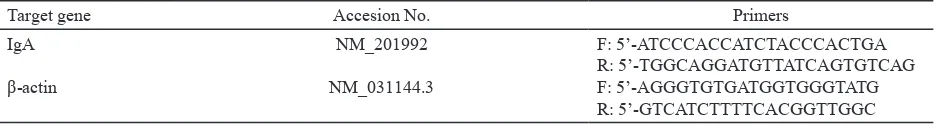
Primer Design. Primer sequences specific for IgA and β-actin encoding genes were designed using the NetPrimer program (Table 1). An mRNA sequence
encoding the Rattus norvegicus Fc fragment of the
IgA gene was used as a template to design the primer. The housekeeping gene, β-actin, was used as the reference gene, and the mRNA sequence encoding R. norvegicus β-actin gene was used to design the primer. Full sequences of the IgA and β-actin genes were taken from the GenBank database (http://www. ncbi.nlm.nih.gov).
RNA Extraction. Total rat mRNA was extracted from ileum tissue samples using the RNeasy Mini Kit (Qiagen, US) according to the manufacturer’s instructions. The quality and quantity of RNA were further evaluated by measuring A260/230 and A260/280 values using NanoDrop 2000 (Thermo Fisher Scientific, USA).
Assessment of mRNA Gene Expression.
Quantitative real-time polymerase chain reactions (qRT-PCR) were performed in an iQ5 Real-Time Detection System (BioRad, US) using KAPA SYBR FAST One-Step qRT-PCR Kit (Kapa Biosystem, USA). The master mix for qRT-PCR consisted of 10 µL 2x KAPA SYBR FAST qPCR master mix,
1 µL forward primer (10 pmol µl-1), 1 µL reverse
primer (10 pmol µL-1), 0.4 µL dUTP (10 mM), 0.4
µL KAPA RT Mix , 6.2 µL nuclease-free water, and
1 µL RNA template (100 ng µL-1). After 5 min at
42 °C for reverse transcription and 5 min at 95 °C for RT inactivation, further amplification reaction was carried out through 45 cycles of denaturation (3
Table 1. Primer sequences used for qRT-PCR
Target gene Accesion No. Primers
sec at 95 °C) and annealing (20 sec at 58 °C). The housekeeping gene, β-actin, was used as the reference gene. Gene expression levels were calculated based on the cycle threshold (Ct) value using the following formulas:
∆Ct (treatment) = Ct (treatment) – Ct (β-actin) ∆Ct (control) = Ct (control) – Ct (β-actin)
∆∆Ct = ∆Ct (treatment) - ∆Ct (control) Respective gene expression level = 2-∆∆Ct
IgA Quantification using ELISA Method.
Ninety-six well polystyrene plates (Nunc Maxisorp, Wiesbaden, Germany) were coated with standard IgA (Abcam, UK) and ileum lysates (after 4 month at -20 °C) in phosphate buffer saline (PBS) at 4 °C overnight. The remaining binding sites were blocked using PBS-Tween-5% skim milk for 1 hour at 37 °C, and then the plates were washed 4 times with PBS-Tween 0.01%. Plates were incubated 1 h at 37 °C with goat anti-mouse IgA horseradish peroxidase (Santa Cruz Biotechnology, Texas, USA) at 1:1000 dilution. Subsequently, IgA was detected by adding the substrate (3,3’,5,5’-Tetramethylbenzidine) diluted in phosphate citrate buffer (Sigma). The enzymatic
reaction was stopped using H2SO4 2N and absorbance
was measured at 450 nm. Data were interpolated into the IgA standard curve, and IgA concentration was expressed as ng/mg in tissue.
RESULTS
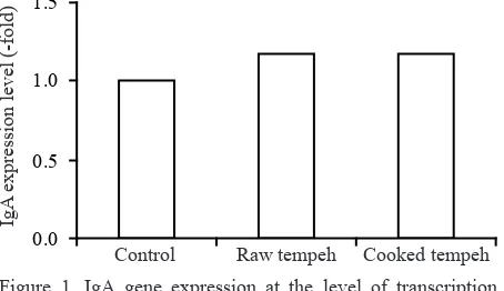
IgA expression at the level of transcription and of translation were compared between rats groups supplemented with raw and cooked tempeh, and without tempeh as a control during 28 days of treatment. IgA gene transcription results, as shown in Figure 1, indicated that mRNA expression of IgA genes as a result of the transcription process, in rats supplemented with raw and cooked tempeh, were 1.18 and 1.17 fold higher, respectively, compared to the control group. The greater expression of IgA gene at the transcription level was also followed by higher secretion of IgA protein as a result of the protein translation process in the small intestine. Such gene expression and protein secretion was higher by 2.46 and 2.08 fold, respectively, compared to the control group as shown in Figure 2. The secretion of IgA protein in ileum tissue from rats in the two treatment groups –one receiving raw tempeh and one given cooked tempeh– was higher compared to the control group, i.e. 3815 and 3225 ng/mg respectively, vs. 1500 ng/mg. This result indicates that tempeh supplementation may promote IgA secretion in small intestine.
DISCUSSION
Tempeh is fermented using a mixed culture of molds, yeasts, lactic acid bacteria, and different
Gram-negative bacteria. Rhizopus oligosporus is
the most dominant fungus in tempeh, although some
other molds are also present, such as Rhizopus oryzae
or Mucor spp. In addition, bacteria species such as Acetobacter indonesiensis, Klebsiella pneumoniae, Bacillus subtilis, and Flavobacterium sp. have been reported as the dominant bacteria found in EMP
tempeh; whilst Klebsiella sp. and Pseudomonas
putida were the dominant bacteria found in WJB
tempeh (Barus et al. 2008). Moreno et al. (2002)
and Nout and Kiers (2005) showed previously that the process of cooking soybeans reduced microbial populations in large numbers. These differences in bacteria communities between both types of tempeh consequently influences the nutrients in tempeh.
For example, bacteria belong to phylum of
Proteobacteria and Firmicutes were recently reported
to produce vitamin B12 in tempeh (Seumahu 2012).
comparing treatment groups– supplemented with
raw tempeh, and cooked tempeh-- and the control group.
Control Raw tempeh Cooked tempeh
6000
Control Raw tempeh Cooked tempeh
Mucosal IgA/gut weight (ng/mg tissue)
Figure 2. The product of IgA translation, defined as IgA protein
concentration in ileum lysate per ng/mg ileum tissue. Protein concentration was compared between
Subsequent study of vitamin B12 - producing bacteria
showed that K. pneumoniae found in tempeh, has a
different genetic profile than pathogenic isolates of
the same bacteria (Ayu et al. 2014). The presence of
these microorganism communities in tempeh might contribute to higher secretion of IgA.
Ingestion of probiotics has been proven to stimulate the secretion of IgA –the major antibody in mucosal immune system– in the small intestine,
(Tuohy et al. 2003). The mucosal epithelium is
the first line defense against pathogens and when breached by microorganisms will stimulate immediate innate immune response, such as engulfment by macrophages. Meanwhile, an adaptive immune response will be developed if the innate system is not able to overcome the invasion. Constituents of the bacterial cell, wall like lipopolysaccharides, peptidoglycan, and oligosaccharides, are powerful inducers of immune response. These substances will be recognized by antigen receptors which induce B-cell activation and stimulate IgA secretion in the intestine. This immunological memory will provide lifelong protection against pathogens (Adams 2010).
Probiotics are commonly described as live, non-pathogenic microorganisms which, in adequate doses, confer health benefits to the host. However, the results of this study showed that both raw and cooked tempeh can stimulate similar increases in IgA secretion, despite the fact that cooked tempeh does not include live microorganisms. Although they performed similarly, raw tempeh supplementation did produce slightly higher IgA levels compared to cooked tempeh. Viable probiotic cells may induce higher numbers of antibodies than non-viable cells because of their protein properties. Heat treatment to inactivate probiotic cells may result in modifying the bacterial properties by denaturing and changing the conformation of proteins, potentially leading to altered interaction of bacterial cell surface components with the host immune cells (Taverniti & Guglielmetti 2011). Many studies have showed the same beneficial result when hosts are administered non-live microorganisms.
The results of this study showed that both raw and cooked tempeh supplementation can stimulate IgA secretion in gut mucosal tissue. IgA gene expression at the transcription level in rats supplemented with raw and cooked tempeh were higher compared to the control group. Furthermore, this IgA gene result at transcription was also followed by higher IgA protein secretion at the translation level, in the groups supplemented with tempeh compared to the control group. Previous studies have found that IgA protein secretion in the small intestine of untreated rats was
around 1500-1600 ng/mg ileum tissue (Cladera et
al. 2012).
Since the study of tempeh as an immunomodulatory agent has not previously been done, further studies are needed to evaluate the precise mechanisms by which tempeh supplementation confers health benefits. Adding non-fermented soybean supplementation as a control treatment may help to differentiate and clarify the effect of tempeh supplementation in the gut immune system. Further analysis of tempeh as an agent of immune modulation at the levels of transcription and translation might show the specific process of stimulation tempeh has in the IgA gene expression system.
This research confirmed that both live and dead cells can confer a health benefit by stimulating IgA expression in gut mucosal tissue.
REFERENCES
Adams CA. 2010. The probiotics paradox: live and dead cells
are biological response modifiers. Nutr Res Rev 23:37-46. Ayu E, Suwanto A, Barus T. 2014. Klebsiella pneumoniae from
Indonesian Tempeh were genetically different from that of pathogenic isolates. Microbiol Indones 8:9-15.
Babu PD, Bhakyaraj R, Vidhyalakshmi R. 2009. A low cost nutritional food “tempeh”- a review. World J Dairy Food Sci 4:22-27.
Barus T, Suwanto A, Wahyudi AT, Wijaya H. 2008. Role of bacteria in tempeh bitter-taste formation and molecular biological analysis based on 16S rRNA gene. Microbiol Indones 2:17-21.
Binns N. 2013. Probiotics, prebiotics and the gut microbiota. In: Howlett J (ed). ILSI Europe Concise Monograph Series. Brussels (BE): ILSI Europe. p 1-32.
Cladera MM, Berezo TP, Franch A, Castell M, Perez-Cano FJ. 2012. Cocoa modulatory effect on rat faecal microbiota and colonic crosstalk. Arch Biochem Biophys 527:105-112. Collins MD, Gibson GR. 1999. Probiotics, prebiotics, and
synbiotics approaches for modulating the microbial ecology of the gut. Am J Clin Nutr 69:1052S-1057S.
Dixit AK, Antony JIX, Sharma NK, Tiwari RK. 2011. Soybean
Constituents and Their Functional Benefits. Kerala (IN): Research Signpost.
Egounlety M. 1994. Production, properties and utilization of mould fermented foods from soybean (Glycine max Merr.), cowpea (Vigna unguiculata L. Walp) and groundbean (Macrotyloma geocarpa Harms) [Dissertation]. Ibadan (NG): University of Ibadan.
Kataria J, Li N, Wynn JL, Neu J. 2009. Probiotic microbes: do
they need to be alive to be beneficial? Nutr Rev 67:546-550. Moreno MRF, Leisner JJ, Tee LK, Ley C, Radu S, Rusul G,
Vancanneyt M, de Vuyst LT. 2002. Microbial analysis of Malaysian tempeh, and characterization of two bacteriocins produced by isolates of Enterococcus faecium. J Appl Microbiol 92:147-157.
Nout MJR, Kiers JL. 2005. Tempe Fermentation, innovation and functionality: update into the third millennium. J Appl Microbiol 98:789-805.
Reyna-Garfias H, Miliar A, Jarillo-Luna A, Rivera-Aguilar V,
Pacheco-Yepez J, Baeza I, Campos-Rodriguez R. 2010. Repeated restraint stress increases IgA concentration in rat small intestine. Brain Behav Immun 24:110-118.
Robertfroid M, Slavin J. 2000. Nondigestable oligosaccharide. Crit Rev Food Sci Nutr 40:461-480.
Schrenzenmeir J, Vrese M. 2001. Probiotics, prebiotics, and synbiotics: approaching a definition. Am J Clin Nutr 73:316S-4S.
Scott KP, Duncan SH, Flint HJ. 2008. Dietary fibre and the gut
microbiota. Nutr Bull 33:201-211.
Seumahu CA, Suwanto A, Rusmana I, Solihin DD. 2012. Comparison of DNA extraction methods for microbial community analysis in Indonesian tempe employing
amplified ribosomal intergenic spacer analysis. Hayati J Biosci 19:93-98.
Taverniti V, Guglielmetti S. 2011. The immunomodulatory properties of probiotic microorganisms beyond their viability (ghost probiotics: proposal of paraprobiotic concepts). Genes Nutr 6:261-274.
Teran FR, Owens JD. 1999. Fate of oligosaccharides during production of soybean tempeh. J Sci Food Agric 79:249-252. Tuohy KM, Probert HM, Smejkal CW, Gibson GR. 2003.