Early neurological deterioration represents recurrent attack in acute
small non-lacunar stroke
Noriko Matsumoto
a,*, Kazumi Kimura
a, Chiaki Yokota
b, Kiminobu Yonemura
a,
Kuniyasu Wada
a, Makoto Uchino
c, Kazuo Minematsu
aaCerebrovascular Division, Department of Medicine, National Cardiovascular Center, 5-7-1 Fujishirodai, Suita, Osaka 565-8565, Japan bCerebrovasucular Laboratory, National Cardiovascular Center, Research Institute, 5-7-1 Fujishirodai, Suita, Osaka 565-8565, Japan
c
Department of Neurology, Kumamoto University School of Medicine, 1-1-1 Honjo, Kumamoto 860-0811, Japan
Received 19 February 2003; received in revised form 11 September 2003; accepted 12 September 2003
Abstract
The aim of this study was to identify the frequency and possible pathogenic mechanisms of early neurological deterioration in patients with acute small non-lacunar infarction. We studied 46 patients (35 men, 11 women; age, 70.3F10.4 years) with acute small non-lacunar infarction.
Small non-lacunar infarction was diagnosed using diffusion-weighted magnetic resonance imaging (DWI) as being < 15 mm in diameter and located in the cortex and centrum ovale in the middle cerebral artery (MCA) territory. The patients were divided into two groups; Group D (n= 6) had neurological deterioration within 7 days after symptom onset, while Group N (n= 40) did not have any neurological deterioration. In Group D, the interval from symptom onset to clinical deterioration was 3.3F1.5 days (range 2 – 6 days). Blood pressure on admission was
higher in Group D than in Group N (p< 0.05). In Group D, four of these five patients with follow-up DWI had new acute small ischemic lesions in addition to the initial lesions, indicating recurrent attacks of brain infarction. Neurological deterioration occurred within 7 days after symptom onset in 13% of patients. Neurological deterioration was frequently caused by recurrent infarction detected by DWI.
D2003 Elsevier B.V. All rights reserved.
Keywords:Acute stroke; Small non-lacunar infraction; DWI; Neurological deterioration; Recurrent stroke
1. Introduction
Early neurological deterioration is a common event in acute stroke, being 20 – 40% in frequency [1]. In stroke patients with moderate-to-severe neurological deficits, Eu-ropean Cooperative Acute Stroke Study (ECASS) I reported that the incidence of early and late progressing stroke was 37.5% and 20.3% [2]. The cause of the progressing stroke is often explained by the development of brain edema associated with an brain infarct [2]. In lacunar infarction with mild neurological deficits, the incidence of a progressive course during the acute phase of stroke is also observed in 24 – 36% of patients [3 – 5].
The impairment of the microcirculation in penetrating artery may play a major role in this phenomenon. How-ever, there is still no precise knowledge of the cause of progression and we are unable to predict patients at risk. Therefore, it is important to advance the search for the underlying pathogenic mechanisms of neurological deteri-oration in acute stroke patients.
Recently, we reported that symptomatic small non-lacunar infarcts (small centrum semiovale infarcts and cortical infarcts) were more frequently associated with large vessel disease and cardioembolism than lacunar infarcts[6,7]. We concluded that the mechanism of stroke in this form of infarction was often embolic from artery or heart, and should be differentiated from lacunar infarction, which is a small vessel disease.
Recurrent infarction must be considered as one potential cause of neurological deterioration following stroke. How-ever, it is often difficult to distinguish progressing stroke from a recurrent attack. Neuroimaging may help the
diag-0022-510X/$ - see front matterD2003 Elsevier B.V. All rights reserved. doi:10.1016/j.jns.2003.09.003
* Corresponding author. Department of Neurology, Kumamoto Uni-versity School of Medicine, 1-1-1 Honjo, Kumamoto 860-0811, Japan. Tel: +81-96-373-5893; fax: +81-96-373-5895.
E-mail address:[email protected] (N. Matsumoto).
nosis of a recurrent attack. If neuroimaging can display new lesions separately to the initial lesions, we can diagnose the patient as having a recurrent attack.
Diffusion-weighted imaging (DWI) is a powerful tool for detecting recent small ischemic lesions, particularly in the centrum ovale or cortex [6,8]. We experienced a patient with neurological deterioration in small non-lacu-nar infarcts, whose follow-up DWI study revealed new small infarcts in addition to the initial infarcts, indicating recurrent attacks. Therefore, we hypothesized that neuro-logical deterioration during the acute phase in patients with small non-lacunar infarcts might be caused by recurrent attacks.
To the best of our knowledge, the frequency and mech-anisms of neurological deterioration in patients with small non-lacunar infarcts remain unclear. We studied consecutive patients with small non-lacunar infarcts to solve the above-mentioned problems using DWI.
2. Materials and methods
The aim of this study was firstly to examine the frequency of neurological deterioration within 7 days after symptom onset in patients with small non-lacunar infarcts. Second, we compared clinical characteristics between patients with and without neurological deterioration. Fur-thermore, in patients with neurological deterioration, we performed a follow-up DWI study to detect new small ischemic lesions in addition to the initial lesions after deterioration.
We enrolled consecutive patients with small non-lacunar infarction admitted to our division of National Cardiovas-cular Center within 7 days of symptom onset between January 2000 and February 2002 into the present study. DWI was performed within 7 days of symptom onset to detect acute ischemic lesions.
Small non-lacunar infarcts were defined as follows: (1) lesions on DWI were acute; (2) diameter of the lesions was smaller than 15 mm; and (3) lesions were located in the cortex or centrum ovale[9]. An infarct situated in the corona radiata, putamen, globus pallidus, and internal capsule, which are supplied by the deep perforating arteries of the middle cerebral artery (MCA), were excluded from this study[9,10].
We assessed the neurological severity on admission using National Institutes of Health Stroke Scale (NIHSS) score
[11]and handicap at discharge using modified Rankin scale (mRS)[12]. Neurological deterioration was diagnosed when NIHSS score increased z2 points from the baseline NIHSS score during the 7 days after symptom onset. The mode of deterioration was classified into four subgroups; abrupt, stepwise, fluctuating and linear slope-like. Patients were classified into two groups; patients displaying neurological deterioration (Group D), and those without any neurological deterioration (Group N).
MR imaging studies were conducted for all patients within 7 days after symptom onset. When patients had neurological deterioration, a follow-up DWI study was conducted. MR imaging was performed using a Siemens MAGNETOM Vision 1.5-T MR unit with echo-planar capability. DWI was performed simultaneously using a multislice, single-shot, spin echo planar imaging sequence in all patients within 7 days of symptom onset. Diffusion gradients were applied in each of the x-, y-, and z-axes with two b values (0 and 1000 s/mm2). Fluid-attenuated inversion recovery (FLAIR, TR/TE, 9000/105) images was carried out simultaneously with DWI. The criterion for the diagnosis of acute infarcts on DWI was focal hyperinten-sity, judged not attributable to normal anisotropic diffusion or magnetic susceptibility artifacts.
Vascular risk factors were identified as follows: (1) use of antihypertensive agents, or systolic blood pressure >160 mm Hg or diastolic blood pressure >95 mm Hg on admission for hypertension; (2) use of oral hypoglycemic agents or insulin, or glycosylated hemoglobin (HbA1C) >6.4% for diabetes mellitus; (3) use of antihyperlipi-demic agents or serum cholesterol level >220 mg/dl for hypercholesterolemia.
We carried out color-flow duplex carotid ultrasonogra-phy, conventional cerebral angiograultrasonogra-phy, and magnetic resonance angiography (MRA) to evaluate arterial diseases in the carotid system. Color-flow duplex carotid ultraso-nography (Toshiba SSA 270A, Toshiba Inc., Tokyo, Japan, or Ultramark 9 HDI, ATL, Bothel, WA) was performed in all patients as a routine test. The grade of arterial stenosis was determined according to the criteria used in the North American Symptomatic Carotid Endar-terectomy Trial (NASCET) [13]. Arterial diseases were considered significant when stenosis >70%, occlusion, or ulceration were evident in the ipsilateral carotid system. In addition, the peak systolic blood flow velocity in the internal carotid artery (ICA) greater than 200 cm/s on ultrasonography was considered equivalent to ICA steno-sis >70% for the NASCET criteria [14].
To detect an emboligenic cardiac disease, all patients were examined using 12-lead electrocardiography (ECG), 24-h ECG monitoring, and transthoracic echocardiogra-phy (TTE). Additionally, we conducted transesophageal echocardiography (TEE) to evaluate patent foramen ovale (PFO) and atherosclerosis of the aortic arch (aortic complicated plaque). Emboligenic cardiac diseases includ-ed non-valvular atrial fibrillation (NVAF), mitral stenosis, left ventricular aneurysm, prosthetic cardiac valves, dilat-ed cardiomyopathy, and PFO. An aortic complicatdilat-ed plaque was considered significant as a plaque z4 mm or mobile plaque in the aortic arch visualized on TEE
[15].
WhitneyU-test between the two groups. Values ofp< 0.05 were considered statistically significant.
3. Results
A total of 404 patients with acute ischemic stroke or TIA were admitted to our division within 7 days after symptom onset, and 356 (88%) patients were performed DWI within 7 days after symptom onset. Out of them, 46 patients (35 men, 11 women) with a small non-lacunar infarct were enrolled into the present study. Age (meanFS.D.) of the patients was 70.3F10.4 years (median 72-years-old, range 38 – 87).
An initial DWI study showed a small ischemic lesion in 14 patients, and multiple lesions in 32 patients. In 14 patients with a single lesion, the lesion was located in the
cortex in seven patients, and in the subcortex in the other seven patients. In the 32 patients with multiple lesions, the lesions were located only in the cortex in seven patients, only in the subcortex in seven patients, and in both the cortex and subcortex in the remaining 18 patients.
We performed conventional cerebral angiography in six patients (13%), MRA in 27 patients (59%), and both assess-ments in 12 patients (26%). Twenty-three patients (50%) had a significant arterial disease. The following arterial lesions were observed; MCA occlusion in one patient, more than 50% stenosis of the horizontal portion of the MCA in three, ICA occlusion in four, more than 70% ICA stenosis in 10, less than 70% ICA stenosis but with ulceration in two, more than 70% ICA stenosis and the anterior cerebral artery occlusion in one, and more than 70% stenosis of the ICA and horizontal portion of the MCA in two.
Table 1
Demographic and clinical feature of patients of Group D
Age/Sex Day of Mode of NIHSS score Site of lesion (distribution, number) Potential embolic deterioration deterioration On
admission Before deterioration
After deterioration
Initial DWI Follow-up DWI (after deterioration)
source
Initial lesion Additional lesion 77/M 2 abrupt 7 7 9 Lt MCA (cortex and subcortex),
multiple
no change Lt ICA 50% stenosis with ulceraion, Ao. 57/M 6 abrupt 7 7 13 Rt MCA (subcortex), Lt MCA
(cortex), multiple
Rt MCA (subcoetex), single
Ao.
66/M 2 stepwise 5 5 9 Rt MCA (subcortex), Lt MCA (cortex, subcortex), multiple
Lt MCA (subcoetex), single
Bilateral ICA 70% stenosis, Ao. 78/M 3 stepwise 6 4 9 Rt MCA (cortex and subcortex),
multiple
Rt MCA (cortex and subcortex), multiple
Rt.ICA 90% stenosis
81/F 3 stepwise 2 2 6 Lt MCA (subcortex), multiple NA NVAF, Ao. 74/M 4 fluctuating 6 6 8 Lt MCA (subcortex), multiple Lt MCA (subcortex),
single
>50% MCA stenosis
Ao.: aortic complicated plaque, NVAF: non valvular atrial fibrillation, NA: not available.
We conducted the TEE in 30 patients. Emboligenic cardiac diseases were detected in 22 (48%) patients; only NVAF in 10, only PFO in seven, left ventricular aneurysm with thrombus in two, both AF and a prosthetic mitral valve in one, and both NVAF and PFO in two. Aortic complicated plaques were detected in 17 patients. Overall, 41 patients (89%) had a potential embolic source.
Six patients (13%; Group D) had neurological deteriora-tion, and 40 patients (87%; Group N) did not have any deterioration. The interval from symptom onset to neuro-logical deterioration was 3.3F1.5 days (range 2 – 6 days). No patients had neurological deterioration before the first MRI study. The mode was abrupt in two patients, fluctuat-ing in one and stepwise in three. The demographic and clinical features of the six patients with neurological dete-rioration are summarized in Table 1. All patients had a
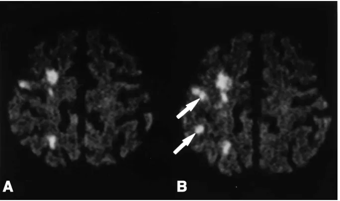
potential embolic source. In the Group D, a follow-up MRI study was performed on all the patients except one, who declined an MRI test. Four of the five patients who underwent follow-up imaging had new acute ischemic lesions surrounding the initial lesions (Fig. 1). While the remaining one patient had no new lesions. In the Group N, a follow-up DWI study was performed in only one patient within 7 days of symptom onset. The patient did not have any new lesions except for initial lesions.
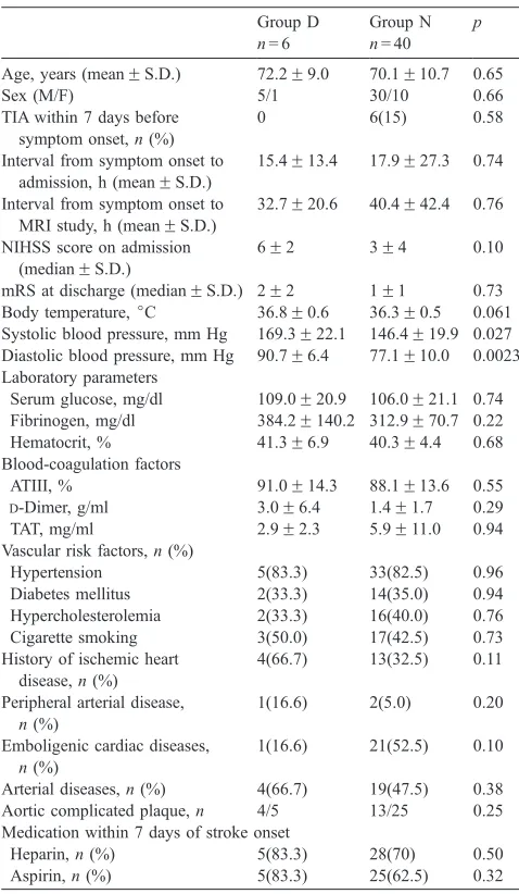
Table 2 shows the clinical characteristics of the two
groups. Systolic and diastolic blood pressures on admission were higher in Group D than in Group N. No statistically significant differences in age, sex, TIA within 7 days before symptom onset, the interval from symptom onset to admis-sion, and to initial DWI study, NIHSS score on admisadmis-sion, body temperature on admission, laboratory parameters, blood-coagulation factors, vascular risk factors, history of ischemic heart disease, peripheral arterial disease, arterial and cardiac diseases, aortic complicated plaques and use of medication were observed between the two groups. An mRS score at discharge was not different.
4. Discussion
Our study demonstrated that the frequency of neurolog-ical deterioration in patients with small non-lacunar infarcts was 13% during 7 days of symptom onset. In lacunar stroke, frequency of neurological progression was 24 – 36%[3 – 5]. Nakamura et al. [4] reported that diabetes mellitus and the severity of motor deficits on admission might predict pro-gression of motor deficits. Lodder et al. [5] showed that progression of symptom was associated with a large infarct volume. Lacunar infarction was caused by occlusion of deep perforators from the horizontal portion of MCA. On the other hand, small non-lacunar infarction was due to occlusion of the MCA branches and the medullary arteries originating from superficial branches of MCA. Furthermore, the fre-quency of arterial and cardiac disease was different between non-lacunar and lacunar infarction[6,7]. In fact, 89% of our patients had a potential embolic source. We suspect that a small non-lacunar infarct may be caused by embolism from a large artery and heart [6,7]. Therefore, the discrepancy in frequency of neurological deterioration between lacunar and small non-lacunar infarction was explained by the difference in the pathogenic mechanism of stroke.
High serum glucose levels, history of diabetes, stroke severity on admission, and early focal hypodensity and brain swelling on the initial CT scan have been associated with neurological deterioration in acute stroke[2,16 – 19]. Above-mentioned factors might result in insufficient collateral blood supply, expanding brain edema, or metabolic deterioration during acute phase of stroke. Infarct size in our patients was always too tiny for initial CT findings to represent an important factor. In the present study, no differences in serum glucose levels and history of diabetes were observed between
Table 2
TIA within 7 days before symptom onset,n(%)
0 6(15) 0.58
Interval from symptom onset to admission, h (meanFS.D.)
15.4F13.4 17.9F27.3 0.74
Interval from symptom onset to MRI study, h (meanFS.D.) Body temperature,jC 36.8F0.6 36.3F0.5 0.061 Systolic blood pressure, mm Hg 169.3F22.1 146.4F19.9 0.027 Diastolic blood pressure, mm Hg 90.7F6.4 77.1F10.0 0.0023 Laboratory parameters
Serum glucose, mg/dl 109.0F20.9 106.0F21.1 0.74 Fibrinogen, mg/dl 384.2F140.2 312.9F70.7 0.22 Hematocrit, % 41.3F6.9 40.3F4.4 0.68 Blood-coagulation factors
ATIII, % 91.0F14.3 88.1F13.6 0.55
D-Dimer, g/ml 3.0F6.4 1.4F1.7 0.29 TAT, mg/ml 2.9F2.3 5.9F11.0 0.94 Vascular risk factors,n(%)
Hypertension 5(83.3) 33(82.5) 0.96 Diabetes mellitus 2(33.3) 14(35.0) 0.94 Hypercholesterolemia 2(33.3) 16(40.0) 0.76 Cigarette smoking 3(50.0) 17(42.5) 0.73 History of ischemic heart
Arterial diseases,n(%) 4(66.7) 19(47.5) 0.38 Aortic complicated plaque,n 4/5 13/25 0.25 Medication within 7 days of stroke onset
Heparin,n(%) 5(83.3) 28(70) 0.50 Aspirin,n(%) 5(83.3) 25(62.5) 0.32 NIHSS: National Institutes of Health Stroke Scale, mRS: modified Rankin scale.
the two groups. The number of our patients might be too small to find differences between the two groups.
Systolic and diastolic blood pressures on admission were higher in Group D than Group N. Da´valos et al.[17]reported that high systolic blood pressure on admission was indepen-dently related with early deterioration after ischemic stroke. Whereas, Jørgensen et al. [18] showed that high systolic blood pressure on admission decreased risk for early pro-gression. In the present study, the exact relationship between high blood pressure and neurological deterioration is un-known. A further study will be needed to solve the issue.
In the present study, NIHSS score and the body tem-perature was higher in Group D than in Group N, but these difference was not significant. Patients with high NIHSS score at admission were likely to have neurological deterio-ration in acute phase of ischemic stroke [2,19]. A few investigators reported that patients with higher temperature had a worse stroke outcome[20 – 22]. However, it has still unknown whether higher temperature is associated with early deterioration at acute phase of ischemic stroke.
In our study, the follow-up DWI study in all the patients but one with neurological deterioration revealed new small infarcts addition to the initial infarcts. Therefore, we con-cluded that recurrence of small infarcts resulted in neuro-logical deterioration. In patients with small non-lacunar infarcts, prevention of recurrent infarcts is important for avoiding neurological deterioration.
A number of problems were present in this study. Firstly, there was a small sample size of deterioration patients with a follow-up DWI study. Therefore, statistic analysis was weak. Secondly, we could not conduct follow-up DWI studies in many patients without neurological deterioration. Therefore, we could not exclude the possibility that new but asymptom-atic lesions appeared if follow-up DWI in those patients is performed.
In conclusion, our study demonstrated that the frequency of neurological deterioration in patients with small non-lacunar infarcts was 13% within 7 days after symptom onset. Neurological deterioration in these patients was frequently accompanied by recurrent infarction visualized with DWI.
Acknowledgements
This study was supported in part by Research Grants for Cardiovascular Disease (12A-4, 14C-1) from the Ministry of Health, Labor and Welfare of Japan and by Special Coordinating Funds for Promoting Science and Technology (Strategic Promotion System for Brain Science) from the Science and Technology Agency of Japan.
References
[1] Da´valos A, Castillo J. Progressing stroke. In: Fisher M, Bogousslavsky J, editors. Current Review of Cerebrovascular Disease. Philadelphia (PA): Current Medicine Inc; 1999. p. 149 – 60.
[2] Da´valos A, Toni D, Iweins F, Lesaffre E, Bastianello S, Castillo J, et-al., for the ECASS Group. Neurological deterioration in acute ischemic stroke. Potential predictors and associated factors in Euro-pean cooperative acute stroke study (ECASS) I. Stroke 1999;30: 2631 – 6.
[3] Serena J, Leira R, Castillo J, Pumar JM, Castellanos M, Da´valos A. Neurological deterioration in acute lacunar infarctions: the role of excitatory and inhibitory neurotransmitters. Stroke 2001;32: 1154 – 61.
[4] Nakamura K, Saku Y, Ibayashi S, Fujishima M. Progressive motor deficits in lacunar infarction. Neurology 1999;52:29 – 33.
[5] Lodder J, Gorsselink EL. Progressive stroke caused by CT-verified small deep infarcts; relation with the size of the infarct and clinical outcome. Acta Neurol Scand 1985;71:328 – 30.
[6] Yonemura K, Kimura K, Minematsu K, Uchino M, Yamaguchi T. Small centrum ovale infarcts on diffusion-weighted magnetic reso-nance imaging. Stroke 2002;33:1541 – 4.
[7] Wada K, Kimura K, Minematsu K, Uchino M, Yamaguchi T. Spotty cortical enhancement detected by magnetic resonance imaging: a sign of embolic transient ischemic attack and stroke? J Stroke Cerebrovasc Dis 2001;10:19 – 22.
[8] Warach S, Gaa J, Siewert B, Wielopolski P, Edelman RR. Acute human stroke studied by whole brain echo planar diffusion-weighted magnetic resonance imaging. Ann Neurol 1995;37:231 – 41. [9] Tatu L, Moulin T, Bogousslavsky J, Duvernoy H. Arterial
territo-ries of human brain. Cerebral hemispheres. Neurology 1998;50: 1699 – 708.
[10] Bogousslavsky J, Regli F. Centrum ovale infarcts: subcortical infarc-tion in the superficial territory of the middle cerebral artery. Neurol-ogy 1992;42:1992 – 8.
[11] Brott T, Adams Jr HP, Olinger CP, Marler JR, Barsan WG, Biller J, et al. Measurements of acute cerebral infarction: a clinical examina-tion scale. Stroke 1989;20:864 – 70.
[12] Swieten JC, Koudstaal PJ, Visser MC, Schouten HJA, Gijn J. Inter-observer agreement for the assessment of handicap in stroke patients. Stroke 1988;19:604 – 7.
[13] North American Symptomatic Carotid Endarterectomy Trial Collab-orators. Beneficial effect of carotid endarterectomy in symptomatic patients with high-grade carotid stenosis. N Engl J Med 1991;325: 445 – 53.
[14] Koga M, Kimura K, Minematsu K, Yamaguchi T. Diagnosis of in-ternal carotid artery stenosis greater than 70% with power doppler duplex sonography. AJNR Am J Neuroradiol 2001;22:413 – 7. [15] Amarenco P, Cohen A, Tzourio C, Bertrand B, Hommel M, Besson
G, et al. Atherosclerotic disease of the aortic arch and the risk of ischemic stroke. N Engl J Med 1994;331:1474 – 9.
[16] Toni D, Fiorelli M, Gentile M, Bastianello S, Sacchetti ML, Argentito C, et al. Progressing neurological deficit secondary to acute ischemic stroke. A study on predictability, pathogenesis, and prognosis. Arch Neurol 1995;52:670 – 5.
[17] Da´valos A, Cendra E, Teruel J, Martinez M, Genı´s D. Deteriorating ischemic stroke: risk factors and prognosis. Neurology 1990;40: 1865 – 9.
[18] Jørgensen HS, Nakayama H, Raaschou HO, Olsen TS. Effect of blood pressure and diabetes on stroke in progression. Lancet 1994;344: 156 – 9.
[19] DeGraba TJ, Hallenbeck JM, Pettigrew KD, Dutka AJ, Kelly BJ. Progression in acute stroke. Value of the initial NIH Stroke Scale Score on patient stratification in future trials. Stroke 1999;30:1208 – 12. [20] Azzimondi G, Bassein L, Nonino F, Fiorani L, Vignatelli L, Re G,
et al. Fever in acute stroke worsens prognosis: a prospective study. Stroke 1995;26:2040 – 3.
[21] Castillo J, Da´valos A, Marrugat J, Noya M. Timing for fever-related brain damage in acute ischemic stroke. Stroke 1998;29:2455 – 60. [22] Wang Y, Lim LL, Levi C, Heller RF, Fisher J. Influence of admission