Acute Kidney Injury Following Cardiac Surgery
- Incidence, Risk Factors, Association With Other
Perioperative Complications, Survival, and Renal Recovery
Solveig Helgadottir
Thesis for the degree of Doctor of Philosophy
Supervisor and advisor:
Tomas Gudbjartsson
Doctoral committee:
Arnar Geirsson, Gisli H. Sigurdsson, Martin I. Sigurdsson,
Runolfur Palsson
Bráður nýrnaskaði eftir hjartaskurðaðgerðir
- tíðni, áhættuþættir, tengsl við aðra fylgikvilla við og eftir
skurðaðgerð, lifun og langtímaáhrif á nýrnastarfsemi
Sólveig Helgadóttir
Ritgerð til doktorsgráðu
Umsjónarkennari og leiðbeinandi:
Tómas Guðbjartsson
Doktorsnefnd:
Arnar Geirsson, Gísli H. Sigurðsson,
Martin I. Sigurðsson og Runólfur Pálsson
Thesis for a doctoral degree at the University of Iceland. All rights reserved. No part of this publication may be reproduced in any form without the prior permission of the copyright holder.
© Sólveig Helgadóttir 2017 ISBN 978-9935-9319-5-5 Printing by Háskólaprent ehf. Reykjavik, Iceland 2017
Ágrip
Framfarir í skurðlækningum og gjörgæslu hafa leitt til bættrar meðferðar sjúklinga sem þjást af hjarta- og æðasjúkdómum. Í dag eru opnar hjartaaðgerðir algengar á Vesturlöndum og útlit er fyrir að þeim muni fjölga á komandi áratugum, samfara auknum fjölda aldraðra. Sjúklingar sem gangast undir opnar hjartaaðgerðir eru oft með flókin heilsufarsvandamál sem eykur tíðni fylgikvilla. Á meðal algengustu fylgikvilla eftir opnar hjartaaðgerðir eru hjartsláttaróregla, einkum gáttatif, og blæðingar. Bráður nýrnaskaði (BNS) er einnig algengur fylgikvilli eftir hjartaaðgerðir og tengist verri horfum sjúklinga eftir aðgerð. Skortur á gögnum um grunnstarfsemi nýrna hefur þó verið galli á mörgum fyrri rannsóknum sem auk þess hafa oft á tíðum skoðað mjög sérhæfða sjúklingahópa.
Árlega eru framkvæmdar um 250 opnar hjartaaðgerðir á Íslandi og eru um 2/3 þeirra kransæðahjáveituaðgerðir. Markmið þessarar doktorsritgerðar var að meta tíðni og áhættuþætti BNS eftir hjartaaðgerðir á Íslandi en skort hefur á slíkar rannsóknir fram til þessa. Auk þess voru könnuð tengsl BNS og annarra fylgikvilla eftir opnar hjartaaðgerðir. Loks var samband BNS og langtíma skerðingar á nýrnastarfsemi rannsakað, þar með talið þörf á nýrnaskilunarmeðferð, sem og langtímalifun. Að lokum var sérstaklega lagt mat á hvaða áhrif bati á nýrnastarfsemi hefur á horfur sjúklinga sem hljóta BNS.
Við rannsóknirnar var stuðst við gagnagrunna með ítarlegum upplýsingum um alla sjúklinga sem gengust undir hjartaaðgerð á Íslandi frá 2001-2015. Ber þar sérstaklega að nefna nákvæma skráningu á nýrnastarfsemi sjúklinga, bæði fyrir og eftir aðgerð, en þær upplýsingar eru forsenda til að geta lagt mat á tíðni og áhættuþætti BNS.
Rannsóknirnar leiddu í ljós að tíðni BNS eftir hjartaaðgerðir er heldur lægri hér á landi samanborið við nágrannalönd okkar. Líktog aðrar rannsóknir hafa sýnt er BNS tengdur undirliggjandi ástandi sjúklinga og voru aldur, skerðing á nýrnastarfsemi fyrir aðgerð, hærri áhættustigun fyrir aðgerð, umfangsmeiri skurðaðgerð og hærri líkamsþyngdarstuðull sjálfstæðir áhættuþættir.
Bráður nýrnaskaði tengdist verri horfum sjúklinga eftir aðgerð sem meðal annars endurspeglaðist í hærri tíðni ýmissa fylgikvilla. Gáttatif reyndist algengasti fylgikvillinn og greindist í nærri helmingi sjúklinga sem gengust
þess kom í ljós að tíðni gáttatifs var marktækt aukin hjá sjúklingum sem fengu BNS. Eins og fyrir BNS, reyndust hærri aldur, umfang aðgerðar og hærri áhættustigun fyrir aðgerð vera sjálfstæðir áhættuþættir fyrir greiningu gáttatifis.
Greining BNS tengdist einnig aukinni þörf á skammtíma nýrnaskilunarmeðferð og versnun á langtíma nýrnastarfsemi. Aukinheldur höfðu sjúklingar sem fengu BNS marktækt lakari lifun eftir aðgerð og reyndist langtímalifun í öfugu hlutfalli við alvarleika upphaflegs nýrnaskaða. Meirihluti sjúklinga virtist hinsvegar endurheimta nýrnastarfsemi eftir BNS í kjölfar aðgerðar. Bati á nýrnastarfsemi spáði ekki fyrir um eins árs lifun sjúklinga en hinsvegar lifðu sjúklingar sem náðu bata marktækt lengur en samanburðarhópur sem ekki náði bata.
Með því að notast við yfirgripsmikla og nákvæma gagnagrunna, sem ná til heillar þjóðar, reyndist unnt að meta tíðni BNS og gáttatifs eftir opnar hjartaaðgerðir á Íslandi um leið og lifun og forspárþættir lifunar eftir þessar aðgerðir voru kannaðir. Flestir sjúklingar sem hlutu BNS endurheimtu nýrnastarfsemi og fáir hlutu endastigs nýrnabilun. Samt sem áður virðist BNS tengjast verri útkomu eftir hjartaaðgerð og ljóst er að úrræði skortir þegar greining BNS liggur fyrir. Snemmbúin greining BNS og kortlagning áhættuþátta á þó vonandi eftir að auka skilning okkar á BNS og hvernig fyrirbyggja má sjúkdóminn og að bæta horfur þessa sjúklingahóps.
Lykilorð:
Abstract
In line with advances in surgery, perioperative care, and intensive care, the outcome in patients treated for cardiovascular disease has improved. Open-heart surgery is common in most western countries and, in line with the general ageing of populations, the frequency of these procedures is expected to increase in the coming decades. Patients who undergo cardiac surgery often have complex medical problems that predispose them to a range of surgical complications. Among the most common postoperative complications are arrhythmias, most often atrial fibrillation, and bleeding. Acute kidney injury (AKI) is also a common and often serious complication―as well as being a strong risk factor for worse postoperative outcome and increased mortality after surgery. However, many previous studies have suffered from a lack of data on baseline kidney function before AKI as well as selective patient cohorts.
Approximately 250 open-heart surgeries are performed annually in Iceland. However, there has been a lack of data on the incidence and risk factors for AKI following heart surgery. The aim of the studies in this thesis was to evaluate the incidence of AKI using internationally validated criteria. Furthermore, we wanted to examine the association between AKI and other postoperative complications, and also long-term outcome, mainly regarding kidney function and survival. In that context, we particularly examined the effect of recovery of renal function on long-term outcome.
Data were gathered from comprehensive nationwide databases that contained detailed information on all patients who underwent heart surgery in Iceland from 2001 through 2015. Information on kidney function, both pre- and postoperatively, was recorded in a thorough manner, as it is a prerequisite for reliable evaluation of AKI.
The studies showed that the incidence of AKI after cardiac surgery in Iceland was relatively low compared to neighboring countries. In line with previous studies, the preoperative condition of patients was shown to be associated with the incidence of AKI. Advanced age, reduced preoperative kidney function, higher preoperative risk assessment scores, a higher body mass index, and more complex surgery were found to be independent predictors of the development of AKI.
reflected by higher rates of various postoperative complications when compared to patients with normal kidney function postoperatively. Atrial fibrillation, diagnosed in almost half of the patients, was the most common postoperative complication following surgical myocardial revascularization and aortic valve replacement in Iceland. Furthermore, the incidence of atrial fibrillation was higher in patients who sustained AKI than in those who did not. As in the case of AKI, the independent predictors of atrial fibrillation were advanced age, more complex surgery, and higher preoperative risk assessment scores.
The diagnosis of AKI was found to be associated with an increased need for renal replacement therapy and worse long-term kidney function. Also, AKI patients had significantly higher postoperative mortality and worse long-term survival, with survival being inversely correlated to the severity of AKI. Importantly, the majority of patients recovered their renal function after AKI. Although renal recovery was not a significant predictor of one-year survival, it was found to be associated with more favorable long-term survival.
By using extensive comprehensive nationwide databases, it was possible to determine the incidence of AKI after open-heart surgery in Iceland and to define risk factors. Most patients who sustained AKI recovered their renal function and the rate of progression to end-stage renal disease was low. Although AKI appears to be associated with a worse postoperative outcome in patients, early detection of AKI and gaining a better understanding of the risk factors remain important steps in reducing the morbidity and mortality of patients diagnosed with AKI. It is to be hoped that a better understanding of these factors will help to improve the long-term prognosis in this patient group, leading to increased rates of renal recovery and reduced costs of treatment.
Key words:
Acute kidney injury, incidence, long-term outcome, postoperative atrial fibrillation, renal recovery.
Acknowledgements
Many people deserve my sincere gratitude for their help on this long journey: Tomas Gudbjartsson, my main supervisor, for his support, guidance, and valuable friendship. For taking me on as a fifth-year medical student and leading me down this path, with all its twists and turns, and ups and downs.
My doctoral committee and colleagues, Arnar, Gisli, Martin, and Runolfur, for their helpful tips and encouragement. I am especially grateful to my friend Martin, with whom I have had the privilege to work on several studies and in clinical practice. He has tirelessly helped me with his vast knowledge in fields ranging from statistics to pop music and has spurred me on when it was most needed.
All the medical students and colleagues whom I have come to know and admire throughout the arduous process of data collection and recording; especially you, Thorir E. Long and Dadi Helgason. Special thanks also go to my colleagues and friends Inga Lara Ingvarsdottir and Gudbjorg Jonsdottir for their valuable support in pulling me through.
Gunnhildur Johannsdottir and Ingibjorg Richter are due heartfelt thanks for all their secretarial help over the years.
I am thankful to editors and proofreaders, and also for the statistical help from my friend and excellent scientist, Sigrun Helga Lund.
My colleagues and supervisors at the ANIVA clinic of the Akademiska Hospital in Uppsala, Sweden.
The University of Iceland Research Fund, the Landspitali University Hospital Science Fund, and the Memorial Fund of Helga Gudmundsdottir and Sigurlidi Kristjansson for financial support.
My amazing family, especially my dad and moms for teaching me the importance of endurance, patience, and hard work mixed with the finer things in life.
My siblings and in-laws, for being among the finer things in life, and for stimulating discussions on subjects that had nothing to do with medicine. I would like to thank Oddny, my sister, especially, for being an inspiration.
when I´ve been hopelessly buried in piles of textbooks and Excel files and so absent minded that I’ve forgotten everything else, even what it means to be a proper friend. Sunna Arnarsdottir has been especially supportive through her ongoing interest in scientific research.
I would like to thank my children, Helga Maria and Daniel Loki, for bearing with me and all the precious time I have spent away from them. Takk elsku hjartagullin mín, þið gerið alla daga svo óendanlega mikið betri.
Above all, I would like to thank my husband, Boas, who has given me endless love, support, and encouragement throughout this process. If not for him, I don’t think I would have set out on―let alone completed―this journey.
Finally, I want to express my gratitude and love to my dear ones who passed away before the final steps of this long journey.
Contents
Ágrip ... i
Abstract ... iii
Acknowledgements ... v
Contents ...vii
List of abbreviations ...xi
List of figures ...xiii
List of tables ...xv
List of original papers ...xvii
Declaration of contribution ...xviii
1 Introduction ... 1
1.1 The kidneys ... 1
1.2 Assessment of renal function ... 3
1.3 Estimation of GFR ... 5
1.4 Acute renal failure ... 6
1.5 Acute kidney injury ... 6
1.5.1 Defining AKI – the RIFLE, AKIN, and KDIGO criteria... 7
1.5.2 Ascertainment of baseline SCr ...10
1.5.3 The pathophysiology and etiology of AKI ...10
1.5.4 Risk factors for AKI ...11
1.6 Postoperative AKI ...12
1.6.1 AKI following cardiac surgery ...12
1.7 AKI in the ICU setting ...13
1.8 Prevention and treatment of AKI ...13
1.8.1 Renal replacement therapy ...14
1.9 Recovery of renal function ...15
1.10 Chronic kidney disease ...18
1.11 Cardiorenal syndrome ...19
1.12 Cardiovascular disease ...20
1.13 Postoperative atrial fibrillation following cardiac surgery ...23
1.13.1 Risk factors for POAF ...24
1.13.2 Treatment of POAF ...25
2.1 Study I ...29
2.2 Study II ...29
2.3 Study III ...29
2.4 Study IV ...29
2.5 Study V ...29
3 Materials and methods ...31
3.1 Registers ...31
3.1.1 Centralized registers ...31
3.1.2 The National Patient Register ...31
3.1.1 The Icelandic End-Stage Renal Disease Registry...33
3.1.2 Statistics Iceland and the Icelandic Cause of Death Register ...33 3.2 Study population ...33 3.2.1 Study I ...33 3.2.2 Study II ...33 3.2.3 Study III ...34 3.2.4 Study IV ...34 3.2.5 Study V ...35 3.3 Data collection ...36
3.4 Risk scores and preoperative comorbidity classification ...36
3.5 Laboratory methods ...36
3.6 Classification of AKI ...37
3.7 Classification of POAF ...37
3.8 Classification of complications and mortality ...37
3.9 Outcome measures ...38 3.9.1 Study I ...38 3.9.2 Study II ...38 3.9.3 Study III ...38 3.9.4 Study IV ...38 3.9.5 Study V ...38 3.10 Statistical analysis ...38 3.10.1 Calculation of incidence ...39
3.10.2 Univariate and multivariate analysis of risk factors...40
3.10.3 Receiver operating characteristic curve ...41
3.10.4 Follow-up ...41
3.10.6 Propensity score matching ...42
4 Results...45
4.1 Study I – Identification of baseline kidney function ...45
4.1.1 Patient characteristics ...45
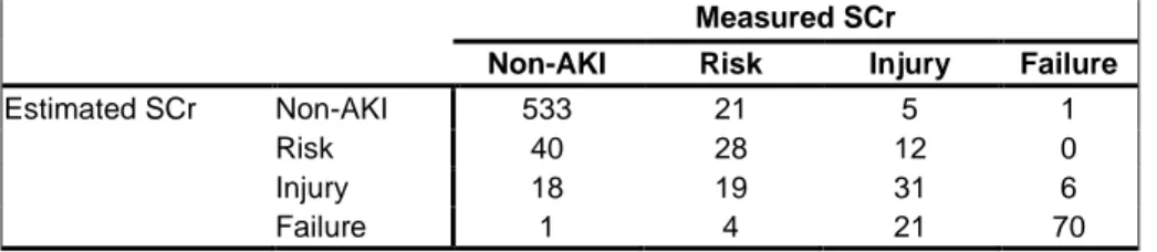
4.1.2 Estimated versus measured baseline SCr ...46
4.1.3 Risk factors for AKI ...46
4.1.4 Complications and survival ...47
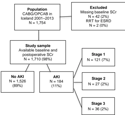
4.2 Study II – AKI following CABG ...48
4.2.1 Patient characteristics ...48
4.2.2 Operative factors and postoperative complications ...51
4.2.3 Risk factors for AKI ...53
4.2.4 Renal recovery and long-term kidney function ...53
4.2.5 Survival ...54
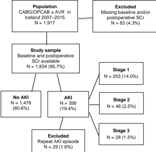
4.3 Study III – AKI following AVR ...55
4.3.1 Baseline patient characteristics ...55
4.3.2 Intraoperative characteristics and postoperative complications ...57
4.3.3 Risk factors for AKI ...58
4.3.4 Survival ...59
4.4 Study IV – Recovery of renal function following AKI ...59
4.4.1 Assessment of criteria for renal recovery ...60
4.4.2 Renal recovery ...62
4.4.3 Survival ...64
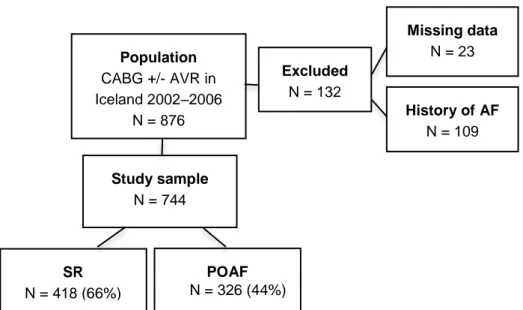
4.5 Study V – Postoperative atrial fibrillation ...67
4.5.1 Postoperative complications and survival ...67
4.5.2 Risk factors for POAF, and probability assessment ...69
5 Discussion ...71
5.1.1 Study I – Identification of baseline kidney function in AKI studies ...71
5.1.2 Study II – AKI following CABG ...72
5.1.3 Study III – AKI following AVR ...73
5.1.4 Study IV - The importance of renal recovery following AKI ...75
5.1.5 Study V – POAF, the most common complication of heart surgery ...76
References ...83 Original publications ...107 Paper I ...109 Paper II ...123 Paper III ...139 Paper IV ...149 Paper V ...163
List of abbreviations
ADQIAFib
Acute Dialysis Quality Initiative atrial fibrillation
AKI AKIN
acute kidney injury
Acute Kidney Injury Network APACHE
ARDS
acute physiology and chronic health evaluation acute respiratory distress syndrome
ARF AS ASA
acute renal failure aortic stenosis
American Society of Anesthesiology physical status classification
ATN AUC
acute tubular necrosis area under the curve AVR
BMI
aortic valve replacement body mass index
CABG coronary artery bypass grafting
CAD coronary artery disease
CHF CI
congestive heart failure confidence interval
CKD chronic kidney disease
CKD-EPI CO COPD
Chronic Kidney Disease-Epidemiology Collaboration cardiac output
chronic obstructive pulmonary disease
CPB cardiopulmonary bypass
CRRT CRS
continuous renal replacement therapy cardiorenal syndrome
CS-AKI cardiac surgery-associated acute kidney injury CVD DM ECG eGFR cardiovascular disease diabetes mellitus electrocardiogram
estimated glomerular filtration rate ESKD
ESRD EuroSCORE GFR
Hb
end-stage kidney disease end-stage renal disease
European System for Cardiac Operative Risk Evaluation
glomerular filtration rate hemoglobin
HF heart failure
ICD-10 International Classification of Diseases, tenth revision
ICU intensive care unit
IHD ischemic heart disease
LVEF KDIGO KDOQI
left ventricular ejection fraction
Kidney Disease: Improving Global Outcomes Kidney Disease Outcome Quality Initiative MDRD Modification of Diet in Renal Disease MI
MOF
myocardial infarction multiple organ failure
NSR normal sinus rhythm
OPCAB off-pump coronary artery bypass OR P odds ratio pressure PB PC PCI
hydrostatic pressure in Bowman‘s capsule hydrostatic pressure of the glomerular capillaries percutaneous coronary intervention
POAF PPV PSM RBC
postoperative atrial fibrillation positive pressure ventilation propensity score match red blood cell
RA RE RCT RenR RIFLE ROC RRT SCr SD SR TAVI TIA πc
renal efferent arteriolar resistance renal blood flow
randomized controlled trial renal recovery
Risk, Injury, Failure, Loss of kidney function, and End-stage kidney disease
receiver operating characteristic renal replacement therapy serum creatinine
standard deviation sinus rhythm
transcatheter aortic valve insertion transient ischemic attack
List of figures
Figure 1. Anatomy of the kidney. ... 1
Figure 2. The nephron. ... 2
Figure 3. Factors governing glomerular filtration rate. ... 3
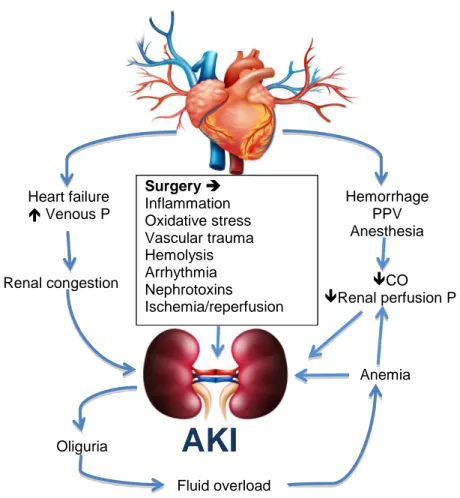
Figure 4. A schematic representation of factors that contribute to acute kidney injury in the perioperative period ... 11
Figure 5. A schematic overview of extracorporeal hemodialysis. ... 15
Figure 6. The relationship of the estimated glomerular filtration rate to serum creatinine... 17
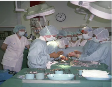
Figure 7. Cardiothoracic surgery performed at Landspitali University Hospital ... 21
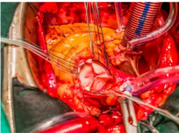
Figure 8. Aortic valve surgery. ... 23
Figure 9. The first open-heart operation performed in Iceland with the aid of cardiopulmonary bypass technique ... 26
Figure 10. Aortic stenosis in a bicuspid aortic valve and a tricuspid aortic valve ... 26
Figure 11. Flow chart of the study population in study II ... 34
Figure 12: Population flow chart in study IV ... 35
Figure 13. Flow chart of the study population in study V ... 36
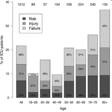
Figure 14. The absolute proportion of patients admitted to the intensive care unit who met the RIFLE criteria for acute kidney injury ... 45
Figure 15. One-year survival of patients admitted to Icelandic intensive care units based on classification according to the RIFLE criteria ... 48
Figure 16. Annual percentage of acute kidney injury in patients in study II. ... 49
Figure 17. Kaplan-Meier plot of recovery of renal function in patients in study II ... 54
Figure 18. Kaplan-Meier curve showing comparison of survival of patients with no acute kidney injury and those with acute kidney injury in study II ... 54
Figure 20. Graphic representation of ROC analysis of predictive value of renal recovery on one-year mortality of patients in
study IV. ... 61
Figure 21. Renal recovery rates for patients in study IV shown
according to the initial stage of postoperative AKI ... 63
Figure 22. Comparison of long-term survival of individuals who achieved renal recovery and a propensity score-matched control group of individuals who did not recover renal
function. ... 66
Figure 23. A Kaplan-Meier survival curve comparing patients with postoperative atrial fibrillation and normal sinus rhythm following CABG/OPCAB or AVR ± CABG in Iceland, 2002‒
List of tables
Table I: The RIFLE criteria for acute kidney injury. ... 8
Table II: The AKIN criteria for acute kidney injury ... 9
Table III: The KDIGO criteria for acute kidney injury. ... 9
Table IV: The KDOQI classification of chronic kidney disease ... 19
Table V: Cardiorenal syndrome. ... 20
Table VI: Overview of materials and methods. ... 32
Table VII: Classification of intensive care unit patients into the RIFLE subgroups of acute kindey injury ... 46
Table VIII: The frequency of comorbid diseases and contributory causes of acute kidney injury according to the RIFLE classification ... 47
Table IX: Baseline characteristics of patients in study II ... 50
Table X: Intraoperative and postoperative characteristics of patients in study II ... 52
Table XI: Independent risk factors for acute kidney injury in study II. ... 53
Table XII: Independent risk factors for long-term mortality in study II. ... 55
Table XIII: Baseline patient characteristics of patients in study III. ... 56
Table XIV: Intraoperative characteristics and postoperative complications of patients in study III ... 58
Table XV: Renal recovery of individuals with acute kidney injury at postoperative days 10, 20, and 30 in study IV. ... 60
Table XVI: Comparison of different renal recovery criteria at 10, 20, and 30 days after surgery, and their relation to one-year survival of patients in study IV. ... 61
Table XVII: Univariate Cox analysis of different renal recovery criteria in patients in study IV ... 62
Table XVIII: Comparison of preoperative and intraoperative characteristics of patients who did and did not recover renal function in study IV ... 64
Table XIX: Risk factors for reduced one-year survival after surgery of patients in study IV. ... 65
Table XX: Multivariate Cox proportional hazard analysis of risk factors of long-term mortality in study IV. ... 66
Table XXII: Intraoperative characteristics of patients in study V, and
postoperative complications. ... 68
Table XXIII: Independent risk factors for POAF in study V... 70
List of original papers
This thesis is based on the following original publications, which are referred to in the text by their Roman numerals (I‒V):
I. Acute kidney injury in intensive care units according to RIFLE classification: a population-based study. Martin I Sigurdsson, Iris O Vesteinsdottir, Kristinn Sigvaldason, Solveig Helgadottir, Olafur S Indridason, and Gisli H Sigurdsson. Acta Anaesthesiol Scand 2012;56:1291-1297.
II. Renal recovery and long-term survival following acute kidney injury after coronary artery surgery: a nationwide study. Solveig Helgadottir, Martin I. Sigurdsson, Runolfur Palsson, Dadi Helgason Gisli H. Sigurdsson, and Tomas Gudbjartsson. Acta Anaesthesiol Scand 2016;doi:10.1111/aas/12758.
III. Acute kidney injury and outcome following aortic valve replacement for aortic stenosis. Dadi Helgason, Solveig Helgadottir, Sindri A. Viktorsson, Andri W. Orrason, Inga L Ingvarsdottir, Arnar Geirsson, and Tomas Gudbjartsson. Interact CardioVasc Thorac Surg 2016; doi:10.1093/icvts/ivw117.
IV. Renal recovery following postoperative acute kidney injury – evaluation of definitions, clinical risk factors, and survival. Solveig Helgadottir (equally contributing first authors), Thorir E. Long (equally contributing first authors), Dadi Helgason, Gisli H. Sigurdsson, Runolfur Palsson, Olafur S Indridason, Tomas Gudbjartsson, and Martin I. Sigurdsson. Manuscript.
V. Atrial fibrillation following cardiac surgery: risk analysis and long-term survival. Solveig Helgadottir, Martin I. Sigurdsson, Inga L. Ingvarsdottir, David O. Arnar, and Tomas Gudbjartsson. J Cardiothorac Surg 2012 Sept 19;7:87.
In addition, some unpublished data has been presented. All papers are reprinted by kind permission of the publishers.
The doctoral student, Solveig Helgadottir, planned the research work for Papers II, IV, and V, of which she is the first author. She also applied for the appropriate ethical and research approval and performed the statistical analyses in cooperation with the co-authors. She participated in planning and writing of the manuscripts of Papers I and III in cooperation with the co-authors. She wrote this thesis under the guidance of her supervisor and doctoral committee.
1
Introduction
Acute kidney injury (AKI) is a complex condition that is seen in diverse settings. The term covers a broad range of renal dysfunction, ranging from a slight decrease in kidney function to extensive renal injury. The aim of this thesis is to provide some insight into AKI after cardiac surgery, with emphasis on AKI following either coronary artery bypass grafting (CABG) surgery or aortic valve replacement (AVR). Normal anatomy and kidney function will be explained briefly before any further discussion on the mechanisms underlying AKI. Then the importance of baseline and postoperative kidney function will be examined and the effect of recovery of renal function following AKI assessed. Lastly, postoperative atrial fibrillation (POAF), the most common complication of open-heart surgery, and its relationship to AKI, will be examined.
1.1 The kidneys
The kidneys are a pair of bean-shaped, retroperitoneal organs that play an important role in various homeostatic mechanisms that are essential for survival (Figure 1). They serve in regulating fluid, pH, and electrolyte balance, filter and eliminate waste products of metabolism from the circulation, aid in the regulation of blood pressure via the renin-angiotensin-aldosterone system, and are important producers of hormones (Hall & Guyton, 2011; Wein et al., 2012).
Each kidney normally has around 800,000 to 1.5 million nephrons, the kidney’s urine-producing functional units (Brenner, 2005). The nephron consists of an initial filtering component (the renal corpuscle) and an associated tubule. The renal corpuscle is made up of the glomerulus, a network of capillaries that receives blood from the afferent renal arteriole and is responsible for ultrafiltration of blood. The glomerulus is covered by a simple layer of squamous epithelium, called the Bowman’s capsule (Figure 2) (Wein et al., 2012).
The kidneys usually weigh around 115-170 g in an adult human and make up only 1‒2% of the total body mass (Wein et al., 2012). However, every minute, between 20% and 25% of the total cardiac output passes through the kidneys and is filtered by them (Janson & Tischler, 2012). In the glomeruli, primary urine is produced at an approximate rate of 125 mL/min, amounting to roughly 180 L on a daily basis (Hall & Guyton, 2011). The tubuli serve to further modify the initial corpuscular filtrate before it progresses to the collecting system of the kidneys and is excreted from the body in the form of urine.
Figure 2. The nephron.
Glomerular filtration rate (GFR) is the term used to describe the rate of flow of filtered fluid through the kidney, and it is measured in mL/min. Glomerular filtration is a passive process governed by Starling forces that force fluid to filter along a pressure gradient. GFR depends on the filtration coefficient (KF) and the balance between factors driving and opposing
filtration (Figure 3) (Brenner, 2005). The main factor that drives filtration is the hydrostatic pressure inside the glomerular capillaries (Pc), whereas the
hydrostatic pressure in the Bowman’s capsule (PB) and the mean colloid
osmotic pressure of the glomerular capillaries (πc) oppose filtration (Guyton &
Hall, 2006). Thus, GFR can be expressed by the following equation: GFR = KF × ((Pc − PB) − πc)
Physiologically, GFR is primarily modified through changes in pressure inside the glomerular capillaries, which is in turn dependent on renal afferent arteriolar resistance (RA) and efferent arteriolar resistance (RE), as well as on
systemic arterial pressure. It should, however, be noted that due to an autoregulation of GFR and the renal blood flow, the pressure in the afferent arterioles in the glomerulus remains constant over a wide range of systemic arterial pressure (Guyton & Hall, 2006).
Figure 3. Factors governing glomerular filtration rate. Modified from http://pathway medicine.org/glomerular-filtration-rate.
The condition that arises when the kidneys become incapable of maintaining GFR, and even of excreting nitrogenous waste, regulating electrolytes and intravascular volume, and in assisting in maintenance of the acid-base balance, is termed renal failure. Renal failure may be acute or chronic. Before any further explanation of the complex condition of renal failure, another key topic will be covered in the following sections, namely how renal function is assessed in clinical practice.
1.2 Assessment of renal function
Renal function can be estimated in several ways. For instance, urine output (diuresis), routinely monitored in patients in intensive care units (ICUs), is an indirect marker of renal function. Diuresis is, however, affected by several other mechanisms, e.g. the normal physiological response of increased
vasopressin secretion due to volume depletion and administration of diuretics. Diureses can therefore be an unreliable marker of renal function in the setting of AKI (Bellomo et al., 2004).
The most accurate method of determining GFR, and thereby assessing renal function, is by performing repeated blood measurements of intravenously administered exogenous filtration markers (e.g. inulin, iothalamate, or iohexol), which, however, is economically and temporally impractical in clinical practice (Stevens & Levey, 2005, 2009).
In the clinical setting, the most widely used method of estimation of GFR (eGFR) is measurement of serum levels of the endogenous filtration marker creatinine (SCr) (Brenner, 2005). Creatinine is an amino acid compound derived from the conversion of creatine to phosphocreatinine in skeletal muscle and subsequent metabolism in the liver. It is mainly excreted through glomerular filtration and secreted in an unchanged form (Miller, 2003; Taylor, 1989). Measurement of SCr permits estimation of creatinine clearance, which in turn has an inverse correlation with GFR (Stevens & Levey, 2005). The normal range of eGFR is 90‒130 mL/min/1.73 m2, but around the fifth decade of life it begins to decrease (by approximately 10 mL/min/1.73 m2 per decade) (Brenner, 2005; Feehally et al., 2007). However, estimation of GFR by measurement of SCr has several important limitations (Bagshaw & Gibney, 2008; Shemesh et al., 1985; Waikar et al., 2009):
i. Release of creatinine varies with age, sex, exercise, diet, and muscle mass.
ii. Up to 10‒40% of creatinine is secreted by the proximal tubular cells of the kidneys, leading to overestimation of GFR, especially in states of renal dysfunction.
iii. SCr values become abnormal only when there is a more than a 50% reduction in GFR.
iv. SCr is a functional marker rather than an injury marker. Thus, it does not reflect kidney injury in the early stages, it may take up to 24 hours to rise following abrupt damage to the kidneys, and it has low predictive value regarding injury. Furthermore, critical illness itself may also cause large fluctuations in SCr due to factors such as deranged volume status and catabolic metabolism.
The search for biomarkers that are more sensitive in the recognition of AKI is ongoing, but none are currently well enough established to replace creatinine as a biomarker of renal function (de Geus et al., 2012; Dedeoglu et al., 2013; Susantitaphong et al., 2013b). In the meantime, to overcome some
of the obstacles in using SCr and to increase the accuracy of GFR and creatine clearance estimations in clinical practice, equations adding demographic and clinical data to SCr values have been formulated. It should, however, be emphasized that all these equations are based on SCr, which, as already mentioned, has various limitations―especially in the AKI setting (Waikar et al., 2009).
1.3 Estimation of GFR
Despite efforts to correct for differences regarding age, sex, race, and body mass, it can be difficult to produce a single equation that can accurately predict creatine clearance and GFR in all clinical settings. The following three formulas have been in most extensive use:
i. The Cockcroft-Gault formula was published in 1973, and provides estimated creatinine clearance values, which in turn provide an estimation of GFR. It is still widely used, but recommended drug doses are commonly based on Cockcroft-Gault values (Cockcroft & Gault, 1976). The equation was developed based on 249 males with steady-state SCr, and women were assigned an adjustment factor of 0.85, based on a theoretically lower muscle mass (by 15%). Lean body weight is included in the numerator of the formula to adjust for muscle mass. That may, however, adversely affect the performance of the formula in patients where weight change is not a result of changes in muscle mass (e.g. pregnancy, obesity, and edematous states) (Coresh & Stevens, 2006). Furthermore, due to tubular secretion of creatinine, the formula will lead to overestimation of GFR (Botev et al., 2009). Since publication of the formula, SCr measurement has been standardized. However, samples from the original study are no longer available for measurement and there is no version of the equation with standardized creatinine values. ii. The Modification in Diet and Renal Disease (MDRD) formula for
estimation of GFR was published in 1999, and has been extensively validated in populations between the ages of 18 and 70 with chronic kidney disease (CKD) (eGFR < 60 mL/min/1.73 m2) (Levey et al., 1999). The equation has, however, been criticized for the fact that the study that it was based on was conducted on a population that was not representative of the general population―but rather a population with reduced kidney function (with a mean measured GFR of 40 mL/min/1.73 m2). The MDRD equation thus overestimates eGFR at values < 30 mL/min/1.73 m2, but underestimates eGFR at
values from 30 to 90 mL/min/1.73 m2 (Levey et al., 1999; Stevens et al., 2006).
iii. The Chronic Kidney Disease-Epidemiology Collaboration (CKD-EPI) formula for estimation of GFR was developed in 2009 and uses the same premises as the MDRD formula, but is more accurate in patients with eGFR > 60 mL/min/1.73 m2 (Levey et al., 2009).
1.4 Acute renal failure
Acute renal failure (ARF) involves an abrupt loss of kidney function (Webb & Dobb, 2007). It can have various causes, e.g. renal tissue damage, reduced renal blood flow, urinary tract obstruction, or exposure to nephrotoxins (Brenner, 2005).
At the turn of the 21st century, inter-study comparison of research on the subject of ARF suffered from a lack of consensus diagnostic criteria (Eknoyan, 2002). Indeed, at the time, over 35 different definitions of ARF were reported, leading to wide variations in reported ARF rates (Chertow et al., 1997; Kellum et al., 2002). In ARF, the decrease in renal function occurs abruptly, usually within hours or days, and results in imbalance in fluid and electrolyte states (Webb & Dobb, 2007). Many cases of ARF are reversible due to the kidney’s unique ability to recover from almost complete loss of function. So for a long time, ARF, in its less severe form was considered to be a relatively minor event with little clinical impact. However, there is mounting evidence to indicate that even a slight rise in SCr can negatively affect patient outcome, be it morbidity, short-term complications, or long-term mortality (Chertow et al., 1998; Lassnigg et al., 2004; Mangano et al., 1998). Changes in SCr could simply coincide with unmeasured variables that lead to increased mortality, but even after controlling for other clinical variables, a consistent linear relationship between reduced kidney function and worse outcome has been observed. Consequently, research has undergone much change in recent years―with the term AKI largely replacing the term ARF.
1.5 Acute kidney injury
Acute kidney injury is defined as a sudden and sustained decrease in kidney function that develops within seven days and leads to a rapid reduction in GFR (Webb & Dobb, 2007). Decreases in GFR stem from a decline in the number of nephrons, a decline in single nephron GFR, or both, and reflect various etiologies, including specific renal disease and extra-renal pathology (Brenner, 2005). This may result in extensive derangement of normal homeostasis, e.g. acid-base disturbances, electrolyte imbalance, retention of nitrogenous waste products, and fluid overload, and it has also been shown
to hasten progression of CKD and impair innate immunity (De Waele et al., 2008; Rossaint et al., 2011; Thakar et al., 2003).
As mentioned, AKI is a known complication in various clinical settings and it may develop as a consequence of complex interactions between an insult and subsequent activation of pro-inflammatory and coagulatory responses. However, AKI may occur with only minimal histological signs of tissue damage (Kosaka et al., 2016).
AKI is a large and costly problem for patients and healthcare systems (Chertow et al., 2005). In a recent meta-analysis involving 312 studies and almost 50 million hospitalized patients worldwide, AKI was diagnosed in one in five adults and in one in three children (Susantitaphong et al., 2013a). The most common underlying causes for hospital-diagnosed AKI are sepsis and cardiac surgery (Susantitaphong et al., 2013a; Zakeri et al., 2005; Zarbock et al., 2014). One concern is the fact that studies have indicated that the incidence of AKI is rising, and the same holds true for incidence of AKI requiring dialysis (Hsu, 2010; Hsu et al., 2013).
1.5.1 Defining AKI – the RIFLE, AKIN, and KDIGO criteria
The use of varying definitions of AKI and the heterogeneity of different patient populations has led to large differences in reported AKI rates in previously published studies (Kaufman et al., 1991; Metcalfe et al., 2002). This in turn has made studies of the epidemiology and outcome of AKI unnecessarily complex (Gottlieb et al., 2002). In an effort to provide evidence-based guidelines, several interdisciplinary groups have proposed standardized criteria for the diagnoses and staging of AKI in the last decade. In the coming chapters, the three most frequently used criteria will be discussed further: the RIFLE, AKIN, and KDIGO criteria (see List of abbreviations).
1.5.1.1 The RIFLE criteria
The RIFLE criteria, shown in Table I, were published in 2004 (Bellomo et al., 2004). The criteria are multi-level and consist of three graded levels of kidney injury (Risk, Injury, and Failure) and outcome measures (Loss of kidney function and End-stage kidney disease). They are based on a sudden increase in SCr and decrease in GFR or urine output (known or presumed to have developed over seven days or less) compared to baseline levels. Since they were published, various studies of different populations have shown the criteria to be useful in determining the incidence of AKI as well as being predictive of complications and perioperative mortality (Hoste et al., 2006; Kuitunen et al., 2006).
Table I: The RIFLE criteria for acute kidney injury (Bellomo et al., 2004).
Stage SCr criteria Urine output criteria
Risk 1.5-fold increase in SCr or decrease in eGFR of > 25% from baseline
< 0.5 mL/kg/h for > 6 h Injury 2-fold increase in SCr or decrease in eGFR
of > 50% from baseline
< 0.5 mL/kg/h for > 12 h Failure 3-fold increase in SCr, decrease in eGFR of
> 75% from baseline, SCr ≥ 354 μmol/L or acute rise in SCr ≥ 44 μmol/L
< 0.3 mL/kg/h for > 24 h or anuria for > 12 h Loss Complete loss of kidney function for > 4 weeks
ESKD End-stage kidney disease (> 3 months)
eGFR, estimated glomerular filtration rate; ESKD, end-stage kidney disease; SCr, serum creatinine.
A major point of criticism of the RIFLE criteria is the requirement for baseline SCr values, which are often missing in the clinical setting. The criteria recommend assuming baseline GFR to be 75 mL/min/m2 when there is no history of CKD, and calculation of SCr by using the MDRD equation. However, studies have shown this to be too rough an approximation in many cases (Bagshaw et al., 2009; Pickering & Endre, 2010).
1.5.1.2 The AKIN criteria
The Acute Kidney Injury Network later modified the original RIFLE criteria, in an effort to increase both sensitivity and specificity (Table II) (Mehta et al., 2007). The AKIN criteria differ from the RIFLE criteria mainly regarding the time component and the fact that not outcome measures are described. While the RIFLE criteria are defined as change within seven days, the AKIN criteria propose using 48 hours (Mehta et al., 2007). The AKIN criteria also include less severe kidney dysfunction in the diagnosis of AKI, and do not use GFR as a marker, as GFR has been shown to be unreliable in the setting of AKI. Lastly, the AKIN criteria propose that the diagnoses of AKI should only be made after optimization of fluid balance and exclusion of urinary tract obstructions. However, studies have failed to reliably show superior prognostic value of the AKIN criteria over the original RIFLE criteria (Englberger et al., 2011; Ostermann & Chang, 2011).
Table II: The AKIN criteria for acute kidney injury (Mehta et al., 2007).
Stage SCr criteria Urine output criteria
1 Increase in SCr of ≥ 26.4 μmol/L in ≤ 48 h or 1.5- to 2-fold increase in baseline SCr
< 0.5 mL/kg/h for > 6 h 2 Increase in SCr to > 2- to 3-fold from
baseline SCr
< 0.5 mL/kg/h for > 12 h 3 Increase in SCr to > 3-fold from baseline
or baseline SCr ≥ 354 μmol/L with an acute increase of ≥ 44 μmol/L in ≤ 48 h
< 0.3 mL/kg/h for > 24 h or anuria for > 12 h
eGFR, glomerular filtration rate; SCr, serum creatinine.
1.5.1.3 The KDIGO criteria
Efforts have been made to further homogenize and improve the definition of AKI―with the KDIGO definition, combining the RIFLE and AKIN criteria, as a recent example (Table III) (Khwaja, 2012). The three classifications of AKI mentioned above have been compared in several studies, but no conclusive evidence of superiority of one system over the others has yet emerged (Bastin et al., 2013; Joannidis et al., 2009; Luo et al., 2014).
Table III: The KDIGO criteria for acute kidney injury (Khwaja, 2012).
Stage SCr criteria Urine output
criteria 1 SCr ≥ 1.5‒1.99 times baseline in ≤ 7 days or ≥ 26.5
μmol/L increase occuring in ≤ 48 h
< 0.5 mL/kg/h for 6‒12 h 2 SCr 2.0‒2.99 times baseline in ≤ 7 days < 0.5 mL/kg/h
for ≥ 12 h 3 SCr ≥ 3.0 times baseline in ≤ 7 days or
SCr ≥ 354 μmol/L with either rise of ≥ 26.5 μmol/L in ≤ 48 h or SCr ≥ 1.5 times from baseline ≤ 7 days or
initiation of renal replacement therapy or
in patients < 18 years a decrease in eGFR to < 35 mL/min per 1.73 m2
< 0.3 mL/kg/h for ≥ 24 h or anuria for ≥12 h
1.5.2 Ascertainment of baseline SCr
Although standardized definitions of AKI, based on changes in SCr and urine output, have facilitated epidemiological studies, some controversy remains regarding the diagnosis of AKI. All the aforementioned criteria are based on relative deviance from baseline SCr, and they can be significantly affected by the choice of reference SCr. An important hurdle is choosing a suitable baseline SCr, i.e. one that accurately reflects kidney function in the steady state, prior to onset of AKI (Siew & Matheny, 2015). Studies have indicated that in patients for whom data on SCr is missing, the eGFR 75approach, suggested in the RIFLE criteria, is likely to provide too rough an estimation of baseline kidney function in a study cohort (Bagshaw et al., 2009; Pickering & Endre, 2010). Knowledge of actual kidney function is preferable, and pre-hospital SCr values may be useful in determining baseline SCr (Siew & Matheny, 2015).
1.5.3 The pathophysiology and etiology of AKI
To aid in diagnosis and management, the causes of AKI have been divided into three main categories (Brenner, 2005):
i. Pre-renal: This accounts for approximately 55% of cases. It represents a physiological response to renal hypoperfusion (e.g. hypovolemia or low cardiac output) and―by definition―is reversible upon correction of renal blood flow, as no induction of ischemic injury to renal parenchyma has taken place.
ii. Intrinsic: This comprises approximately 40% of cases, and it may be a complication of diverse diseases of the renal parenchyma. Most intrinsic injury is triggered by renal ischemia or nephrotoxins (such as radio-contrast or aminoglycosides). As such, intrinsic injury has been shown to go hand in hand with major cardiovascular surgery, trauma, hemorrhage, or sepsis.
iii. Post-renal: This represents about 5% of cases and is caused by urinary tract obstruction, usually distally in the ureter or urethra. It has, however, been pointed out that this may be an oversimplification for underlying pathological mechanisms that frequently overlap, as evident in cases where hypoperfusion of renal tissue (pre-renal) may cause sufficient harm to lead to ischemic cellular injury and acute tubular necrosis (intrinsic) (Case et al., 2013). A schematic representation of postoperative AKI is shown in Figure 4. It highlights some of the complex interplay of various factors that affect the process.
Figure 4. A schematic representation of factors that contribute to AKI in the perioperative period (Brenner, 2005; Legrand & Payen, 2013). AKI, acute kidney injury; CO, cardiac output; P, pressure; PPV, positive pressure ventilation.
1.5.4 Risk factors for AKI
Risk factors for AKI can be divided into patient- or exposure-specific. Individual clinical factors known to increase a patient’s susceptibility include advanced age, diabetes mellitus, and congestive heart failure (CHF) (Long et al., 2016). As already mentioned, sepsis and cardiac surgery are the leading extrinsic causes of AKI (Susantitaphong et al., 2013a). Other reported risk factors include trauma, exposure to nephrotoxic drugs (e.g. radio-contrast and aminoglycosides), and burns (Bagshaw et al., 2008; Rewa & Bagshaw, 2014). Lastly, pre-existing kidney disease is an established risk factor for the development of AKI, and the risk has been found to be proportional to the
Surgery Inflammation Oxidative stress Vascular trauma Hemolysis Arrhythmia Nephrotoxins Ischemia/reperfusion CO Renal perfusion P Fluid overload Anemia Heart failure Venous P Renal congestion Oliguria Hemorrhage PPV Anesthesia
AKI
respective stage of CKD (Hsu et al., 2008; Khosla et al., 2009).
1.6 Postoperative AKI
Postoperative AKI is believed to account for approximately 20‒50% of hospital-acquired AKI, and is a result of complex pathophysiological mechanisms (Carmichael & Carmichael, 2003). According to a large prospective multicenter study that examined ARF in almost 30,000 ICU patients, 41% of ARF cases were defined as postoperative (Uchino et al., 2005). Cardiovascular surgical procedures were by far the most common surgical causes of postoperative AKI, comprising more than twice as many cases as the second most common surgical procedure, i.e. gastrointestinal surgery (56.4% vs. 27.8%). In contrast to cardiac surgery, considerably less emphasis has been placed on AKI following non-cardiac surgical procedures, perhaps due to a significantly lower incidence of AKI (Long et al., 2016). Nevertheless, there is some evidence that AKI may complicate up to one-third of all major non-cardiac surgeries (Bihorac et al., 2009)..
1.6.1 AKI following cardiac surgery
Patients who undergo cardiac surgery are faced with a unique combination of possible renal stressors, related to both the surgery itself and, in some cases, the cardiopulmonary bypass (CPB) (Rosner & Okusa, 2006). Reported rates of AKI following open-heart surgery vary widely, depending on factors such as the patient population and the type and extensivness of surgery, but most are within the range of 15‒60% (Machado et al., 2009; Roh et al., 2012; Rosner & Okusa, 2006). The incidence of cardiac surgery-associated AKI (CS-AKI) is lowest after CABG, followed by valvular surgery, and it is highest after combined CABG and valvular procedures (Rosner et al., 2008).
Multiple risk factors for AKI following cardiac surgery have been reported but among the most prominent are: emergency surgery, chronic kidney disease, and female gender (Bagshaw et al., 2008; Chertow et al., 1997; Mangano et al., 1998; Rosner & Okusa, 2006). Advanced age and diabetes have also been shown to be risk factors for AKI―a fact that has increasing implications considering constantly ageing patient populations and the growing incidence of diabetes (Wild et al., 2004).
The pathophysiology of CS-AKI is complex and is believed to be multifactorial (Rosner & Okusa, 2006). It has, however, been proposed that the occurrence of CS-AKI may be explained by six different but
synergistically acting mechanisms: endogenous and exogenous toxins, metabolic mechanisms, neurohormonal activation, ischemia-reperfusion, oxidative stress, and inflammation (Bellomo et al., 2008).
The latter two pathways are one of the reasons that lipid-lowering statins have been suggested as a possible prophylactic treatment for AKI, due to their anti-inflammatory and anti-oxidative properties (Morgan et al., 2009). This also holds true for another well-known complication of cardiac surgery, POAF (Liakopoulos et al., 2009). However, this possible pleiotropic effect of statin therapy has been disputed, and a recent randomized studyexamining the effects of treatment with rosuvastatin in patients undergoing cardiac surgery indicated no effect on rates of POAF. Also, rates of AKI were higher in the treatment group (Zheng et al., 2016).
1.7 AKI in the ICU setting
Most studies on the overall incidence of AKI in the ICU have found rates in the 40‒70% range, with mortality as high as 80% (Case et al., 2013; Hoste & Kellum, 2006). Furthermore, the incidence of AKI in the ICU setting has been on the rise worldwide (Bagshaw et al., 2007; Case et al., 2013). This trend appears not only to be due to the fact that older patients with more comorbidities and higher risk of AKI are being admitted to ICUs, as the incidence of AKI also seems to be on the rise in the less severely ill patients. However, studies have shown that acute physiology and chronic health evaluation (APACHE) scores and simplified acute physiology scores have not changed in line with changes in incidence (Bagshaw et al., 2007; Lameire et al., 2006). It has therefore been speculated that the increase in incidence of AKI may be due to the more rigorous diagnostic interventions in later years (Lameire et al., 2006). The highest incidence has been seen in patients with sepsis, but the incidence of AKI is generally lower in surgical patients than in medical patients in the ICU (Case et al., 2013). Even so, and reassuringly, recent studies have indicated that mortality in patients with AKI has decreased (Bagshaw et al., 2007; Waikar et al., 2006). This may stem from various factors such as the overall improvement in ICU treatment, a reduction in the use of nephrotoxic agents, and increased awareness of AKI (Waikar et al., 2006).
1.8 Prevention and treatment of AKI
Despite our improved knowledge of the pathophysiology of AKI, and advances in surgical techniques together with improved ICU management
(including dialysis therapies), effective therapeutic interventions for AKI have yet to emerge. Furthermore, randomized studies on various pharmacological agents and renal replacement therapy (RRT) have not shown any conclusive effect in reducing mortality (Bagshaw et al., 2013; Jo et al., 2007; Schneider et al., 2013). Emphasis is therefore placed on prevention. Although no single preventive measure reaches the highest levels of evidence, current recommendations are that one of the key targets for amelioration of AKI should be strict maintenance of hemodynamic stability (Prowle et al., 2014). Optimally, the goal should be to ensure that there is adequate renal perfusion and oxygen delivery to the kidneys, without fluid overload, as several studies have shown that overhydration increases the likelihood of adverse events and possibly contributes to AKI (Payen et al., 2008; Prowle et al., 2014; Stein et al., 2012). What is more, restrictive fluid management has been associated with reduced incidence of intra-abdominal hypertension (IAH), a well-defined risk factor for the development of postoperative AKI, especially following abdominal surgery (Dalfino et al., 2008).
1.8.1 Renal replacement therapy
When preventive measures fail and patients progress to end-stage renal disease (ESRD), RRT ensues. RRT may be indicated in patients with symptomatic CKD (e.g. electrolyte disturbances) (Lameire & Van Biesen, 2010) or in patients with AKI (Ronco et al., 2015). In AKI, the most common indications are acidemia, electrolyte abnormalities, fluid overload that is unresponsive to diuretic treatment, complications of uremia, and―in rare cases―intoxication with dialyzable substances (Ronco et al., 2015).
RRT includes conventional dialysis (both hemodialysis and peritoneal dialysis), hemofiltration and hemodiafiltration, and also kidney transplantation (Ronco et al., 2015). The first three treatment modalities take on the blood filtration role of the kidneys, but they lack other aspects of kidney function and are not curative (de Francisco & Pinera, 2006). They can be performed using arteriovenous or venovenous modes, either in a continuous manner (so-called continuous renal replacement therapy (CRRT)) or intermittently (Ronco et al., 2015) (Figure 5). In the ICU setting, patients are often selected for CRRT rather than intermittent hemodialysis, as they have lower MAP and are deemed too hemodynamically unstable to tolerate intermittent dialysis (Ronco et al., 2015).
Figure 5. A schematic overview of extracorporeal hemodialysis.
1.9 Recovery of renal function
There are more and more studies examining how patients who survive critical illness fare later on (Goldstein, 2014; Palevsky et al., 2005). An estimated 8‒ 22% of critically ill patients suffer an AKI episode from which they fail to completely recover, which may even necessitate ongoing intermittant renal dialysis after discharge (Bell et al., 2007; Uchino et al., 2005).
Animal studies have indicated that there is a causal relationship between AKI and CKD (Basile et al., 2001). Furthermore, a growing number of studies in humans also point to an association between AKI and progression of―or to―CKD (Amdur et al., 2009; Hsu et al., 2009; Ishani et al., 2011; Waikar & Winkelmayer, 2009). Following AKI, incomplete regeneration of the tubulointerstitial epithelium, characterized by fibrosis and persistent inflammation, may occur and correlate with loss of kidney function. However, the condition may remain subclinical for prolonged periods (Gueler et al., 2004).
Importantly, several studies on the long-term effect of AKI have suggested that early recovery of kidney function may be associated with better clinical
outcome after AKI (National Kidney, 2012; Rosner & Okusa, 2006). Furthermore, recovery of renal function following AKI has been shown to be an independent determinant of morbidity and mortality in patients (Bagshaw, 2006; Pannu et al., 2013; Swaminathan et al., 2010). Factors that affect the long-term prognosis after AKI are not completely understood, but the process leading to development of CKD may be at least partially modifiable, e.g. by tighter regulation of blood pressure and diabetes (Murugan & Kellum, 2011).
No agreed-upon criteria exist for the definition of recovery of renal function following an episode of AKI, making comparison of studies on the subject cumbersome. The majority of previously employed definitions fall into one of three categories:
i. Freedom from dialysis (Bagshaw et al., 2005; Schiffl & Fischer, 2008).
ii. Absolute cut-off values of follow-up eGFR (Cantarovich et al., 2004; Macedo et al., 2012).
iii. Relative changes in SCr or eGFR (Macedo et al., 2008; Pannu et al., 2013).
These definitions of renal recovery (RenR) vary widely, are based on functional improvement, and do not take into consideration the complex structural effect of acute injury on the renal tissue (Srisawat et al., 2010). This is an important point, as complete functional RenR may be noted even in the setting of residual structural damage, which in turn can have implications for progression to CKD (Gueler et al., 2004; Hsu et al., 2009). Furthermore, current definitions of RenR lack a relationship with the time course of recovery of renal function, and insufficiently define optimal length of follow-up of patients’ renal function following AKI. The RIFLE criteria propose a cut-off of 3 months after AKI prior to diagnosis of CKD in patients who do not have recovered renal function (Bellomo et al., 2004). However, some studies have revealed that the RenR process may go on for up to 18 months (Macedo et al., 2012).
RenR, defined as liberation from dialysis, only covers those with the most severe forms of AKI. However, milder forms of AKI have also been shown to affect morbidity and mortality (Zakeri et al., 2005). Thus, definitions have emerged that outline absolute and relative changes in eGFR and SCr. However, a disadvantage of basing definitions of RenR on SCr and/or eGFR is the non-linear relationship between SCr and GFR (Figure 6). Another problem with definitions based on SCr might also be the confounding factor
of decreased muscle mass during severe illness (Srisawat et al., 2010). In addition, cut-off levels for criteria of renal recovery may in fact be over baseline in patients with advanced CKD, limiting the usefulness of the definition in that patient group (Macedo et al., 2012). On the other hand, the disadvantage of basing definitions of RenR on relative changes in SCr or eGFR is the dependency on availability of baseline SCr.
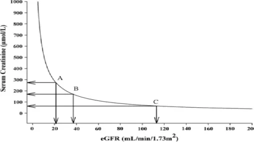
Figure 6. The relationship of the estimated glomerular filtration rate (eGFR) to serum creatinine (SCr). The graph shows how a substantial decline in eGFR only leads to slight elevations in SCr on the flat part of the curve, meaning that a substantial decrease in eGFR may lead to a very subtle increase in SCr. However, on the steep part of the curve a very slight decrease in eGFR leads to a significant increase in SCr (Damman et al., 2012).
The Acute Dialysis Quality Initiative (ADQI) Group sought to provide a consensus definition of AKI recovery, and suggested that complete RenR should be defined as a return of SCr to below the definition of RIFLE-R, or within 50% of baseline SCr. Furthermore, partial RenR was defined as freedom from RRT, but not to within 50% of baseline SCr (Bellomo et al., 2004).
A 2008 study with 5-year follow-up provided important insight into the course of AKI after critical illness. In the study, only 57% achieved full renal recovery (defined as eGFR within 10% of baseline) before hospital discharge (Schiffl & Fischer, 2008). The study also confirmed the poorer survival of patients diagnosed with AKI, even during long-term follow-up. This was most evident in patients with more extensive comorbidities and in those who did not achieve full recovery of kidney function. Furthermore, the study showed that if patients failed to reach normal renal function 6‒12 months after the index insult, no further improvement could be expected. Importantly, around
10% of the group that had only partially recovered renal function had a further decline in renal function during subsequent long-term follow-up. Lastly, the study highlighted that a further kidney insult in a patient recovering from AKI leads to a reduced likelihood of complete RenR (Schiffl & Fischer, 2008). Similarly, other studies have shown that more than one episode of AKI is an independent predictor of poorer survival (Guerin et al., 2000). This further drives home the message of the importance of scrupulous follow-up in this patient population, both during hospitalization and after discharge.
Research on factors that affect RenR is in progress. Loss of autoregulation of renal blood flow and hypotension have been singled out as key factors in delayed RenR (Conger & Hammond, 1992). Fluid overload has also been shown to negatively affect renal recovery (Bouchard et al., 2009; Heung et al., 2012). Furthermore, it has been shown that the degree of recovery from AKI requiring dialysis is associated with the initial dialysis modality used (Sun et al., 2014). This is in line with the results of several other studies, with higher rates of RenR in patients treated with CRRT as compared to intermittent hemodialysis (Bell et al., 2007; Lin et al., 2009; Uchino et al., 2007). However, the effect of factors such as inflammation and oxygen delivery to the kidneys on renal recovery has yet to be fully determined (Srisawat et al., 2010).
1.10 Chronic kidney disease
During the acute phase of kidney injury, mechanisms such as hyperfiltration and hypertrophy may aid in ensuring the kidney‘s ability to secrete waste products. In the long run, however, they are believed to factor in as causes of progressive renal dysfunction due to propagation of increased glomerular capillary pressures, ultimately leading to glomerulosclerosis, destruction of renal parenchyma, and CKD (Helal et al., 2012; Wald et al., 2009). Whatever the underlying cause of CKD, patients usually experience progressive kidney dysfunction when the loss of functional nephrons reaches a certain threshold, as irreversible sclerosis occurs in the remaining, strained functional units of the kidneys (Hsu et al., 2009). Subsequently, the rate of progression depends on factors such as age, underlying etiology, and successful treatment of exacerbating factors (Gullion et al., 2006). There are many components that may exacerbate the process, i.e. systemic hypertension, hyperlipidemia, obesity, nephrotoxic substances (e.g. non-steroidal anti-inflammatory drugs, and various antibiotics), sub-optimally treated diabetes mellitus (DM), and smoking (Bash et al., 2009; Brenner, 2005; Chang et al., 2016; Thakar et al., 2011). In a recent study, a validated risk calculator was reported, which is applicable to adult populations and based upon routine laboratory tests and baseline characteristics. The study showed that lower eGFR, albuminuria, young age, and male sex were indicative of higher risk of progression to
end-stage kidney disease, the final end-stage of CKD. Furthermore, the results suggested that lower albumin, calcium, and bicarbonate levels and higher phosphate levels were predictive of elevated risk of disease progression (Tangri et al., 2011).
However, the term CKD covers a wide range of kidney dysfunction, and in 2002 the National Kidney Foundation’s Kidney Disease Outcome Quality Initiative (KDOQI) presented guidelines for classification of CKD ("KDOQI Clinical Practice Guideline for Diabetes and CKD: 2012 Update," 2012). They defined CKD as deranged kidney function and albuminuria with or without a decreased GFR, persisting for over three months. Furthermore, they divided CKD into stages based on the degree of the decrease in GFR (Table IV).
Table IV: The KDOQI classification of chronic kidney disease ("KDOQI Clinical Practice Guideline for Diabetes and CKD: 2012 Update," 2012).
Stage GFR Description
1 ≥ 90 Kidney damage (albuminuria) with normal GFR
2 60‒89 Kidney damage (albuminuria) with mild reduction of GFR 3 30‒59 Moderate reduction of GFR
4 15‒20 Severe reduction of GFR 5 < 15 Kidney failure
GFR, glomerular filtration rate.
Importantly, studies have consistently shown a relationship between CKD and the development of vascular disease, be it coronary artery disease or peripheral vascular disease (Coresh et al., 2003; Gullion et al., 2006). Rates of hospitalization in patients with CKD are elevated compared to the general population, especially due to infections and cardiovascular diseases (Bash et al., 2009; Foley et al., 2005). Furthermore, all-cause mortality rates increase as kidney function decreases and mortality rates are up to six times higher in patients with stage-5 CKD compared to the general population (Go et al., 2004). Interestingly, the leading cause of death in this group of patients is cardiovascular disease (CVD) (Neovius et al., 2014; Perazella & Khan, 2006; Tonelli et al., 2006).
1.11 Cardiorenal syndrome
The heart and kidneys cooperate in maintaining hemodynamic stability and organ perfusion, and are connected through interdependent relationships, e.g. neuronal and hormonal control (Brenner, 2005). A number of bidirectional interactions exist between the heart and kidney functions e.g.
regulation of electrolytes and fluid status (Ronco et al., 2008). The term cardiorenal syndrome (CRS) has been used to define the relationship in cases of dysfunction as: “a disorder of the heart and kidneys whereby acute or chronic dysfunction in one organ may induce acute or chronic dysfunction in the other” (Ronco & Ronco, 2012). CRS has been divided into five types (Ronco & Ronco, 2012) (Table V).
Table V: Cardiorenal syndrome. Type Initial
dysfunction
Result Example
1 Acute heart dysfunction
Acute kidney injury or dysfunction Cardiogenic shock, acute decompensated heart failure
2 Chronic heart dysfunction
Progressive kidney dysfunction Chronic heart failure 3 Acute kidney
dysfunction
Acute cardiac dysfunction (e.g. arrhythmia or heart failure)
Acute kidney injury 4 Chronic kidney
disease
Cardiac dysfunction or
hypertrophy and/or higher risk of adverse cardiovascular events
Chronic glomerular disease
5 Systemic condition
Heart and kidney dysfunction Diabetes mellitus, sepsis, hypertension It has since been argued that this might be too simplistic a definition, but the consensus remains that a pathophysiological condition may occur in which the combination of heart and kidney dysfunction can amplify organ dysfunction through induction of pathological mechanisms affecting both organ systems (Braam et al., 2014). In line with this are studies showing increa