RESVERATROL DIMERS FROM STEM BARK OF
Hopea gregaria
AND THEIR CYTOTOXIC PROPERTIES
Sahidin1,*,Euis Holisotan Hakim2, Yana Maolana Syah2, Lia Dewi Juliawaty2,Sjamsul Arifin Achmad2, Laily Bin Din3, and Jalifah Latip3
1
Department of Chemistry, Faculty of Mathematics and Natural Sciences, Haluoleo University, Kampus Baru Anduonohu,
Kendari, South East Sulawesi, Indonesia. 2
Natural Products Research Group, Departement of Chemistry, Institut Teknologi Bandung, Jalan Ganeca 10 Bandung 40132, Indonesia
3
School of Chemical Sciences & Food Technology, Faculty of Science and Technology, Universiti Kebangsaan Malaysia, 43600 UKM Bangi, Selangor D.E., Malaysia
Received 19 Februari 2008; Accepted 28 Februari 2008
ABSTRACT
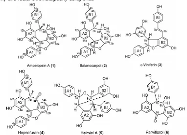
Six resveratrol dimers have been isolated from the stem bark of H. gregaria, ampelopsin A (1), balanocarpol (2), ε-viniferin (3), hopeafuran (4), heimiol A (5), and parviflorol (6). The structures of these compounds were determined based on spectroscopic evidence such as UV, IR, 1-D, 2-D NMR and comparison with the reported data. This compound inventions are strengthen conclusion that Hopea tends to produce resveratrol dimers. Biological activity of those compounds against murine leukemia P-388 cells showed that ε-viniferin (3) is the most active compound with IC50 value 5.1 ± 0.3 μg/mL.
Keywords: H. gregaria, resveratrol dimers, murine leukemia P-388 cells.
INTRODUCTION
Dipterocarpaceae has local name “meranti, keruing or selangan”, is a big family plant which comprises about 600 species and 16 genera. Major genera of this family are Shorea (150 sp.), Hopea (100 sp.), Dipterocarpus
(75 sp.) and Vatica (60 sp.) [1]. Phytochemically, main secondary methabolites of Dipterocarpaceae family plant is resveratrol oligomers [2].
Study on resveratrol dimers from stem bark of
Hopea gregaria carries on phytochemical research of Dipterocarpaceaous plants especially Hopea genus. Chemical contents of Indonesian Hopea plants have been reported that are H. sangal [3], H. bancana [4], H. dryobalanoides [5], and H. mengarawan [6]. Chemical data of those plants complete previously information of secondary methabolites of Hopea plants i.e. H. odorata
[6], H. cardifolia [7], H. jucunda [8], H. malibato [9], and
H. parviflora [10]. Based on the reported chemical data,
Hopea tends to produce resveratrol dimers.
Some resveratrol oligomers showed interesting activities such as ε-viniferin (dimer) active toward
Staphylococcus oxford and Entamoeba coli [11], S. aureus [12], cytotoxic against HL-60 cells [13] and inhibitor rat liver 5α-reductase [14].
ε
-Viniferin (trimer) revealed activities toward S. aureus [12], and antiinflamatory [15]. Hopeaphenol (tetramer) proved activities against Microbacterium smegmatis and S. aureus [16] antiinflamatory toward leukotriena B4 cells [17]. Moreover, the bigger size of resveratrol oligomers, the lower their cytotoxic properties toward murin tyrosinase cells [18]. The diversity of resveratrololigomer compounds encourage to determine biological activities of isolated resveratrol dimers from H. gregaria
toward murin leukemia P-388 cells.
This paper will explain isolation and structure elucidations of resveratrol dimers from stem bark of H. gregaria. In addition, cytotoxic properties against murine leukemia P-388 cells and biogenetic relationship of those compounds will be discussed.
EXPERIMENTAL SECTION
General Experimental Procedure
Optical rotations were determined using a Perkin-Elmer 341 polarimeter. UV spectra were measured with a Cary Varian 100 Conc. Spectrophotometer. IR spectra were evaluated by Perkin Elmer Spektrum One FT-IR spectrometer. 1H and 13C NMR were recorded with JEOL LTD, ECP400 DELTA_NMR spectrometer, operating at 400 MHz (1H) and 100 MHz (13C). Vacuum Liquid Chromatography (VLC) and column chromatography were carried out using silica gel G60 (230-400 mesh) (Merck), while radial chromatography was performed using silica gel PF254 (Merck). For TLC analysis, precoated silica gel plates (Merck Kiesel-gel GF254 0.25 mm) were used.
Plant Material
Sample of the stem bark of H. gregaria was collected during June 2004 from Pohara Forest Kendari South East Sulawesi. The plant was identified by staff at the Herbarium Bogoriense, Bogor Botanical Garden,
* Corresponding author. Tel/Fax : 0062-081341888268 Email address : HTSahidin02@yahoo.comTH
Bogor, and a voucher specimen has been deposited at the herbarium.
Extraction and Isolation
The dried powdered of stem bark of H. gregaria
(2.5 kg) was macerated with acetone 3 x 5 L @ 24 hours, and concentrated by vacuum rotary evaporator (280 g extract). A part of acetone extract (25 g) was fractionated by vacuum liquid chromatography (VLC) (silica gel, hexane-etilacetate to etilacetate 100 %) to give 5 fractions that are F1 (1.8 g), F2 (2.6 g), F3 (4.3 g), F4 (7.3 g) and F5 (5.4 g). F1 fraction is a non polar fractions which usually contains terpenoids. Moreover, F2 (0.5 g) was fractionated and purified by radial chromatography (silica gel, n-hexane:CHCl3:MeOH = 4:5:1) to yield compounds 1 (0.10 g) and 2 (0.15 g). Using the same way as F2, F3 (0.5 g) produced compound 3 (0.15 g). Compounds 4 (0.1 g), 5 (0.2 g), and 6 (0.05 g) were isolated by radial chromatography (silica gel, eluen: CHCl3 : MeOH = 1.5:8.5) from F4 (0.5 g).
Biological Activity Test
Biological activities of the isolated compounds were determined toward murin leukemia P-388 cells using Alley methode [19].
Compound 1, yellow, amorf, mp. 186-189 oC, [α]D
20
-171o (c 0.1 MeOH), UV (MeOH) λmaks (log ) 205 (5.23), 231 (4.96), 283 nm (4.35), (MeOH+NaOH) λmaks (log ) 211 (5.46), 246 (4.92), 293 nm (4.43). IR (KBr) υ (cm-1) 3367 (OH), 2920 (CH-aliphatic), 1613, 1514, 1451 (C=C aromatic), and 834 (para-disubstituted benzene). 1 13b), 159.2 (C-13a), 158.9 (C-4a), 157.5 (C-11a), 156.2 (C-4b), 143.4 (C-9a), 139.8 (C-9b), 133.2 (C-1a), 131.0 (C-1b), 130.1 (C-2/6a), 129.0 (C-2/6b), 119.9 (C-10b), 119.2 13a), 116.2 3/5a), 115.7 3/5b), 110.8 (C-14b), 105.4 (C-14a), 101.6 (C-12a), 97.6 (C-12b), 89.2 (C-7a), 71.7 (C-8b), 49.5 (C-8a), and 44.1 (C-7b). 3366 (OH), 1613, 1512, 1451 (C=C aromatic), and 834 (para-disubstituted benzene). 1H NMR (Me2CO-d6, 400 113.9 (C-3(5)a), 155.5 (C-4a), 50.0 (C-7a), 72.9 (C-8a), 140.6 9a), 113.6 10a), 159.5 11a), 94.8 (C-12a), 158.9 (C-13a), 104.2 (C-14a), 133.4 (C-1b), 130.3 2/6a), 116.2 3/5b), 158.3 4b), 93.3 (C-7b), 58.3 (C-(C-7b), 142.6 (C-9b), 120.2 (C-10b), 157.2 11b), 101.8 12b), 156.5 13b), and 106.5 (C-14b). and 832 (para-disubstituted benzene). 1H NMR (Me2CO-d6, 400 MHz) H (ppm) 7.14 (2H, d,J=8.4 Hz, (5.10), 223 (4.96), 396 nm (4.16), (MeOH+NaOH) λmaks (log ) 209 (5.41), 245 (4.92), 441 nm (4.15). IR (KBr) υ (cm-1) 3365 (OH), 1694 (C=O), 1613, 1512, 1451 (C=C aromatic), and 833 (para-disubstituted benzene). 1H NMR (Me2CO-d6, 400 MHz) H (ppm) 7.70 (2H, d, (5.03), 229 (4.82), and 283 nm (4.06), (MeOH+NaOH)
λmaks (log ) 206 (5.32), 249 (4.65), and 287 nm (4.09).
IR (KBr) υ (cm-1) 3365 (OH), 2891 (C-H aliphatic), 1612, 1512, 1456 (C=C aromatic), and 838 (para -disubstituted benzene). 1H NMR (Me2CO-d6, 400 MHz) (C-13a), 157.6 (C-13b), 157.5 (C-4a), 156.6 (C-4b),
147.5 11b),155.1 11a), 147.5 9b), 143.1 (C-9a), 137.4 (C-1a), 137.7 (C-1b), 130.5 (C-2/6a), 128.4 (C-2/6a), 117.3 (C-10b), 116.6 (C-10a), 115.8 (C-3/5b), 115.7 3/5a), 107.7 14a), 105.1 14b), 102.7 12b), 102.5 12a), 81.9 8b), 81.8 7a), 51.3 (C-7b), and 47.4 (C-8a).
Compound 6, yellow, amorf, mp. 172-176 oC, [α]D
20
+122o (c 0.1 MeOH). UV (MeOH) λmaks (log ) 222 (4.99), 339 nm (4.27), (MeOH+NaOH) λmaks (log ) 206 (5.36), 374 nm (4.78). IR υ(cm-1) 3412 (OH), 1649 (C=O), 1615, 1511, 1461 (C=C aromatic), 836 (para -disubstituted benzene). 1H NMR (Me2CO-d6, 400 MHz)
H (ppm) 7.41 (1H, d, J=2.5 Hz, H-6b), 6.85 (2H, d,
J=8.8 Hz, H-2/6a), 6.75 (1H, br d, J=2.8 Hz, H-14a), 6.72 (1H, d,J=2.5 Hz, H-4b), 6.48 (2H, d, J=8.8 Hz, H-3/5a), 6.08 (1H, d, J=2.5 Hz, H-12a), 5.30 (1H, br s, H-8a), 5.15 (1H, br s, H-7a), -OH {14.13 (1H, br s, C-3b), 9.17 (1H, br s ), 8.66 (1H, br s), 8.49 (1H, br s ), and 7.89 (1H, br s)}. 13C NMR (Me2CO-d6, 100 MHz) C (ppm) 195.1 8a), 168.5 11b), 164.1 13b), 156.8 (C-13a), 155.7 (C-4b), 155.5 (C-11a), 148.4 (C-9b), 141.1 (C-9a), 130.7 (C-1b), 130.0 (C-2/6b), 121.7 (C-10a), 114.5 3/5b), 111.2 10b), 110.0 14a), 107.7 (C-12a), 106.7 (C-14b), 101.7 (C-12b), 74.3 (C-8b) and 47.9 (C-7b).
RESULT AND DISCUSSION
Isolates (1-6) were purified from the acetone-soluble part of stem bark of H. gregaria by column chromatography and radial chromatography using silica
gel as adsorbent. Structure elucidations of 1-6 were made by analysis of the spectral data and by comparison with literatures. All of the compounds showed at Fig 1.
UV and IR spectra of compound 1 indicated that this compound has phenolic chromophor and a substituent at para position. Moreover, 13C NMR spectra (Table 1) showed 24 carbon signals which represented 28 carbon atoms including 6 C-oxyaryls, 18 C-aromatics and 4 C-aliphatics. This fact proved that compound 1 is a resveratrol dimer. It was supported by 1H NMR spectra which revealed two sets of ortho-coupled protons at δH 7.01, 6.68, 6.82, and 6.57 ppm (2H each, d, J=8.4 Hz and 8.8 Hz), symbolized two units of para-hydroxyphenyl, two sets of meta-coupled protons at δH 6.31, 6.11, 6.11, and 6.52 ppm (1H each, d, J=2.0 Hz), characterized two units of 1,2,3,5-tetrasubstituted benzene, and four methine protons at δH 5.69 and 4.02 ppm (1H each, d,
J=11.6) for a cycloheptadiene system, two protons at
δH 5.38 ppm (2H, d, J=4.8) corresponded to a dihydrofuran ring. Based on the data, compound 1 has molecular formula C28H22O7, and comparison of
1 H and 13
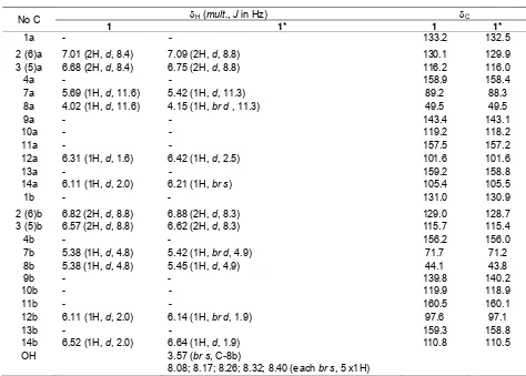
C NMR data between compound 1 and literature at Table 1, showed that those data have highly similarity parameters with ampelopsin A [20]. As a result, compound 1 can be proved as ampelopsin A which was also supported by HMBC data (figure 2 and 3).
Fig 1. The structure of resveratrol dimers from stem bark of H. gregaria
Figure 2. HMBC Spectrum of ampelopsin A (1)
Table 1. NMR spectra of ampelopsin A (1)
δH (mult., J in Hz) δC
No C
1 1* 1 1*
1a - - 133.2 132.5
2 (6)a 7.01 (2H, d, 8.4) 7.09 (2H, d, 8.8) 130.1 129.9
3 (5)a 6.68 (2H, d, 8.4) 6.75 (2H, d, 8.8) 116.2 116.0
4a - - 158.9 158.4
7a 5.69 (1H, d, 11.6) 5.42 (1H, d, 11.3) 89.2 88.3
8a 4.02 (1H, d, 11.6) 4.15 (1H, br d , 11.3) 49.5 49.5
9a - - 143.4 143.1
10a - - 119.2 118.2
11a - - 157.5 157.2
12a 6.31 (1H, d, 1.6) 6.42 (1H, d, 2.5) 101.6 101.6
13a - - 159.2 158.8
14a 6.11 (1H, d, 2.0) 6.21 (1H, br s) 105.4 105.5
1b - - 131.0 130.9
2 (6)b 6.82 (2H, d, 8.8) 6.88 (2H, d, 8.3) 129.0 128.7
3 (5)b 6.57 (2H, d, 8.8) 6.62 (2H, d, 8.3) 115.7 115.4
4b - - 156.2 156.0
7b 5.38 (1H, d, 4.8) 5.42 (1H, br d, 4.9) 71.7 71.2
8b 5.38 (1H, d, 4.8) 5.45 (1H, d, 4.9) 44.1 43.8
9b - - 139.8 140.2
10b - - 119.9 118.9
11b - - 160.5 160.1
12b 6.11 (1H, d, 2.0) 6.14 (1H, br d, 1.9) 97.6 97.1
13b - - 159.3 158.8
14b 6.52 (1H, d, 2.0) 6.64 (1H, d, 1.9) 110.8 110.5
OH 3.57 (br s, C-8b)
8.08; 8.17; 8.26; 8.32; 8.40 (each br s, 5 x1H)
Fig 3. Interpretation of HMBC spectrum of ampelopsin A (1)
Table 2. Chemical shifts of aliphatic protons of ampelopsin A (1) and balanocarpol (2)
No. C Ampelopsin A (1) Balanokarpol (2)
7a 5.69 (1H, d, 11.6) 5.69 (1H, d, 9.5) 8a 4.02 (1H, d, 11.6) 5.15 (1H, d, 9.5) 7b 5.38 (1H, d, 4.8) 4.89 (1H, br s) 8b 5.38 (1H, d, 4.8) 5.38 (1H, br s)
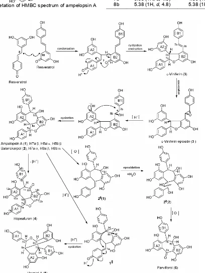
Fig 4. Biogenetic pathway of resveratrol dimers from H. gregaria
Furthermore, the structures of 2-6 were determined by interpreting of spectroscopic data alike compound 1. Compounds 2,3,4,5, and 6 are balanocarpol [21], -viniferin [22], hopeafuran [23], heimiol A [24], and parviflorol [20], respectively. Based on 1H NMR data, the differences between ampelopsin A (1) and balanocarpol (2) were located at aliphatic protons, especially H-7b and H-8b. At ampelopsin A (1), two protons (H-7b and H-8b) are trans-position (d, J=4.8 Hz) while at balanocarpol (2) are cis-position (br s)(Table 2).
ε-Viniferin (3) is a resveratrol dimer which has a
trans-vinylic system as a particular character of this compound at C-7b and C-8b 6.83 (1H, d, J=16.3 Hz), and 6.58 (1H, d, J=16.3 Hz), respectively. Furthermore, spectroscopic data showed that hopeafuran (4) is a resveratrol dimer that has only a proton aliphatic at δH 6.12 (1H, br s, H-7b) as an individual nature of this compound. The difference of spectral data of heimiol A (5) with other resveratrol dimers was displayed by 13C NMR data of aliphatic carbons. This compound has four aliphatic carbons, two of them have identical chemical shifts i.e. C-7a and C-8b (each 81.8 and 81.9 ppm). Conclusion of parviflorol (6) structure was especially based on 13C NMR spectra. This compound has only 21 carbon atoms or a resveratrol dimer that has lost seven carbon atoms. In addition, one of the carbon atom is carbonyl atom which gave chemical shift at C 195,1 ppm (C-8a). Relationship of those compounds could be seen at biogenetic pathway at Fig 4. This biogenetic pathway followed pattern of synthesis of resveratrol and
ε-viniferin [25] and biogenetic reactions on
tetramerstilbenes [26].
ε-Viniferin (3) came from two resveratrol units through cyclization and enolization. Epoxidation, hydrogenation and cyclization of compound 3 yielded ampelopsin A (1) and balanocarpol (2). Dehydrogenation of ampelopsin A (1) or balanocarpol (2) produced hopheafuran (4), whilst oxidation of balanocarpol (2) which was followed by epoxidation, hydration and oxidation to give parviflorol (6). In the meantime, hydrogenation of ampelopsin A (1) and followed by cyclization to produce heimiol A (5).
Cytotoxic properties of isolated resveratrol dimers from stem bark of H. gregaria against murine leukaemia P-388 cells (Table 3) indicated that ε-viniferin (3) was the most active compound which was pursued by balanocarpol (2), ampelopsin A (1), hopeafuran (4), parviflorol (6) and heimiol A (5), respectively. It was predicted that caused by electron distribution of ε -viniferin especially at unit B. Orbital hybrid of all carbon atoms at unit B of ε-viniferin are sp2.
CONCLUSION
Study on resveratrol dimers from acetone extract of
H. gregaria’s stem bark yielded six resveratrol dimers
that were ampelopsin A (1), balanocarpol (2), -viniferin (3), hopeafuran (4), heimiol A (5), and parviflorol (6). Biogenetically, all isolated compounds have closed relationship, and -viniferin (3) is a main precursor. Moreover, invention of those compounds is strengthen conclusion that Hopea tends to produce resveratrol dimers. The cytotoxic properties against murine leukemia P-388 cells indicated that ε-viniferin (3) was the most active compound.
ACKNOWLEDGEMENT
We thank the Ministry of National Education of Republic of Indonesia for Doctor Programme Scholarship (BPPs). We also express thanks the Head of Forestry of South East Sulawesi province for the permission to take the sample. In addition, we show appreciation to the Herbarium Bogoriense, Bogor, Indonesia for identification of the plant specimen.
REFFERENCES
1. Ashton, P. S., 1983, Flora Malesiana:
Spermatophyta I, Martinus Nijhoff, The Hague, 391-436.
2. Sotheeswaran, S., and Pasupathy, V., 1993,
Phytochemistry 32 (5), 1083-1092.
3. Atun, S., 2004, Fitokimia beberapa spesies Dipterocarpaceae Indonesia dari genus Vatica, Anisoptera, Hopea, and Dipterocarpus, Disertasi Program Doktor, Institut Teknologi Bandung, 76-162.
4. Tukiran, 2004, Senyawa Mikromolekul dari
Beberapa Tumbuhan Meranti (Shorea) Indonesia, Disertasi Program Doktor, Institut Teknologi Bandung, 70-112.
5. Sahidin, Hakim, E. H., Juliawaty, L. D., Syah, Y. M., Din, L. B., Ghisalberti, E. L., Latip, J., Said, I. M., and Achmad, S. A., 2005, Z. Naturforsch. C, 60c: 718-723
6. Sahidin, Hakim,E.H., Syah, Y.M., Juliawaty,L.D., Achmad, S.A., Din, L.B., Latip, J., and Ghisalberti, E.L., 2006, Berita Biologi 8 (2), 107-114.
7. Coggon, P., McPhail, A.T., and Wallwork, S.C. J., 1970, J. Chem. Soc. (B), 884-897.
8. Sotheeswaran, S., Sultanbawa, M.U.S.,
Surendrakumar, S., Balasubramaniam, S., and Bladon, P., 1983, J. Chem. Soc. I, 159-162.
9. Diyasena, M. N. C., Sotheeswaran, S., and Surendrakumar, S., 1985, J. Chem. Soc. Perkin Trans I, 1807-1809.
10. Dai, J.R., Hallock, Y.F., Cardellina II, J.H., and Boyd, M.R., 1998, J. Nat. Prod., 61, 351-353.
11. Sotheeswaran, S., Sultanbawa, M.U.S.,
Surendrakumar, S., and Bladon, P., 1985, J. Chem. Soc. 4, 159-162.
12. Nitta, T., Arai, T., Takamatsu, H., Inatomi, Y., Murata, H., Iinuma, M., Tanaka, T., Ito, T., Asai, F., Ibrahim, I., Nakanishi, T., and Watanabe, K., 2002,
J. Health Sci., 48(3):273-276.
13. Kang, H. J., Park, H. Y., Choi, S. W., Yang, E. K., and Lee, W. J., 2003, Experimental and Molecular Medicine, 35 (6), 467-474.
14. Hirano, Y., Kondo, R., and Sakai, K., 2003, J. Wood Sci., 49(1), 53-58.
15. Pryce, R. J., and Langcake, P., 1977,
Phytochemistry, 16, 1452-1454.
16. Kitanaka, S., Ikezawa, T., Yasukawa, K.,
Yamanouchi, S., Takido, M., Sung, H. K., and Kim, I. H., 1990, Chem. Pharm. Bull., 38 (2), 432-435. 17. Pols, J. R. Z., Freyer, A. J., Killmer, A. J., and Porter,
J, R, 2002, J. Nat. Prod., 65, 1554-1559.
18. Ohguchi, K., Tanaka, T., Ito, T., Iinuma, M., Matsumoto, K., Akao, Y., and Nozawa, Y., 2003,
Biosci. Biotech. Biochem., 67 (7), 1587-1589.
19. Alley, M. C., Scudiero, D. A., Monks, A., Hursey, M. L., Czerwinski, M. J., Fine, D. L., Abbott, B. J., Mayo,
J. G., Shoemaker, R. H., and Boyd, M. R. , 1988,
Cancer Res., 48: 589-601.
20. Tanaka, T., Ito, T., Ido, Y., Son, T. K., Nakaya, K., Iinuma, M., Ohyama, M., and Chelladurai, V., 2000,
Phytochemistry, 53, 1009-1014.
21. Ito, T., Tanaka, T., Ido, Y., Nakaya, K., Iinuma, M., and Riswan, S., 2000, Chem. Pharm. Bull., 48 (12), 1001-1005.
22. Oshima, Y., and Ueno, Y., 1993, Phytochemistry, 33 (1), 179-182.
23. Tanaka, T., Ito, T., Ido, Y., Nakaya, K., Iinuma, M., and Chelladurai, V., 2001, Chem. Pharm. Bull., 49 (6), 785-787.
24. Weber, F. J., Wahab, I. A., Marzuki, A., Thomas, N.F., Kadir, A. A., Hadi, A. H.A., Awang, K., Latiff, A.A., Richomme, P., and Delaunay, 2001,
Tetrahedron Letter, 42, 4895-4897.
25. Sako, M., Hosokawa, H., Ito, T., and Iinuma, M., 2004, J. Org. Chem., 69, 2598-2600.
26. Takaya, Y., Yan, K. X., Tereshima, K., He, Y. H., and Niwa, M., 2002, Tetrahedron, 58, 9265-9721.