α
αα
αα
-Lipoic acid can not prevent effet on
cell viability, collagen synthesis inhibition,
collagen degradation induction in ultraviolet
A irradiated human fibroblast cell
Putu Dyah Ayu Saraswati1*, Soedirman Sastrodiprodjo2, Yohanes Widodo Wirohadidjojo2
1Department of Dermatovenereology, Wangaya Distric General Hospital, Denpasar, Bali,
2Department of Dermatovenereology, Faculty of Medicine, Gadjah Mada University,
Yogyakarta, Indonesia
ABSTRACT
Ultraviolet A (UVA) irradiation on human skin can generate free radical and stimulate matrix metalloproteinase production resulting in collagen degradation and transforming growth factor beta (TGF-β) inhibition. It causes synthesis collagen inhibition and induces cell death. α-Lipoic acid (ALA) is an universal antioxidant and a powerful scavenger of free radicals. In this study we investigated the effect of ALA on viability, collagen synthesis and degradation in UVA irradiated human fibroblasts. Normal human skin fibroblasts cell culture were irradiated with UVA for three times with each dose of 3000 mJ/cm2 UVA. α-Lipoic acid in various concentration was added to the culture following UVA irradiation and incubated for 48 hours. The cell viability was determined by MTT-assay while collagen synthesis and degradation were determined by Sirius red binding assay. The difference of cell viability and collagen synthesis and degradation between fibroblasts cell after and without UVA irradiation were analyzed using paired-t test with 95% confidence interval (p<0.05). The results showed that UVA irradiation decreased cell viability, inhibited collagen synthesis and induced collagen degradation in fibroblasts cell. However, ALA was not sufficient to increase viability, to increase collagen synthesis and to inhibit collagen degradation in fibroblasts cell due to UVA irradiation. In conclusion, ALA can not prevent UVA irradiation effect on human skin fibroblasts cell.
Key words: UVA – irradiation - human skin fibroblasts – antioxidants – α-lipoic acid
ABSTRAK
Pajanan sinar ultraviolet A pada kulit dapat menghasilkan radikal bebas dan memicu produksi matrik metaloproteinase sehingga menghambat sintesis kolagen dan transforming growth factor beta (TGF-β). Hal ini menghambat sintesis kolagen dan memicu kematian sel. Asam α-lipoat (ALA) merupakan antioksidan dengan kemampuan kuat menangkap radikal bebas. Dalam penelitian ini telah dikaji pengaruh ALA terhadap viabilitas, sintesis dan degradasi kolagen sel fibroblas kulit yang dipajan sinar UVA. Kultur sel fibroblas kulit manusia normal dipajan sinar UVA sebanyak tiga kali dengan dosis masing-masing 3000 mJ/cm2. Asam α-lipoat pada berbagai konsentrasi ditambahkan pada kultur fibroblas diikuti pajanan sinar UVA selanjutnya diinkubasikan selama 48 jam. Viabilitas sel ditetapkan dengan MTT
assay sedangkan sintesis dan degradasi kolagen ditetapkan dengan Sirius binding assay. Perbedaan viabilitas sel, sintesis dan degradasi kolagen sel fibroblas setelah dan tanpa pajanan UVA dianalisis dengan uji t pasangan dengan tingkat kepercayaan 95% (p<0.05). Hasil penelitian menunjukkan pajanan sinar UVA menurunkan viabilitas, menghambat sintesis dan memacu degradasi kolagen sel fibroblas. Namun demikian, ALA tidak cukup kuat meningkatkan viabilitas, memacu sintesis dan menghambat degradasi kolagen sel fibroblas akibat pajanan sinar UVA. Dengan demikian dapat disimpulkan bahwa ALA tidak dapat mencegah efek pajanan sinar UVA pada sel fibroblas kulit manusia.
INTRODUCTION
Fibroblasts are important component of skin. Fibroblast cells produce and organize extracellular matrix in dermis which consists of collagen type I type III, elastin, proteoglycans and glycosaminogly-cans, and play an important role in tensile strength of the human skin.1 Meanwhile, ultraviolet A (UVA)
irradiation in skin fibroblasts cell up regulates collagen-degrading matrix metalloproteinase (MMPs) and inhibits collagen synthesis.2,3 UVA
Irradiation also induces cell damage through generation of reactive oxygen species (ROS) which causing deoxyri-bonucleic acid (DNA) mutation, lipid peroxidation and finally cell death.4 These conditions
may lead to functional changes of cell as shown in the photoaging process.3
Human cells have antioxidants property designed to detoxify harmful effect of reactive oxygen species (ROS) molecules before DNA and other cell components damages occurred. However, UVA-irradiation suppresses the cell’s defense against ROS damage by reducing antioxidants activity.4 In
this case, supplementation of exogenous antioxidant may support the endogenous antioxidants and prevent generation of free radical.5
α-Lipoic acid (ALA) and dihydrolipoic acid (DHLA) have been acknowledged as universal antioxidant since they have solubility both in lipid and water resulting in their ability to reach all portion of the cell to provide antioxidants protection.6 ALA
prevents stress-induced lipid peroxidation7,
scavenges ROS and prevents cellular senescence.3
This study was conducted to investigate the effect of ALA on collagen degradation and synthesis and also on cellular viability on UVA-irradiated human skin fibroblasts cells.
MATERIALS AND METHODS
Cell culture
Fibroblasts sub cultured passage 3 were established from foreskin of 2 healthy human donors aged 11 years and were grown in Dulbecco’s modified Eagle’s Medium (DMEM) supplemented with 10% bovine serum, 1000 µg/100 unit Penstrep-Gibco™/mL, amphotericine B-Gibco™ 1000 µg/
mL, and 100 mg/mL ceftriaxone at 37°C in 5% CO2. Fibroblasts used were 2 × 10⁴ cells for each column.
Irradiation of fibroblasts with UVA
The cultures were divided into two groups. In the first groups, fibroblasts cell culture was irradiated with UVA (PAST Sun B-LIPITM)for three times
with each dose of 3000 mJ/cm2. α-Lipoic acid in
various concentration was added to the culture following UVA irradiation and incubated for 48 hours before the culture was given another UVA irradiation. This procedure was repeated until the fibroblasts cultures were irradiated with UVA 9000 mJ/cm2. In the second group, fibroblasts cell culture
was added with ALA of various concentration similar to first group but without following UVA irradiation.
α αα
αα-Lipoic acid
α-Lipoic acid (Mecola Forte® -PT. Lapi.
Indonesia) was dissolved in dimethyl sulfoxide (DMSO) and added to the medium in final concentration of 50, 100, 200 and 400 µM. The highest concentration of organic solvent (DMSO) used in this study was no more than 0.2%. α-Lipoic acid was added on fibroblasts cell culture immediately after UVA irradiation.
Cell viability, collagen deposition and collagen degradation
Forty-eight hours after fibroblasts cell culture and ALA incubated, cell viability was determined by 3,-(4,5-dimethyl-thiazol-2-yl)-2,5-diphenyltetra-zoliumbromide (MTT) assay according to method used by Freshney.8 Collagen degradation and
synthesis were detemined using Sirius red binding assay according to method used by Heng et al.9 and
Taskiran et al.10, respectively. Statistical analysis
RESULTS
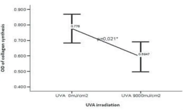
The effect of UVA irradiation on cell viability of human skin fibroblasts is presented in FIGURE 1. Cell viability of human skin fibroblasts decreased after UVA irradiation as expressed by the significant decrease of OD of fibroblasts cell after UVA irradiation compared with those without UVA irradiation (p<0.05). More over, UVA irradiation also
significantly decreased collagen synthesis (p<0.05). The mean of OD of collagen synthesis after UVA irradiation was lower than those without UVA irradiation (FIGURE 2). In ralated to the decrease of collagen synthesis, the collagen degradation significantly increased after UVA irradiaton (p<0.05). The mean of OD of collagen degradation after UVA irradiation was higher than those without UVA irradiation (FIGURE 3).
FIGURE 1. Effect of UVA irradiation on cellular viability of human skin fibroblasts
FIGURE 3. Effect of UVA irradiation on collagen degradation of human skin fibroblasts
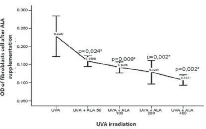
The cellular viability of human fibroblasts cell after ALA supplementation in various doses and UVA irradiation resulted in significant decrease of
fibroblasts viability compared with the control (p<0.05) as shown on FIGURE 4.
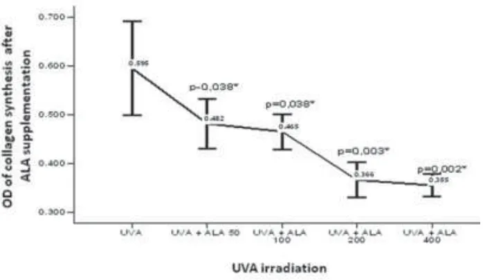
Supplementation of ALA on UVA irradiated human fibroblasts also resulted in significant decrease of collagen synthesis compared with the control (FIGURE 4) and resulted in increase of collagen degradation compared with the control (FIGURE
5). However enhancement doses of ALA resulted in decreasing cell viability and collagen synthesis and increasing collagen degradation as shown in FIGURE 3, 4 and 5.
FIGURE 5. Effect of ALA on collagen synthesis of UVA-irradiated human fibroblasts
DISCUSSION
UVA irradiation generates free radicals resulting in collagen degradation, inhibits collagen synthesis and finally induces cell death.11,12 Cell has an ability
to repair damage caused by UVA exposure. Reduction of cell’s ability to repair generates damage and accumulation of cell death that creates functional changes as shown in the photoaging process.4,13 The
decrease of cell viability on UVA-irradiated human fibroblasts cell is also seen in FIGURE 1.
α-Lipoic acid and DHLA are universal antioxidant and act as a scavenger of ROS.6,14
However, several studies showed that ALA and DHLA also have pro-oxidant effect.15,16 The effect
of ALA supplementation in various doses among UVA-irradiated human fibroblasts cell resulted in decrease of cell viability compared with human fibroblasts cell without UVA irradiation or the control (p<0.05) as presented in FIGURE 3, decrease in collagen synthesis (FIGURE 4) and increase in collagen degradation (FIGURE 5). These results did not in accordance with the ALA properties as universal antioxidant that could repair oxidative damage of cell due to UVA irradiation.14
The pro-oxidant properties of ALA may play role in the inability to repair oxidative damage. As pro-oxidant, ALA can induce mitochondrial permeability transition (MPT) resulting in rupture of cell membrane and release proapoptotic factors.17 α-Lipoic acid and DHLA can also lead to cell death through internucleosomal DNA fragmentation and induce caspase 3 cleavage that causes apoptosis of cells with higher cytotoxicity found in DHLA compared with ALA.18 In addition, level of
intracellular oxidant of ALA increased in parralel with ALA concentrations.19
UVA irradiation causes alternations in dermal collagen through two primary pathways: 1). stimulation of collagen breakdown and 2). inhibition of procollagen synthesis, resulting in loss of collagen content.20 UVA stimulates collagenase directly and
causes tissue damage through induction on protein kinase C (PKC) on fibroblast.21 UVA also activates
the activator protein 1 (AP-1) and nuclear factor kappa beta (NF-κβ) which stimulates the production of MMPs that play a role in photoaging.4,11
Transforming TGF-β is also inhibited by over
of procollagen expression.3,12 In this study, UVA irradiation
inhibits collagen synthesis and increases collagen degradation as shown on FIGURE 2 and 3.
Evaluation of collagen synthesis and cell viability of UVA-irradiated human fibroblast was in 48 hours. This evaluation time was based on study by Fisher et al.12 which stated expression of
procollagen of UVA-irradiated returns to their original level 48-72 hours.However, supplementation of ALA among UVA-irradiated human fibroblasts was not able to up-regulate collagen synthesis (FIGURE 4). The collagen synthesis analysis has not been optimized yet due to lack of follow up evaluation and required longer evaluation time.
The photo protective effect of topical antioxidants applied on human skin before UV irradiation has been recognized. Topical application of antioxidants, such as vitamin E significantly reduces acute skin responses such as erythema and edema, sunburn cell formation, lipid peroxidation, DNA adduct formation, immunosuppression as well as UVA-induced binding of photosensitizers.22,23
Application of various antioxidants such as vitamin C, vitamin E, and melatonin 30 minutes before UV-irradiation protects skin from UV-induced erythema.23 The effect of antioxidants administered
after irradiation is less obvious. Diminished erythema formation was reported when antioxidants were topically administered after UVA irradiation.24
However, these findings were in contrast to other human studies which were not able to demonstrate any diminished UV-related skin damage when antioxidants were applied to UV-irradiated in humans.25 In this study no significant effect of ALA
was observed when ALA was supplemented immediately to UVA irradiated human fibroblasts.
CONCLUSION
ALA has no impact when it is supplemented among UVA irradiated human fibroblasts.
ACKNOWLEDGMENT
The authors would like to thank Head of Department of Dermatovenerology, Faculty of Medicine, Gadjah Mada University/Dr. Sardjito General Hospital, Yogyakarta for his permission to perform this study.
REFERENCES
1. Uitto J, Chu M, Gallo R, Eisen AZ. Collagen, elastic fibers and extracellular matrix. In: Wolff K, Goldsmith LA, Katz SI, Gilchrest BA, Paller AS, Leffel DJ editors. Fitzpatrick’s dermatology in general medicine. New York: McGraw Hill Book Co, 2008: 517-42.
2. Fisher GJ, Wang ZQ, Datta SC, Varani J, Kang S, Voorhees JJ. Pathophysiology of premature skin aging induced by ultraviolet light. N Engl J Med 1997; 337: 1419-28
3. Fisher GJ, Kang S, Varani J, Bata-Csorgo Z, Wan Y, Datta S, et al. Mechanism of photoaging and chronological skin aging. Arch Dermatol 2002; 138: 1462 70.
4. Berlett BS, Stadtman ER. Protein oxidation in aging , disease, and oxidative stress. J Biol Chem 1997; 272: 20313-16.
5. Jurkiewicz BA, Bissett DL, Buettner GR. Effect of topically applied tocopherol on ultraviolet radiation-mediated free radical damage in skin. J Invest Dermatol 1995; 104(4): 484-8.
6. Packer L, Witt EH, Tritschler HJ. α-Lipoic acid as a biological antioxidant. Free Radic Biol Med 1995;19:227– 50.
7. Akpinar D, Yargiçoðlu P, Derin N, Alicigüzel Y, Aðar A. The effect of lipoic acid on antioxidant status and lipid peroxidation in rats exposed to chronic restraint stress. Physiol Res 2008;57: 893-901
8. Freshney RI. Cytotoxicity. Culture of animal cell: a manual of basic technique. 4th ed. New York: Wiley-Liss, 2000:329-44.
9. Heng ECK, Huang Y, Black Jr SA, Trackman PC. 2006. CCN2, connective tissue growth factor, stimulates collagen deposition by gingival fibroblast via module 3 and α6-and α1 integrins. J Cell Biochem 2006;89(2): 409- 20. 10. Taskiran D, Taskiran E, Yercan H, Kutay FZ.
Quantification of total collagen in rabbit tendon by the Sirius red method. Tr J Med Scie 1999; 29 :7–9 11. Petersen MJ, Hansen C, Craig S. Ultraviolet A irradiation
stimulates collagenase production in cultured human fibroblasts. J Invest Dermatol 1992; 99: 440-4.
12. Fisher GJ, Datta S, Wang ZQ, Li XY, Quan T, Chung JH, et al. c-Jun-dependent inhibition of cutaneous procollagen transcription following ultraviolet irradiation is reversed by all-trans retinoic acid. J Clin Invest 2000; 106: 663-70.
13. Moriwaki S, Takahashi Y. Photoaging and DNA repair. J Dermatol Sci 2008;50: 169-76.
14. Biewenga GP, Haenen GRMM, Bast A. The pharmacology of the antioxidant lipoic acid. Gen Pharmacol 1997;29: 315–31.
15. Cardoso do Vale O, Sa Roriz Fonteles D, Cabral FR, Fonteles MC. A dual action of α-lipoic acid in the brain: an electrophysiological evaluation. Arq Neuropsiquiatr 2003; 61: 738- 45.
16. Cakatay U, Kayah R, Sivas A, Tekeli F. Prooxidant activities of alpha-lipoic acid on oxidative protein damage in the aging rat heart muscle. Arch Gerontol Geriatr 2005; 40:231-40.
17. Morkuanite-Haimi S, Kruglov AG, Teplova VV, Stolze K, Gille L, Nohl H, et al. Reactive oxygen species are involved in the stimulation of the mitochondrial permeability transition by dihydrolipoate. Biochem Pharmacol 2003;65: 43-9.
18. Yamasaki M, Kawabe A, Nishimoto K, Madhyastha H, Sakakibara Y, Suiko M, Okamoto T, et al. Dihydro-α -lipoic acid has more potent cytotoxicity than α--lipoic acid. In Vitro Cell Dev Biol-Animal 2009; 45: 275-80. 19. Moini H, Tirosh O, Park YC, Cho KJ, Packer L. R-α
-Lipoic acid action on cell redox status, the insulin receptor, and glucose uptake in 3T3-L1 adipocytes. Arch Biochem Biophys 2002;397(2): 384-91.
20. Quan T, Qin Z, Xia W, Shao Y, Voorhees JJ, Fisher GJ. Matrix-degrading metalloproteinase in photoaging. J Invest Dermatol 2009;14: 20-4.
21. Matsui MS, DeLeo VA. Induction of protein kinase C activity by ultraviolet radiation. Carcinogenesis 1990; 11: 229-34.
22. Thiele JJ, Dreher F, Packer L. Antioxidants defense systems in skin. In: Elsner P, Maibach H, Rougier A editors. Drugs versus cosmetics: cosmeceuticals. New York: Marcel Dekker Inn, 2000:145-87.
23. Dreher F, Gabard B, Schwindt DA, Maibach HI. Topical melatonin in combination with vitamin E and C protects skin from UV-induced erythema: a human study in vivo. Br J Dermatol 1998;139: 332-9.
24. Montenegro L, Bonina F, Rigano L, Giogilli S, Sirigu S. Protective effect evaluation of free radical scavenger on ultraviolet B induced human cutaneous erythema by skin reflectance spectrophotometry. Int J Cosmet Sci 1995;17: 91-103.


