Abstract
Introduction
Classical epidemiology has established the incremental contribution of the high-density lipoprotein cholesterol (HDL-C) measure in the assessment of atherosclerotic cardiovascular disease (CVD) risk, yet, genetic epidemiology does not support a causal relationship between HDL-C and the future risk of myocardial infarction. Therapeutic interventions directed toward cholesterol loading of the
HDL particle have been based on epidemiological studies that have established HDL cholesterol as a biomarker of atherosclerotic CVD risk. The reference range of HDL-C is 40-50 mg/dL in men and 50-60 mg/dL in women. However, therapeutic interventions such as niacin, cholesterylester transfer protein (CETP) inhibitors increase HDL-C in patients treated with statins, but have repeatedly failed to reduce CVD events.(1) Clinical trials with pharmacological therapies that increase the cholesterol content of HDL particles have failed to establish this convenient metric as
The High Density Lipoprotein Cholesterol Hypothesis Revisited
Anna Meiliana
1,2,, Nurrani Mustika Dewi
2, Andi Wijaya
1,21Postgraduate Program in Clinical Pharmacy, Padjadjaran University, Jl. Eijkman No.38, Bandung, Indonesia 2Prodia Clinical Laboratory, Jl. Cisangkuy No.2, Bandung, Indonesia
Corresponding author. E-mail: [email protected]
Received date: May 17, 2018; Revised date: Jul 20, 2018; Accepted date: Jul 24, 2018
R E V I E W A R T I C L E
B
ACKGROUND: The strong inverse association of plasma levels of high-density lipoprotein cholesterol (HDL-C) with coronary heart disease (CHD) found in human epidemiological studies led to the development of the ‘HDL-C hypothesis’, which posits that intervention to raise HDL-C will result in reduced risk of CHD. However, recent evidence has raised doubts about the hypotheses that elevating HDL-C is necessarily therapeutic. Genetic variations that associate with altered HDL-C do not strongly associate with altered cardiovascular disease risk.CONTeNT: HDL-mediated cholesterol efflux from
macrophage foam cells or measurements of the flux of
cholesterol from macrophages to the liver and feces seem to correlate better with atherosclerotic burden than with HDL-C levels. Thus, it may be time to modify the HDL-C
hypothesis to the ‘HDL flux hypothesis’, where intervention to promote cholesterol efflux and reverse cholesterol
transport will reduce CHD risk, regardless of whether it
affects plasma HDL-C levels. A deeper understanding of the
complex biology of HDL metabolism and its relationship
to reverse cholesterol transport and atherothrombotic events is urgently needed. This might lead to biomarkers of HDL
flux and functionality that are more informative than simple
measurements of HDL-C levels.
SUmmARy: It is now clear from recent clinical trial and genetic studies that some approaches to raising HDL-C levels may have no effect on CHD. This suggests the need to evaluate HDL-C-raising therapies in different clinical
populations, as well as therapies targeted toward HDL flux
and function rather than simply HDL-C elevation. Perhaps moving from a focus on the HDL-C hypothesis to a focus
on the HDL flux hypothesis will permit a biologically
based reassessment of the optimal therapeutic approach to targeting HDL for reduction in cardiovascular risk.
KeywORDS: reverse cholesterol transport, cholesterol
efflux capacity, HDL dysfunction, HDL particle size, HDL
lipidomics, HDL proteomics
an effective strategy for the prevention of CVD events. These therapies have included niacin/niacin-lariproprant and CETP inhibitors.(1)
Atherothrombosis Intervention in Metabolic Syndrome with Low HDL/High Triglycerides: Impact on Global Health Outcomes (AIM-HIGH) and The Heart Protection Study 2–Treatment of HDL to Reduce the Incidence of Vascular Events (HPS2-THRIVE) trials investigated the effect of niacin and niacin/laripoprant, respectively, in coronary heart disease (CHD) patients with low-density lipoprotein cholesterol (LDL-C) levels at the recommended target <70 mg/dL.(2,3) Elevations in HDL-C were not associated with fewer CVD events. Instead, there was harm from niacin treatment as shown by the increased risk of infections.(4) CETP inhibition with multiple agents (torcetrapib, dalcetrapib, and anacetrapib) has failed to reduce CVD events in risk patients treated with high-intensity statin therapy.(5-7) The failure of torcetrapib was ascribed to torcetrapib-induced hypertensive effects via renin-angiotensinaldosterone activation and weak activity of dalcetrapib.(8,9) Early termination of the evacetrapib clinical trial in Assessment of Clinical Effects of Cholesteryl Ester Transfer Protein Inhibition With Evacetrapib in Patients at a High-Risk for Vascular Outcomes (ACCELERATE) (7)
was unexpected, not only based on the LDL-C-lowering efficacy of evacetrapib, but also in the increase in very small HDL particles and macrophage cholesterol efflux capacity.
(8) Based on event rates observed in statin-treated patients, early termination of ACCELERATE may not have allowed
sufficient time to detect a treatment difference in CVD.
HDLs comprise a multitude of discrete subpopulations that differ in composition, metabolism, cellular interactions,
and functional properties.(10,11) Expansion of the core
cholesterol in HDL particles interferes with
apoA-I-mediated cholesterol efflux via ABCA1 and alters the
proteome and lipidome, resulting in species that are less
effective in antioxidant and anti-inflammatory properties.
(12) Of the HDL functional assays, only macrophage
cholesterol efflux has been validated in prospective studies. (11,13-16) Efforts to solve the HDL puzzle require high throughput assays that accurately quantify discrete HDL
species based on their protein and lipid components. HDL
speciation may allow for the identification of specific HDL
populations with functional or dysfunctional properties.
The macrophage cholesterol efflux assay is an established method that has been used to characterize both functional and dysfunctional HDL.(13,14,17,18) Subsequent steps require evaluation of discrete HDL populations in the
prediction of atherosclerosis and cardiovascular events.(19)
HDL-C Hypothesis
The most popular mechanistic hypothesis underlying the HDL-C hypothesis has been the concept of ‘reverse cholesterol transport’, namely that some component of
the HDL system promotes cholesterol efflux from arterial
macrophage foam cells and transports it to the liver for
biliary excretion, leading to a reduction in lesion size
(Figure 1).(20)
However, recent events have raised serious questions
about the validity of the HDL-C hypothesis. Two clinical trials involving therapeutic elevations of HDL-C on individuals with low levels of LDL-C lowering were prematurely terminated on the basis of futility. The AIM-HIGH study was conducted with niacin, the most effective HDL-raising drug currently on the market; the Dalcetrapib in Patients with Recent Acute Coronary Syndrome (dal-OUTCOMES) study involved dalcetrapib, a drug in development that partially inhibits the CETP, which transfers cholesterol from HDL to very-low-density lipoprotein (VLDL) or LDL. Although many issues related to trial design are being actively debated, the hypothesis, based on epidemiological studies, that moderate HDL cholesterol raising would cause a marked reduction in events seems to have been refuted. Furthermore, a causal relationship of HDL-C with CHD has been challenged.(20)
Maturation produces diverse HDL particles that vary in
size, density, and lipid and protein composition. ApoA-I and
apolipoprotein II account for 90% of HDL’s protein content, but close to 100 other HDL proteins have been described as HDL-associated.(21) Measuring HDL-C levels takes only the cholesterol mass into account, but cholesterol accounts for no more than 20% of a particular HDL particle, and its
level may vary up to 10-fold, depending on a particle’s size.
(22) Thus, a confounder in interpreting HDL-C as a measure of circulating HDL concentration is the relative balance between large and small HDL particles.(23) HDL particle concentration, that provides information regarding the actual number of HDL particles in the circulation, can be measured by nuclear magnetic resonance (NMR) (24) or ion mobility analysis (25,26). Calibrated ion mobility analyses further demonstrate that precise measurements of small, medium, and large HDL particles can be obtained from plasma.(26) Therefore, evaluating changes in HDL subpopulations in response to therapy might be a better metric than HDL-C levels for predicting risk.(27)
As well as varying in size, HDL particles vary in
Figure 1. Pathways influencing HDL cholesterol metabolism and flux and potential relationship to atherosclerosis.(20) (Adapted with permission from Springer). HSPC: hematopoietic stem and progenitor cell; LXR: liver X receptor; ABCG1: ATP-binding cassette sub-family G member 1; ABCA1: ATP binding cassette transporter A-1; ApoA-I: apolipoprotein A-I; HDL: high-density lipoprotein; LDL: low-density lipoprotein; VLDL: very low-density lipoprotein; LCAT: lecithin-cholesterol acyltransferase; CETP: cholesterylester transfer protein; SR-BI: scavenger receptor class B type I; LDLR: LDL-receptor; TG: triglyceride.
in protein cargo have been associated with pathological
states, such as inflammation (28), poor response to therapy
(29), autoimmune disease (30), and diabetes (31). Further
improvements in quantitative techniques (32,33) and
their application to clinical studies should identify panels of protein biomarkers that may be pharmacologically
modifiable to reduce risk. Another important variable is HDL’s ability to promote cholesterol efflux from cultured
macrophages with samples derived from serum, termed
cholesterol efflux capacity (CEC). Cholesterol efflux
capacity has been a stronger predictor of prevalent and incident coronary artery disease than HDL-C in multiple clinical trials.(13,14,24) In vitro assays of CEC of HDL may include four possible pathways: aqueous diffusion, scavenger receptor class B type I (SR-B1) receptor, and ATP
binding cassette transporter A-1 (ABCA1), and ATP-binding
cassette sub-family G member 1 (ABCG1) transporters.(27) Genetic epidemiology does not support a causal relationship between HDL cholesterol and future risk of
myocardial infarction. In a Mendelian randomization study
that included 20 studies (12,482 myocardial infarction cases and 95,407 controls), single nucleotide polymorphisms in the endothelial lipase gene (LIPG Asn396Ser) had higher HDL-C levels and similar nonlipid and other lipid risk factors
for myocardial infarction.(34,35) Neither physiological function nor pathological dysfunction is recorded by the measurement of HDL-C. Novel biomarkers are currently searched and validated in clinical or epidemiological studies toward their potential to aid in drug development and improve risk prediction and monitoring of treatment response. Candidate biomarkers include HDL particle numbers, serum amyloid A, apolipoprotein (apo)C-III, sphingosine-1-phosphate (S1P), certain post-translational
modifications of apoA-I, and cholesterol efflux capacity.(36)
It is important that future studies take into consideration the role of HDL function in addition to the level when the association of HDL with risk of cardiovascular disease and outcomes. This is especially true in patients with a high
burden of oxidative stress and inflammation.(37)
HDL Particle Size, Lipidomics and
Proteomics
Despite the spectacular collapse of the HDL-C hypothesis and the paucity of alternative strategies in the clinical space,
the field as a whole has experienced a groundbreaking shift
of HDL. Advances have been made in the areas of HDL functionality as it relates to particle heterogeneity, biogenesis, and variations in lipid and protein cargo. (38) The plasma concentration of HDL particles and their
size distribution can be reliably measured using different approaches to identify subspecies with unique functional and compositional profiles. Currently, there is no consensus
on the relative value of the different methods and on the number, concentration, functional status, and predictive power of the different HDL subparticles.
HDL particles are highly heterogeneous in structure, composition, metabolism, and function. Distinct HDL
subpopulations might, therefore, exert differential effects on atherosclerosis.(39) As a consequence, plasma concentrations of specific HDL subpopulations can be hypothesized to reveal stronger associations with
cardiovascular risk as compared to total HDL particle number. NMR measurements allow distinguish between
large (size 9.4–14.0 nm), medium (8.3–9.3 nm), and
small (7.3–8.2 nm) HDL particles.(24) Among these HDL
subpopulations, levels of large HDL frequently display
inverse relationships with cardiovascular risk in univariate analyses, whereas concentrations of small HDL particles typically reveal positive correlations with the risk.(40-50) Circulating concentrations of large and small HDL particles as well as those of large HDL particles and total LDL particles as measured by NMR are, however, known to be negatively correlated.(51) Available data reveal that diminished HDL particle number can be superior to reduced HDL-C levels in terms of cardiovascular risk prediction. Measurements of circulating concentrations of HDL particles can, therefore, be useful to improve clinical assessment of cardiovascular risk. This approach can also contribute to the evaluation of novel HDL-targeted therapies, which include CETP
inhibitors, peroxisome proliferator-activated receptor
(PPAR) activators, and reconstituted HDL.(51)
HDL can be fractionated by different techniques
into discrete subclasses according to physicochemical
properties and chemical composition. Sequential
ultracentrifugation allows separation of HDL into two main subfractions on the basis of density: large, light, lipid-rich HDL2 (d 1.063–1.125 g/mL) and small, dense, protein-rich HDL3 (d 1.125–1.21 g/mL).(10) HDL2 and HDL3 can be further subfractionated on nondenaturing polyacrylamide
gradient gel electrophoresis into five distinct subpopulations of decreasing size: HDL2b, HDL2a, HDL3a, HDL3b, and HDL3c (10); equivalent subpopulations of increasing density can be quantitatively isolated using isopycnic
density gradient ultracentrifugation (10). Such heterogeneity
of HDL particles directly reflects differences in the relative
content of proteins and lipids in HDL.(52)
HDLs are a class of heterogeneous lipoproteins; their heterogeneity is attributable to a different content
of apolipoproteins, lipids and enzymes and to the remodeling of HDL particles by lipolytic enzymes, lipid transporters and by lipid and apolipoprotein exchange
with other circulating lipoproteins and tissues. Different
HDL subpopulations carry distinct and specific proteins
or lipids, suggesting distinct and characteristic functions.
(39) HDL can be classified on the basis of density, resulting
in the large buoyant HDL2 and the small dense HDL3, which can be further subfractionated into 5 distinct sub- populations (HDL2b, HDL2a, HDL3a, HDL3b and HDL3). Alternatively, HDLs can be separated on the basis of their
electrophoretic mobility in pre-β- particles, α-particles and pre-α-particles. HDLs can also be classified according to
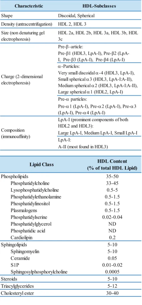
their main apolipoprotein content into particles containing only apolipoprotein A-I (apoA-I, LpA-I) or both apoA-I and apoA-II (LpA- I/A-II) (Table 1).(39,53)
The heterogeneity of HDLs is also reflected in their
functions, since different subpopulations play distinct roles. HDLs possess several antiatherogenic functions. The
best characterized activity of HDL is reverse cholesterol transport, the process by which excess cholesterol is
transported from the peripheral tissues to the liver for
excretion. Different HDL subpopulations interact with different cellular receptors to remove excess cholesterol
from cells. Besides, HDLs, and in particular small dense
HDL3, exert antiinflammatory and antioxidant activities.
HDLs have a protective effect on vascular endothelium, are antithrombotic and antiinfectious and play a role in the modulation of immune responses and the control of glucose homeostasis.(39,54,55)
The protein constituents of HDL in particular have been heavily dissected in recent years, primarily through
liquid chromatography mass spectrometry (LC-MS),
revealing several more proteins additional to the classical apolipoproteins, such as apoA-I, apoA-II, apoE and apoC-III, whose biological activities are diverse, but still encompass the role for HDL as antiatherogenic particle.
Moreover, LC-MS technology has extended well beyond a tool for identification. Changes in apolipoprotein abundance
can now be readily assessed using one or more of a number of strategies including label-free and label-based strategies.(56,57). Quantitative proteomics provides several
innovative workflows that are poised to address many of
Characteristic HDL-Subclasses
Shape Discoidal, Spherical
Density (untracentrifugation) HDL 2, HDL 3
Size (non denaturing gel electrophoresis)
Very small discoidal α-4 (HDL3, LpA-I), Small spherical α3 (HDL3, LpA-I:A-II), Medium spherical α2 (HDL3, LpA-I:A-II), Large spherical α1 (HDL2, LpA-I) Pre-α particles:
Pre-α1 (LpA-I), Pre-α2 (LpA-I), Pre-α3 (LpA-I), Pre-α4 (LpA-I)
LpA-I (prominent components of both HDL2 and HDL3):
Large LpA-I, Medium LpA-I, Small LpA-I LpA-I:
A-II (most found in HDL3) Charge (2-dimensional
electrophoresis)
Composition (immunoaffinity)
Table 1. HDL subclasses and major components of the HDL lipidome(52,53) (Adapted with permission from Karger AG, American Society for Biochemistry and Molecular Biology).
HDL Content (% of total HDL Lipid)
35-50
proteome comprises between 90 and 100 proteins. The HDL proteome is differentially distributed across the HDL
size fractions giving rise to a heterogeneous population
of HDL particles.(58) In addition to proteins consistent with traditionally accepted roles in lipid transport, HDL carries surprising constituents, such as members of the complement pathway, protease inhibitors involved in hemostasis, acute-phase response proteins, immune function mediators, and even metal-binding proteins. This
compositional diversity fits well with hundreds of studies
demonstrating a wide functional pleiotrophy, including
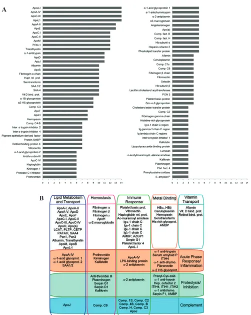
roles in lipid transport, oxidation, inflammation, hemostasis, and immunity.(21) Figure 2A shows the frequency of
detection of HDL-associated proteins in some MS-based proteomic studies, meanwhile Figure 2B shows general functional relationships of HDL proteins. Establishing direct relationships between distinct HDL structure and
composition on the one hand and specific atheroprotective functions on the other requires further study (Table 1).
Such structure-function analysis of HDL particles bears the potential to identify clinically relevant, atheroprotective HDL subpopulations. Furthermore, development of
HDL-based therapies specifically designed to target beneficial
subspecies of the circulating HDL pool can be facilitated
using this approach. HDL lipidomics can equally contribute to the identification of biomarkers of both normal and deficient HDL functionality, which may in turn prove useful
as biomarkers of cardiovascular risk. It remains to be shown whether such novel HDL-based lipidomic biomarkers can be superior relative to HDL-C levels.(52)
Lipid-poor and small HDL displayed both high
capacity for ABCA1-mediated cholesterol efflux and strong anti-inflammatory action, whereas the larger particles showed stronger antioxidant function. This study provides
a novel perspective because it suggests that subspecies of HDL may be involved in different functions, and thus,
the right mix of multiple HDL particle sizes may be most desirable to maximize cardioprotective benefits (Figure 3).
Most importantly, the study offers an approach to generate these particles in vivo.(38)
The HDL mass is equally distributed between lipid
and protein cargo. The HDL lipidome is composed mainly of phospholipids, cholesteryl esters, triglycerides, and free cholesterol, for a total of over 200 individual lipid species in normolipidemic subjects. Some lipid components, such as phospholipids, sphingomyelin, and free cholesterol, have already been investigated as modulators of HDL functions,
such as sterol efflux, vasodilation, and control of oxidation and inflammation.(52)
Regulation of HDL Metabolism
Understanding of the regulation of HDL metabolism has
increased significantly in recent years, although these
B
Figure 2. Relationship of HDL proteins. A: General functional relationships of the HDL proteins; B: Frequency of detection of
HDL-associated proteins in MS-based proteomic studies.(21) (Adapted with permission from American Society for Biochemistry and Molecular Biology).
Figure 3. Overview of the regulation and interaction of traditional and novel high-density lipoprotein (HDL) measures.(38) (Adapted with permission from American Heart Association).
Most types of cells in the body do not express the capability of catabolizing cholesterol, so cholesterol efflux
is essential for homeostasis. For instance, macrophages
possess four pathways for exporting free (unesterified) cholesterol to extracellular HDL. The passive processes include simple diffusion via the aqueous phase and
facilitated diffusion mediated by SR-BI. Active pathways are mediated by ABCA1 and ABCG1, which are membrane
lipid translocases. The efflux of cellular phospholipid and
free cholesterol to apoA-I promoted by ABCA1 is essential for HDL biogenesis.(68)
Recently, S1P, a lipid mediator that acts via G-protein-coupled receptors, has featured prominently in HDL biology. The ability of HDL to protect the endothelium (69), myocardial ischemic injury and vasodilate (70) depends on the S1P cargo. HDL-bound apolipoprotein M binds, carries, and promotes receptor activation in a physiologically relevant manner.(71)
In addition, HDL-bound S1P seems to be distinct from albumin-bound S1P in the inhibition of endothelial
inflammatory processes (72), barrier function (73) and
lymphopoiesis (74), suggesting that chaperone- bound
S1P acts as a biased agonist to evoke specific biological
processes. These observations suggest a major function of S1P in the cardiovascular protection mediated by HDL.(75,76)
increasing burden of CVD worldwide, there is a compelling need to identify strategies and to develop therapeutic agents that can begin to resolve these issues.(59) The heterogeneity
is a consequence of the continual remodeling and
interconversion of HDL subpopulations by multiple plasma factors. Evidence that the remodeling of HDLs may impact on their cardioprotective properties is beginning to emerge. This serves to highlight the importance of understanding not only how the remodeling and interconversion of HDL subpopulations is regulated but also how these processes are affected by agents that increase HDL levels.(59)
The 4 main apolipoproteins in human HDLs, in order of decreasing abundance, are apoA-I, apoA-II, apoA-IV,
and apoE (Figure 4). ApoA-I is synthesized in the liver
and intestine. Hepatic apoA-I is initially generated as a preproprotein that is cleaved intracellularly by a signal peptidase.(60) The resulting propeptide is secreted before cleavage by bone morphogenic protein-1 in a process that is facilitated by procollagen C-proteinase enhancer-2.(61,62)
In vitro studies have found that ≤45% of apoA-I is lipidated
A
Figure 4. The biogenesis of many kind of lipoproteins. A: Biogenesis of apolipoprotein A-I (apoA-I)–containing discoidal high-density lipoproteins (HDLs); B: Biogenesis of spherical high-density lipoproteins (HDLs); C: Remodeling of apolipoprotein E (apoE)– containing high-density lipoproteins (HDLs) by phospholipid transfer protein (PLTP).(59) (Adapted with permission from American Heart Association).
B
There are 3 different transmembrane transporters along the apical membrane of the hepatocyte that actively promotes this process: the heterodimer ABCG5/ABCG8 (which
facilitates cholesterol efflux), ABCB11 and the phospholipid
pump, ABCB4.(78) Moreover, another transporter, ATP8B1, is also necessary for correct secretion of bile. ATP8B1 moves phosphatidylserine in the opposite direction to the transport of phosphatidylcholine by ABCB4 to maintain the
asymmetry of phospholipids required for proper membrane
function.(78,79) Because cholesterol cannot be degraded in the cell, reverse cholesterol transport (RCT) is an essential process to ensure that cholesterol levels are balanced within
the body. Work over the past years has identified miRNAs
as important regulators of HDL-C metabolism. miRNAs control most of the steps of RCT including HDL biogenesis,
cellular cholesterol efflux, hepatic HDL-C uptake, and bile
acid synthesis and secretion.(80) In addition to their role in regulating HDL-C metabolism, HDL-enriched miRNAs
regulate gene expression in recipient cells, thus providing an exciting novel mechanism that could explain a part of the
antiatherogenic effect of HDL (Figure 5).(81,82)
Recent studies have highlighted the close partnership between activation of ecto-F1-ATPase by HDL or their major protein, apoA-I, and P2Y receptor signaling. For instance, on hepatocytes the HDL-apoA-I/ecto-F1-ATPase/
P2Y13 sequence contributes to HDL uptake and would
be atheroprotective.(83-88) On adipocytes, HDL-apoA-I, ecto-F1-ATPase and P2Y signaling are all involved in lipid metabolism.(89-91) On endothelial cells, ecto- F1-ATPase is also activated by HDL-apoA-I and is potentially coupled to P2Y1 or P2Y12 receptors, promoting cell survival and
HDL transcytosis.(92,93) In addition, HDL-apoA-I receptor signalling and P2Y receptor signaling share other common features on endothelium protection, such as the regulation
of NO production and of pro-inflammatory cell adhesion
molecules.(94-96) In many instances, these roles seem to
rely on the enzymatic ATP hydrolase activity of ecto-F1-ATPase, which generates extracellular ADP. Thus, it is
tempting to propose a common framework that would
involve the sequential activation of ecto-F1-ATPase by HDL- apoA-I, modulation of the extracellular ATP/ADP
ratio and of downstream activation/inactivation of P2Y-mediated signalling pathways.(97)
As described, the metabolism of the HDL particle is a multistep process involving several apolipoproteins,
enzymes, and transporters; therefore, genetic variation
in genes regulating each of these steps will greatly affect HDL-C concentrations.(98) Mendelian disorders of high and low HDL-C levels have provided clues about the
biology of HDL.(99) Candidate gene studies have identified
Mendelian causes of low HDL-C levels, including mutations in ABCA1, APOA1, and lecithin-cholesterol acyltransferase (LCAT). Conversely, mutations in CETP and endothelial lipase (LIPG) result in high HDL-C levels. (100) Our microbiota has been linked to intestinal health, immune function, bioactivation of nutrients and vitamins,
and recently, complex disease phenotypes, such as obesity
and insulin resistance.(101) Interestingly, recent studies showed that intestinal microbial processing of dietary
choline to trimethylamine, which is further metabolized to trimethylamineoxide by flavin monoxygenases in human and rodent livers, was significantly correlated with
CVD.(102,103) Furthermore, previous study found that
trimethylamineoxide suppressed RCT via an intestinal
microbiota-dependent mechanism in vivo.(104) These findings suggest a new concept that specific combinations
of intestinal microbiota and host genetics may provide cardiometabolic regulation.(105)showed that intestinal microbial processing of dietary choline to trimethylamine,
which is further metabolized to trimethylamineoxide by flavin monoxygenases in human and rodent livers, was significantly correlated with CVD.(102,103) Furthermore, previous study found that trimethylamineoxide suppressed
RCT via an intestinal microbiota-dependent mechanism in vivo.(104) These findings suggest a new concept that specific
combinations of intestinal microbiota and host genetics may provide cardiometabolic regulation.(105)
Functional and Dysfuctional HDL
HDL-C has direct effects on numerous cell types that influence
CVD and metabolic health. These include endothelial cells, vascular smooth-muscle cells, leukocytes, platelets, adipocytes, skeletal muscle myocytes, and pancreatic cells. The effects of HDL or apoA-I, its major apolipoprotein, occur through the modulation of intracellular calcium,
oxygen-derived free-radical production, numerous kinases, and enzymes, including endothelial nitric-oxide synthase (eNOS). ApoA-I and HDL also influence gene expression, particularly genes encoding mediators of inflammation in
vascular cells.(106) There are several well-documented
HDL functions such as RCT, inhibition of inflammation,
or inhibition of platelet activation that may account for the atheroprotective effects of this lipoprotein. Mechanistically, these functions are carried out by a direct interaction of HDL
particle or its components with receptors localized on the
cell surface followed by generation of intracellular signals. Several HDL-associated receptor ligands such as apoA-I or
S1P have been identified in addition to HDL holoparticles,
which interact with surface receptors such as ABCA1; S1P receptor types 1, 2, and 3 (S1P1, S1P2, and S1P3); or SR-BI and activate intracellular signaling cascades encompassing kinases, phospholipases, trimeric and small G-proteins, and cytoskeletal proteins such as actin or junctional protein such
as connexin. In addition, depletion of plasma cell cholesterol
mediated by ABCA1, ABCG1, or SR-BI was demonstrated
to indirectly inhibit signaling over proinflammatory or
proliferation-stimulating receptors such as Toll-like or growth factor receptors.(107)
Several well-documented functions of HDLs and apoA-I have the potential to protect against cardiovascular
disease. The most extensively studied of these relates to the ability of HDLs to promote efflux of cholesterol from
macrophages in the artery wall.(108) HDLs also inhibit
vascular inflammation (109,110) and has antioxidant (109)
and antithrombotic properties (95). They enhance endothelial function (111), promote endothelial repair (112,114), increase agiogenesis (114), suppress the production and
mobilization of monocytes and neutrophils from bone
marrow (115), and have recently been reported to have
antidiabetic properties (55,116). It has been hypothesized that paraoxonase/arylesterase 1 (PON1) located on HDL possesses the capacity to hydrolyze lipid hydroperoxides and is largely responsible for the antioxidant effect of HDL.
Transgenic animal evidence and clinical epidemiology strongly support an antiatherogenic role for PON1.
Direct in vitro evidence for the PON1 antioxidant
hypothesis has proved controversial, and other HDL components have been proposed to account for the
antioxidant capacity of HDL, such as apoAI and apoM.
These and other HDL components may interact with PON1
to produce its antioxidant effects. The environment provided
for this interaction by HDL may be critical.(117) The
increasing evidence that HDL not only augments hypoxia-mediated angiogenesis but also inhibits inflammatory driven neovascularization. This suggests that the regulation of
angiogenesis by HDL is dependent on the pathophysiological
context. One previous example of this was demonstrated
in a study using the apoA-I mimetic peptide, D-4F. This
peptide significantly increased the vascular expression and activity of haeme oxygenase-1 (HO-1).(118) HO-1 is induced by hypoxia to facilitate angiogenesis in response to ischaemia, but conversely also inhibits leucocyte infiltration by suppressing cytokine expression and consequently reduces inflammation-mediated neovascularization.(119) Studies recently confirmed these observations by directly
comparing the effects of apoA-I/rHDL on angiogenesis
in both hypoxic/ischaemic and inflammatory conditions.
Mechanistically, the key to the conditional regulation of angiogenesis by HDL may be vascular endothelial growth
The antithrombotic properties of native HDL are also related to the suppression of the coagulation cascade and
stimulation of clot fibrinolysis. Furthermore, HDL stimulates the endothelial production of nitric oxide and prostacyclin,
which are potent inhibitors of platelet activation. Thus, HDL’s antithrombotic actions are multiple and therefore, raising HDL may be an important therapeutic strategy to reduce the risk of arterial and venous thrombosis.(121) There is now convincing evidence that HDL modulates glucose metabolism in multiple tissues. These actions have deepened our understanding of the pathophysiology of a variety of disease states associated with low or dysfunctional HDL. While there are still many unanswered
questions relating to the underlying mechanisms and key
HDL component(s) responsible for the metabolic effects, this opens up the possibility of targeting glucose metabolism with HDL therapeutics currently in development. Future preclinical investigations and clinical trials will determine the relevance of HDL-mediated modulation of glucose metabolism to both glycemic control as well as tissue glucose supply to vital organs including the heart and the brain, especially under ischemic conditions.(122)
HDLs and the main HDL apolipoprotein, apolipoprotein (apo)A-I, increase insulin synthesis and secretion in pancreatic b-cells. HDL apolipoproteins increase insulin synthesis and secretion in pancreatic b-cells by a mechanism similar to that of the intestinally derived, endogenous incretins, glucose-dependent insulinotropic peptide (GLP-1) and gastric inhibitory polypeptide (GIP).
(123) Systemic and vascular inflammation has been
proposed to convert HDL to a dysfunctional form that has
impaired anti-atherogenic effects. A loss of anti-inflammatory and antioxidative proteins, perhaps in combination with a gain of proinflammatory proteins, might be another
important component in rendering HDL dysfunctional. The
proinflammatory enzyme myeloperoxidase induces both oxidative modification and nitrosylation of specific residues
on plasma and arterial apoA-I to render HDL dysfunctional, which results in impaired ABCA1 macrophage transport,
the activation of inflammatory pathways, and an increased
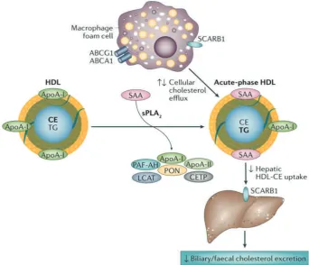
risk of CAD.(16) Figure 6 shows the acute-phase of HDL. The development of monoclonal antibodies that
identify specific forms of dysfunctional apoA-I is a promising
area of investigation for monitoring pathophysiological
Figure 6. Acute-phase HDL. HDL undergoes substantial modification during an acute-phase response.(16) (Adapted with permission
The Changing Face of HDL and The Best
Way to Measure It
It is now well established from large-scale epidemiologic studies that increased plasma levels of HDL-C and LDL-C are associated with decreased and increased cardiovascular
risk, respectively.(124) Consequently, HDL-C and LDL-C
are routinely used as serum biomarkers for assessing an individual’s cardiovascular disease (CVD) risk.(125) Recent
studies have questioned whether HDL-C is only a biomarker
and is not mechanistically linked to cardiovascular protection.(126) While a central focus has been placed on the role of HDL in the RCT process, our appreciation for the other cardioprotective properties of HDL continues to
expand with further investigation into the structure and function of HDL and its specific subfractions. Development
of novel assays is empowering the research community to assess different aspects of HDL function, which at some point may evolve into new diagnostic tests.(127)
The generation of different HDL subfractions
throughout the RCT pathway is demonstrative of the size,
compositional, and functional diversity of HDL. In addition
to altering the size of the HDL subfractions, integration of
cholesteryl esters and triglycerides fundamentally impacts the cardioprotective properties of HDL.(53,128) Further, the makeup of HDL subfractions is dynamic, as different
lipolytic enzymes, lipid transporters, and apolipoprotein exchange mechanisms (with adjacent circulating
lipoproteins and tissues) contribute to the formation and remodeling of HDL subfractions.(65) This also illustrates how simply measuring plasma HDL-C concentrations may
not fully capture the impact of HDL on cholesterol flux
between tissues, or its larger effect on CVD.
A common methodology for assessing the antiatherogenic functionality of HDL is measuring CEC, or the ability of HDL to initiate the RCT pathway by accepting cholesterol from lipid-laden macrophages.(34,129) The
methodology for determining CEC, first validated in a large
clinical trial by Khera, et al., involves quantification of total cholesterol efflux from macrophages with apoB-depleted
processes within the arterial wall and plasma, and for the evaluation of HDL/apoA-I therapies that are directed at mitigating the proatherogenic effects of dysfunctional HDL.
As further research expands our knowledge in this arena,
testing of HDL function could allow for better clinical risk
stratification, optimization of individual clinical treatments,
and the evaluation of novel therapeutics.(16)
serum, and demonstrated CEC as a strong inverse predictor of coronary artery disease status independent of HDL-C
concentration.(34) CEC quantified with macrophages in vitro is a much better predictor of prevalent CVD than is HDL-C.(34,130) In 2 different cohorts, cholesterol efflux
capacity strongly and negatively associated with CVD
status.(34) This relationship remained highly significant
after correction for HDL-C. HDL-C accounted for only 34%
of the variance in efflux capacity, indicating that HDL-C is not a major determinant of cholesterol efflux capacity.
(34,130)
Two recent studies provided strong evidence that impaired CEC is also a strong predictor of incident CVD.
In the Dallas Heart Study, impaired efflux capacity was
the strongest predictor of future CVD events in a large cohort of multiethnic healthy subjects.(13) Similar results were reported in the European Prospective Investigation of Cancer (EPIC)-Norfolk study.(14) In both studies, impaired CEC remained a strong predictor of future events after adjusting for other risk factors, including HDL-C and LDL-C, suggesting that this metric provides clinically valuable information that is independent of traditional lipid risk factors.(131) Given the disappointment so far in developing new HDL-C-targeted drugs for treatment of CVD, there has been great interest in determining whether another HDL metric may better capture its potential antiatherogenic effects. A functional assay assessing
HDL-promoted cholesterol efflux has garnered interest, and may
be a convenient way to assess HDL function in RCT. In this section, we also discuss other potential functions of HDL
and novel qualitative and quantitative assays. HDL is known
to bind over 80 different types of proteins and transports more than 100 different species of lipids, so it likely has other functions outside RCT.(132,133) Finally, the function of HDL is highly associated with its com-osition, so we also discuss methodologies for identifying HDL-associated proteins, lipids, and physical properties.
There is also good evidence that the concentration of HDL, HDL particle number (HDL-P), provides clinically useful information that is distinct from HDL-C. Two methods, NMR spectroscopy (134) and ion mobility (IM)
(135), have been used to quantify HDL-P. However, these
methods give different estimates of HDL-P concentration
and size.(134,135) In future studies, it will be critical for investigators to validate the quantification of HDL-P by
to that of HDL-C in both population studies (136,137) and
randomized, clinical trials of lipid-modifying therapies.
(46,138,139) In the Multi-Ethnic Study of Atherosclerosis (MESA), low HDL-P predicted higher risk of elevated carotid intima-medial thickness regardless of whether the
baseline HDL-C level was high (≥55 mg/dL) or low (<42 mg/dL).(137) In the Justification for the Use of Statins in
Prevention: An Intervention Trial Evaluating Rosuvastatin (JUPITER), HDL-P was a better marker of residual risk in statin-treated patients than chemically measured HDL-C,
apoA-I, or average HDL size.(139)
Evaluation of antioxidant effects of HDL has
encompassed several functional methods that evaluate
the efficiency of HDL particles to protect LDL against oxidative modification. LDL lipid peroxidation by free
radicals the presence of HDL proceeds in a 2-step process that involves a slow rate of conjugated diene accumulation
ascribed to the presence of antioxidants including those
in HDL and a second rapid phase that is principally
dependent on the antioxidative functionality of HDL. (15) An alternative method for evaluating the antioxidant
effects of HDL is the cell-free assay. The cell-free assay
or the HDL-oxidized 1-palmitoyl- 2-arachidonoyl-sn-glycero-3-phosphorylcholine (OxPAPC) assay measures the ability of plasma HDL to reduce formation of oxidized phospholipids.(140-142) The addition of the fluorochrome 2’,7’-dichlorofluorescein produces a fluorescent signal that depends on the concentration of OxPAPC in vitro.
Some candidate biomarkers associated with metrics
that reflect HDL functions have recently emerged. They fall into three categories: size and quantitative features such as HDL particles size and number (51,143), markers
of HDL properties such as HDL-associated sphingosine-1-phosphate that contributes to the cytoprotective activity of
HDL and endothelial nitric oxide production (144-148) and
markers of hepatobiliary RCT such as CEC, HDL turnover and hepatic HDL uptake (13,14,149) Among HDL cellular partners, the ecto-F1-ATPase is a receptor for apoA-I that contributes to several HDL atheroprotective properties, including cellular uptake of HDL and endothelial cell protection.(97) Thus, indicators of ecto-F1-ATPase activity might have the potential to cover several categories of HDL-related biomarkers.(150)
Circulating inhibitory factor 1 (IF1) is strongly and independently associated with mortality in coronary artery disease patients but, in the same population, HDL-C is not. (151) According to our hypotheses, the IF1 level assesses metabolic and vascular HDL protective properties by
reflecting the ecto-F1-ATPase/P2Y pathway activation by
apoA-I, both in hepatic reverse cholesterol transport and vascular endothelium protection. In addition, serum IF1
might also reflect myocardial function, which can explain
its correlation with heart rate and left ventricular ejection fraction (LVEF). F1 thus joins other emerging biomarkers that have proven to be better than HDL-C level for evaluating cardiovascular risk and determining pharmacotherapy, such as HDL CEC or HDL particles number. IF1 can be used in a panel of biomarkers to better stratify cardiovascular risk and to set treatment.
The challenge is to develop laboratory assays that
quantify the various HDL functions that may improve
CVD risk assessment and augment the evaluation of HDL-modifying therapies. Efforts to develop reproducible, cost-effective, validated assays that measure the potentially
protective functions of HDL are now recognized as a major challenge for the cardiovascular field. Currently, there is
no consensus concerning the HDL functions that should be
targeted, nor are there standardized assays to measure HDL
function as a tool to improve either CVD risk assessment or the assessment of therapeutic interventions. Another challenge is to validate measurements of HDL particles
to be able to standardize assays of function with HDL quantification.(15)
In population studies, HDL-C is inversely related to the risk of myocardial infarction and death.(152-155) Of note, in patients fully treated according to current guidelines with intense statin therapy and LDL-C at target levels, HDL-C remains predictive of outcome for major adverse cardiovascular events.(6) Unfortunately, it has
been proven difficult to reduce coronary risk with drugs
increasing HDL-C, such as brates, niacin, or inhibitors of CETP, beyond that achieved with statin therapy alone. (2,5,156,157) Moreover, in several inborn errors of human HDL metabolism and genetic mouse models with altered HDL metabolism, the changes in HDL-C levels were not associated with accompanying changes in cardiovascular
risk or atherosclerotic plaque load, respectively, as has been expected from epidemiological studies.(35,158,159) Thus,
the pathogenic role and, suitability of HDL as a therapeutic
target has increasingly been questioned. In fact, it has
been argued that low HDL-C may only represent a marker for proatherogenic risk factors, rather than HDL being a mediator protecting against atherogenesis (Figure 7).(160)
Figure 7. Proposed direct vascular protective and potentially antiatherogenic effects of normal high-density lipoprotein (HDL).
(160) (Adapted with permission from American Heart Association).
One argument to explain the HDL controversy is
that HDL-C concentration is a poor measure for targeted intervention. HDL-C concentration is considered a surrogate
for the efficiency of cholesterol efflux from tissues.
However, given that macrophage-derived cholesterol represents only a minor proportion of the cholesterol
transported by HDL particles, this may be an inadequate
measure. Moreover, HDL-C concentration is a static measurement, and does not take into account the dynamics of the HDL particle population and HDL functionality, which might differ depending on the metabolic status of individuals. For instance, patients with type 2 diabetes display a higher catabolic rate of HDL-ApoA-I (148) and HDL from coronary patients does not have endothelial
anti-inflammatory effects (161), illustrating the need to identify more precisely the patient subgroups that should benefit from personalized HDL therapies.(162) Therefore, it will be
important to demonstrate that novel drugs not only increase HDL-C plasma levels but also improve HDL function in patients at high cardiovascular risk.(163)
Although RCT was first postulated to be a major
contributor to the causative association between low plasma HDL-C and ischaemic heart disease, numerous other plausible contributing mechanisms have been uncovered. Animal models have been an important vehicle for these discoveries, with more recent investigations providing a
clinical context for several antiatherothrombotic actions of HDL. These include antiplatelet (164,165), antioxidative (166), anti-inflammatory (167), anti-apoptotic (168,169)
and vasodilatory (111,170,171) activities, as well as effects on glucose metabolism (55,172-174). These mechanisms have the potential to act at multiple stages throughout the
development of atherothrombosis. Via these mechanisms, HDL-targeted therapies could both prevent the formation
and reduce the progression of plaques by slowing the
accumulation of cholesterol in the artery wall and also by
stabilizing inflamed plaques that are vulnerable to rupture.
(175)
Several strategies to therapeutically target the metabolism, particle structure and function of HDL are emerging. These include ApoA1-mimetic peptides, agonists of the liver X receptor (LXR), agonists of the farnesoid X receptor (FXR), inhibitors of LIPG, antagonists of microRNAs (miRNAs) and antisense oligonucleotides (ASOs) targeted to genes that are implicated in HDL metabolism, including the CETP and ApoC3 genes.(175)
HDL functions reflect the physiological role of the lipoprotein better than HDL-C quantity, the intake of olive
oil phenolic compounds resulted in an improvement in CEC,
HDL antioxidant defenses, HDL size distribution, and other characteristics related to HDL quality. Olive oil phenolic
compounds bound to HDLs, or surrounding the lipoprotein,
improve their oxidative/inflammatory status which may justify an increase in HDL functionality. Modifications in
HDL composition because of the consumption of virgin
olive oil (VOO) might also explain these changes. However, large-scale, randomized controlled trials with VOO-rich dietary interventions are required to definitively confirm
the protective role of olive oil phenolic compounds in HDL biological functions.(176)
Nevertheless, HDL has not yet been successfully
exploited for therapy. One potential reason for this shortfall is the structural and functional complexity of HDL particles,
than 200 lipid species as well as several microRNAs and other potentially bioactive molecules. This physiological
heterogeneity is further increased in several inflammatory
conditions that increase cardiovascular risk, including coronary artery disease itself but also diabetes mellitus, chronic kidney disease, and rheumatic diseases. The
quantitative and qualitative modifications of the proteome
and lipidome, as well as the resulting loss of functions or gain of dysfunctions, are not recovered by the biomarker HDL-C. As yet the relative importance of the many physiological and pathological activities of normal and dysfunctional HDL, respectively, for the pathogenesis of atherosclerosis
is unknown. The answer to this question, as well as detailed
knowledge of structure-function-relationships of
HDL-associated molecules, is a prerequisite to exploit HDL for
the development of anti-atherogenic drugs as well as of
diagnostic biomarkers for the identification, personalized treatment stratification, and monitoring of patients at
increased cardiovascular risk.(177)
Conclusion
The clinical utility of HDLs has been scrutinized upon the publication of Mendelian randomization studies showing
no effect of HDL-C modifying variants on CVD outcome.
The failures of randomized controlled HDL-C-directed
intervention trials have further fueled this skepticism. This
general criticism originates from oversimplification that has equated ‘HDL-C’ with ‘HDL’ and misconceived both
as the ‘good cholesterol’. HDL particles are heterogeneous and carry hundreds of different lipids, proteins, and microRNAs. Many of them but not cholesterol, that is, HDL-C, contributes to the multiple protective functions of HDLs that probably evolved to manage potentially
life-threatening crises. Quantification of HDL particle numbers, distinct proteins or lipids, and modifications thereof as well as bioassays of HDL functionality are currently explored
toward their diagnostic performance in risk prediction and monitoring of treatment response. Any successful clinical
exploitation of HDLs will depend on the identification of the
most relevant (dys)functions and their structural correlates.
Stringent or prioritized structure-(dys)function relationships
may provide biomarkers for better risk assessment and monitoring of treatment response. The most relevant agonists carried by either functional or dysfunctional HDLs as well as their cellular responders are interesting targets for drug development.
References
1. Rosenson RS. The high-density lipoprotein puzzle. Why classic epidemiology, genetic epifemiology, and clinical trials conflict?
Arterioscler Vasc Thromb Biol. 2016; 36: 777-82.
2. Boden WE, Probsteld JL, Anderson T, Chaitman BR,
Desvignes-Nickens P, Koprowicz K, et al. Niacin in patients with low HDL
cholesterol levels receiving intensive statin therapy. N Engl J Med. 2011; 365: 2255-67.
3. Landray MJ, Haynes R, Hopewell JC, Parish S, Aung T, Tomson J, et al. Effects of extended-release niacin with laropiprant in high-risk
patients. N Engl J Med. 2014; 371: 203-12.
4. Anderson TJ, Boden WE, Desvigne-Nickens P, Fleg JL, Kashyap ML, McBride R, et al. Safety profile of extended-release niacin in
the AIM-HIGH trial. N Engl J Med. 2014; 371: 288-90.
5. Schwartz GG, Olsson AG, Abt M, Ballantyne CM, Barter PJ, Brumm
J, et al. Effects of dalcetrapib in patients with a recent acute coronary
syndrome. N Engl J Med. 2012; 367: 2089-99.
6. Barter PJ, Cauleld M, Eriksson M, Grundy SM, Kastelein JJ, Komajda M, et al. Effects of torcetrapib in patients at high risk for coronary
events. N Engl J Med. 2007; 357: 2109-22.
7. PRNewswire [Internet]. Lilly Provides Update on Evacetrapib Phase 3 Trial [updated 2015 Jul 27; cited 2018 Jan 12]. Available from: https://www.prnewswire.com/news-releases/lilly-provides-update-on-evacetrapib-phase-3-trial-300118811.html
8. Nicholls SJ, Ruotolo G, Brewer HB, Kane JP, Wang MD, Krueger KA, et al. Cholesterol efflux capacity and pre-beta-1 HDL
concentrations are increased in dyslipidemic patients treated with evacetrapib. J Am Coll Cardiol. 2015; 66: 2201-10.
9. Rosenson RS, Brewer HB Jr. New challenges for HDL-modifying therapies as a strategy to lower cardiovascular disease events in statin-treated patients. Cardiovasc Drugs Ther. 2015; 29: 1-3.
10. Rosenson RS, Brewer HB Jr, Chapman MJ, Fazio S, Hussain MM,
Kontush A, et al. HDL measures, particle heterogeneity, proposed
nomenclature, and relation to atherosclerotic cardiovascular events. Clin Chem. 2011; 57: 392-410.
11. Rosenson RS, Brewer HB Jr, Davidson WS, Fayad ZA, Fuster V, Goldstein J, et al. Cholesterol efflux and atheroprotection:
advancing the concept of reverse cholesterol transport. Circulation. 2012; 125: 1905-19.
12. Camont L, Lhomme M, Rached F, Le Goff W, Nègre-Salvayre A, Salvayre R, et al. Small, dense high-density lipoprotein-3 particles
are enriched in negatively charged phospho-lipids: relevance to
cellular cholesterol efflux, antioxidative, antithrombotic, anti-inflammatory, and antiapoptotic functionalities. Arterioscler
Thromb Vasc Biol. 2013; 33: 2715-23.
13. Rohatgi A, Khera A, Berry JD, Givens EG, Ayers CR, Wedin KE,
et al. HDL cholesterol efflux capacity and incident cardiovascular
events. N Engl J Med. 2014; 371: 2383-93.
14. Saleheen D, Scott R, Javad S, Zhao W, Rodrigues A, Picataggi A,
et al. Association of HDL cholesterol efflux capacity with incident
coronary heart disease events: a prospective case-control study. Lancet Diabetes Endocrinol. 2015; 3: 507-13.
15. Rosenson RS, Brewer HB Jr, Ansell B, Barter P, Chapman MJ, Heinecke JW, et al. Translation of high-density lipoprotein function
into clinical practice: current prospects and future challenges. Circulation. 2013; 128: 1256-67.
16. Rosenson RS, Brewer HB Jr, Ansell BJ, Barter P, Chapman MJ, Heinecke JW, et al. Dysfunctional HDL and atherosclerotic
17. Huang Y, DiDonato JA, Levison BS, Schmitt D, Li L, Wu Y, et al. An
abundant dysfunctional apolipoprotein A1 in human atheroma. Nat Med. 2014; 20: 193-203.
18. Shao B, Tang C, Sinha A, Mayer PS, Davenport GD, Brot N, et al.
Humans with atherosclerosis have impaired ABCA1 cholesterol
efflux and enhanced high-density lipoprotein oxidation by myeloperoxidase. Circ Res. 2014; 114: 1733-42.
19. Rader DJ, Alexander ET, Weibel GL, Billheimer J, Rothblat GH. The
role of reverse cholesterol transport in animals and humans and relationship to atherosclerosis. J Lipid Res. 2009; 50(Suppl): S189-94.
20. Rader DJ, Tall AR. Is it time to revise the HDL cholesterol hypothesis?
Nat Med 2012; 18: 1344-6.
21. Shah AS, Tan L, Long JL, Davidson WS. Proteomic diversity of high density lipoproteins: our emerging understanding of its importance in lipid transport and beyond. J Lipid Res. 2013; 54: 2575-85. 22. Toth PP, Barter PJ, Rosenson RS, Boden WE, Chapman MJ, Cuchel
M, et al. High-density lipoproteins: a consensus statement from the
National Lipid Association. J Clin Lipidol. 2013; 7: 484-525.
23. Heinecke JW. Small HDL promotes cholesterol efflux by the ABCA1
pathway in macrophages: implications for therapies targeted to HDL. Circ Res. 2015; 116: 1101-3.
24. Otvos JD. Measurement of lipoprotein subclass profiles by nuclear
magnetic resonance spectroscopy. Clin Lab. 2002; 48 : 171-80.
25. Caulfield MP, Li S, Lee G, Blanche PJ, Salameh WA, Benner
WH, et al. Direct determination of lipoprotein particle sizes and
concentrations by ion mobility analysis. Clin Chem. 2008; 54: 1307-16.
26. Hutchins PM, Ronsein GE, Monette JS, Pamir N, Wimberger J, He Y,
et al. Quantification of HDL particle concentration by calibrated ion
mobility analysis. Clin Chem. 2014; 60: 1393-401.
27. Ronsein GE, Heinecke JW. Time to ditch HDL-C as a measure of
HDL function? Curr Opin Lipidol. 2017; 28: 414-8.
28. Vaisar T, Tang C, Babenko I, Hutchins P, Wimberger J, Suffredini AF, et al. Inflammatory remodeling of the HDL proteome impairs cholesterol efflux capacity. J Lipid Res. 2015; 56: 1519-30.
29. Ronsein GE, Reyes-Soffer G, He Y, Oda M, Ginsberg H, Heinecke
JW. Targeted proteomics identifies paraoxonase/arylesterase
1 (PON1) and apolipoprotein Cs as potential risk factors for hypoalphalipoproteinemia in diabetic subjects treated with
fenofibrate and rosiglitazone. Mol Cell Proteomics. 2016; 15:
1083-93.
30. Marsillach J, Becker JO, Vaisar T, Hahn BH, Brunzell JD, Furlong CE, et al. Paraoxonase-3 is depleted from the high-density lipoproteins
of autoimmune disease patients with subclinical atherosclerosis. J Proteome Res. 2015; 14: 2046-54.
31. Gordon SM, Davidson WS, Urbina EM, Dolan LM, Heink A, Zang H, et al. The effects of type 2 diabetes on lipoprotein composition
and arterial stiffness in male youth. Diabetes. 2013; 62: 2958-67. 32. Ronsein GE, Pamir N, von Haller PD, Kim DS, Oda MN, Jarvik GP,
et al. Parallel reaction monitoring (PRM) and selected reaction monitoring (SRM) exhibit comparable linearity, dynamic range and precision for targeted quantitative HDL proteomics. J Proteomics.
2015; 113: 388-99.
33. Henderson CM, Vaisar T, Hoofnagle AN. Isolating and
quantifying plasma HDL proteins by sequential density gradient
ultracentrifugation and targeted proteomics. Methods Mol Biol. 2016; 1410: 105-20.
34. Khera AV, Cuchel M, de la Llera-Moya M, Rodrigues A, Burke MF, Jafri K, et al. Cholesterol efflux capacity, high-density lipoprotein
function, and atherosclerosis. New Engl J Med. 2011; 364: 127-35. 35. Voight BF, Peloso GM, Orho-Melander M, Frikke-Schmidt R,
Barbalic M, Jensen MK, et al. Plasma HDL cholesterol and risk
of myocardial infarction: a mendelian randomisation study. Lancet. 2012; 380: 572-80.
36. von Eckardstein A, Rohrer L. HDLs in crises. Curr Opin Lipidol. 2016; 27: 264-73.
37. Chang TI, Streja E, Moradi H. Could high-density lipoprotein
cholesterol predict increased cardiovascular risk? Curr Opin
Endocrinol Diabetes Obes. 2017; 24: 140-7.
38. Fazio S, Pamir N. HDL particle size and functional heterogeneity.
Circ Res. 2016; 119: 704-7.
39. Camont L, Chapman MJ, Kontush A. Biological activities of HDL subpopulations and their relevance to cardiovascular disease. Trends Mol Med. 2011; 17: 594-603.
40. Kuller L, Arnold A, Tracy R, Otvos J, Burke G, Psaty B, et al.
Nuclear magnetic resonance spectroscopy of lipoproteins and risk of coronary heart disease in the Cardiovascular Health Study. Arterioscler Thromb Vasc Biol. 2002; 22: 1175-80.
41. Rosenson RS, Otvos JD, Freedman DS. Relations of lipoprotein
subclass levels and low-density lipoprotein size to progression
of coronary artery disease in the Pravastatin Limitation of Atherosclerosis in the Coronary Arteries (PLAC-I) trial. Am J Cardiol. 2002; 90: 89-94.
42. Garvey WT, Kwon S, Zheng D, Shaughnessy S, Wallace P, Hutto A,
et al. Effects of insulin resistance and type 2 diabetes on lipoprotein subclass particle size and concentration determined by nuclear
magnetic resonance. Diabetes Metab Res Rev. 2003; 52: 453-62. 43. Festa A, Williams K, Hanley AJG, Otvos JD, Goff DC, Wagenknecht
LE, et al. Nuclear magnetic resonance lipoprotein abnormalities in
prediabetic subjects in the insulin resistance atherosclerosis study. Circulation. 2005; 111: 3465-72.
44. Goff DC Jr, D’Agostino RB Jr, Haffner SM, Otvos JD. Insulin
resistance and adiposity influence lipoprotein size and subclass
concentrations. Results from the insulin resistance atherosclerosis study. Metabolism. 2005; 54: 264-70.
45. Kathiresan S, Otvos JD, Sullivan LM, Keyes MJ, Schaefer EJ, Wilson PW, et al. Increased small low-density lipoprotein particle number: a prominent feature of the metabolic syndrome in the Framingham Heart Study. Circulation. 2006; 113: 20-9.
46. Otvos JD, Collins D, Freedman DS, Shalaurova I, Schaefer EJ, Mcnamara JR, et al. Low-density lipoprotein and high-density
lipoprotein particle subclasses predict coronary events and are
favorably changed by gemfibrozil therapy in the Veterans Affairs
High-Density Lipoprotein Intervention Trial. Circulation. 2006; 113: 1556-63.
47. Mora S, Szklo M, Otvos JD, Greenland P, Psaty BM, Goff DC, et al. LDL particle subclasses. LDL particle size, and carotid
atherosclerosis in the Multi-Ethnic Study of Atherosclerosis (MESA). Atherosclerosis. 2007; 192: 211-7.
48. Mora S, Otvos JD, Rifai N, Rosenson RS, Buring JE, Ridker PM.
Lipoprotein particle profiles by nuclear magnetic resonance
compared with standard lipids and apolipoproteins in predicting incident cardiovascular disease in women. Circulation. 2009; 119: 931-9.
49. van der Steeg WA, Holme I, Boekholdt SM, Larsen ML, Lindahl C, Stroes ES, et al. High-density lipoprotein cholesterol, high-density lipoprotein particle size, and apolipoprotein A-I: significance for
cardiovascular risk: the IDEAL and EPIC-Norfolk studies. J Am Coll Cardiol. 2008; 51: 634-42.
50. El Harchaoui K, Arsenault BJ, Franssen R, Despres JP, Hovingh GK, Stroes ES, et al. High-density lipoprotein particle size and
51. Kontush A. HDL particle number and size as predictors of
cardiovascular disease. Front Pharmacol. 2015; 6: 218. doi: 10.3389/fphar.2015.00218.
52. Kontush A, Lhomme M, Chapman MJ. Unraveling the complexities
of the HDL lipidome. J Lipid Res. 2013; 54: 2950-63.
53. Pirillo A, Norata GD, Catapano AL. High-density lipoprotein subfractions – What the clinicians need to know. Cardiology. 2013; 123: 116-25.
54. Norata GD, Pirillo A, Ammirati E, Catapano AL. Emerging role of high density lipoproteins as a player in the immune system. Atherosclerosis. 2012; 220: 11–21.
55. Drew BG, Rye KA, Duffy SJ, Barter P, King-well BA. The emerging role of HDL in glucose metabolism. Nat Rev Endocrinol. 2012; 8: 237-45.
56. Singh SA, Andraski AB, Pieper B, Goh W, Mendivil CO, Sacks FM,
et al. Multiple apolipoprotein kinetics measured in human HDL by
high-resolution/accurate mass parallel reaction monitoring. J Lipid Res. 2016; 57: 714-28.
57. Singh SA, Miyosawa K, Aikawa M. Mass spectrometry meets the
challenge of understanding the complexity of the lipoproteome: recent findings regarding proteins involved in dyslipidemia and cardiovascular disease. Exp Rev Proteomics. 2015; 12: 519-32.
58. Singh SA, Aikawa M. Unbiased and targeted mass spectrometry for the HD: proteome. Curr Opin Lipidol. 2017; 28: 68. doi: 10.1097/ mol.0000000000000374.
59. Rye KA, Barter PJ. Regulation of high-density lipoprotein metabolism. Circ Res. 2014; 114: 143-56.
60. Stoffel W, Krüger E, Deutzmann R. Cell-free translation of human
liver apolipoprotein AI and AII mRNA. Processing of primary translation products. Hoppe Seylers Z Physiol Chem. 1983; 364: 227-37.
61. Chau P, Fielding PE, Fielding CJ. Bone morphogenetic protein-1 (BMP-1) cleaves human proapolipoprotein A1 and regulates its activation for lipid binding. Biochemistry. 2007; 46: 8445-50. 62. Zhu J, Gardner J, Pullinger CR, Kane JP, Thompson JF, Francone
OL. Regulation of apoAI processing by procollagen C-proteinase enhancer-2 and bone morphogenetic protein-1. J Lipid Res. 2009; 50: 1330-9.
63. Gillard BK, Lin HY, Massey JB, Pownall HJ. Apolipoproteins A-I, A-II and E are independently distributed among intracellular and newly secreted HDL of human hepatoma cells. Biochim Biophys Acta. 2009; 1791: 1125-32.
64. Kiss RS, McManus DC, Franklin V, Tan WL, McKenzie A, Chimini G,
Marcel YL. The lipidation by hepatocytes of human apolipoprotein A-I occurs by both ABCA1-dependent and -independent pathways. J Biol Chem. 2003; 278: 10119-27.
65. Ji A, Wroblewski JM, Cai L, de Beer MC, Webb NR, van der
Westhuyzen DR. Nascent HDL formation in hepatocytes and role
of ABCA1, ABCG1, and SR-BI. J Lipid Res. 2012; 53: 446-55. 66. Maric J, Kiss RS, Franklin V, Marcel YL. Intracellular lipidation
of newly synthesized apolipoprotein A-I in primary murine
hepatocytes. J Biol Chem. 2005; 280: 39942-9.
67. Nagata KO, Nakada C, Kasai RS, Kusumi A, Ueda K. ABCA1 dimermonomer interconversion during HDL generation revealed by single- molecule imaging. Proc Natl Acad Sci USA. 2013; 110: 5034-9.
68. Phillips MV. Molecular mechanisms of cellular cholesterol efflux. J
Biol Chem. 2014; 289: 24020-9.
69. Kimura T, Sato K, Malchinkhuu E, Tomura H, Tamama K, Kuwabara A, et al. High-density lipoprotein stimulates endothelial
cell migration and survival through sphingosine 1-phosphate and its receptors. Arterioscler Thromb Vasc Biol. 2003; 23: 1283-8.
70. Nofer JR, van der Giet M, Tölle M, Wolinska I, von Wnuck Lipinski K, Baba HA, et al. HDL induces NO-dependent vasorelaxation via
the lysophospholipid receptor S1P3. J Clin Invest. 2004; 113: 569-81.
71. Christoffersen C, Obinata H, Kumaraswamy SB, Galvani S, Ahnström J, Sevvana M, et al. Endothelium-protective
sphingosine-1-phosphate provided by HDL- associated apolipoprotein M. Proc Natl Acad Sci USA. 2011; 108: 9613-8.
72. Galvani S, Sanson M, Blaho VA, Swendeman SL, Obinata H, Conger H, et al. HDL-bound sphingosine 1-phosphate acts as a biased
agonist for the endothelial cell receptor S1P1 to limit vascular
inflammation. Sci Signal. 2015; 8: ra79. doi: 10.1126/scisignal.
aaa2581.
73. Wilkerson BA, Grass GD, Wing SB, Argraves WS, Argraves KM. Sphingosine 1-phosphate (S1P) carrier-dependent regulation of endothelial barrier: high density lipoprotein (HDL)-S1P prolongs endothelial barrier enhancement as compared with albumin-S1P via effects on levels, traf cking, and signaling of S1P1. J Biol Chem. 2012; 287: 44645-53.
74. Blaho VA, Galvani S, Engelbrecht E, Liu C, Swendeman SL, Kono M,
et al. HDL-bound sphingosine-1-phosphate restrains lymphopoiesis and neuroinflammation. Nature. 2015; 523: 342-6.
75. Levkau B. HDL-S1P: cardiovascular functions, disease-associated alterations, and therapeutic applications. Front Pharmacol. 2015; 6: 243. doi: 10.3389/fphar.2015.00243.
76. Galvani S, Hla T. Quality versus quantity. Arterioscler Thromb Vasc
Biol. 2017; 37: 1018-9.
77. Esteller A. Physiology of bile secretion. World J Gastroenterol. 2008; 14: 5641-9.
78. Paulusma CC, Folmer DE, Ho-Mok KS, de Waart DR, Hilarius PM,
Verhoeven AJ, Oude Elferink RP. ATP8B1 requires an accessory protein for endoplasmic reticulum exit and plasma membrane lipid flippase activity. Hepatology. 2008; 47: 268-78.
79. Paulusma CC, Groen A, Kunne C, Ho-Mok KS, Spijkerboer AL, Rudi de Waart D, et al. Atp8b1 deficiency in mice reduces resistance of
the canalicular membrane to hydrophobic bile salts and impairs bile salt transport. Hepatology. 2006; 44: 195-204.
80. Rottiers V, Näär AM. MicroRNAs in metabolism and metabolic disorders. Nat Rev Mol Cell Biol. 2012; 13: 239-50.
81. Michell DL, Vickers KC. Lipoprotein carriers of micrornas. Biochim Biophys Acta. 2016; 1861(12 Pt B): 2069-74.
82. Canfran-Duque A, Lin CS, Goedeke L, Suarez Y,
Fernandez-Hernando C. Micro-RNAs and high-density lipoprotein metabolism. Arterioscler Thromb Vasc Biol. 2016; 36: 1076-84.
83. Martinez LO, Jacquet S, Esteve JP, Rolland C, Cabezón E, Champagne
E, et al. Ectopic beta-chain of ATP synthase is an apolipoprotein A-I
receptor in hepatic HDL endocytosis. Nature. 2003; 421: 75-9.
84. Jacquet S, Malaval C, Martinez LO, Sak K, Rolland C, Perez C, et al.
The nucleotide receptor P2Y13 is a key regulator of hepatic high-density lipoprotein (HDL) endocytosis. Cell Mol Life Sci. 2005; 62: 2508-15.
85. Goffinet M, Tardy C, Boubekeur N, Cholez G, Bluteau A, Oniciu
DC, et al. P2Y13 receptor regulates HDL metabolism and
atherosclerosis in vivo. PLoS One. 2014; 9: e95807. doi: 10.1371/ journal.pone.0095807.
86. Fabre AC, Malaval C, Ben Addi A, Verdier C, Pons V, Serhan N,
et al. P2Y13 receptor is critical for reverse cholesterol transport.
Hepatology. 2010; 52: 1477-83.
87. Serhan N, Cabou C, Verdier C, Lichtenstein L, Malet N, Perret B, et al. Chronic pharmacological activation of P2Y13 receptor in